Review
. 2016 Jul-Aug;61(4):478-97.
doi: 10.1016/j.survophthal.2015.11.006. Epub 2015 Dec 2.

Matrix metalloproteinase 14 modulates signal transduction and angiogenesis in the cornea

Affiliations
Review

Matrix metalloproteinase 14 modulates signal transduction and angiogenesis in the cornea

Jin-Hong Chang et al. Surv Ophthalmol. 2016 Jul-Aug.

Abstract

The cornea is transparent and avascular, and retention of these characteristics is critical to maintaining vision clarity. Under normal conditions, wound healing in response to corneal injury occurs without the formation of new blood vessels; however, neovascularization may be induced during corneal wound healing when the balance between proangiogenic and antiangiogenic mediators is disrupted to favor angiogenesis. Matrix metalloproteinases (MMPs), which are key factors in extracellular matrix remodeling and angiogenesis, contribute to the maintenance of this balance, and in pathologic instances, can contribute to its disruption. Here, we elaborate on the facilitative role of MMPs, specifically MMP-14, in corneal neovascularization. MMP-14 is a transmembrane MMP that is critically involved in extracellular matrix proteolysis, exosome transport, and cellular migration and invasion, processes that are critical for angiogenesis. To aid in developing efficacious therapies that promote healing without neovascularization, it is important to understand and further investigate the complex pathways related to MMP-14 signaling, which can also involve vascular endothelial growth factor, basic fibroblast growth factor, Wnt/β-catenin, transforming growth factor, platelet-derived growth factor, hepatocyte growth factor or chemokines, epidermal growth factor, prostaglandin E2, thrombin, integrins, Notch, Toll-like receptors, PI3k/Akt, Src, RhoA/RhoA kinase, and extracellular signal-related kinase. The involvement and potential contribution of these signaling molecules or proteins in neovascularization are the focus of the present review.

Keywords: MMP-14; VEGF-A; bFGF; corneal neovascularization; signal transduction.


Conflict of interest statement

Disclosures

No conflicts of interest, financial or otherwise, are declared by the authors.

Figures

Figure 1
Figure 1
Three common corneal neovascularization (NV) morphologies are (A) deep NV overlying Descement’s membrane, (B) stromal NV and (C) superficial vascular pannus.
Figure 2
Figure 2
Conserved domains of a typical MMP and MMP-14. (A) Domains of a typical released MMP family member (e.g., MMP-1,-3,-8,-10,-12,-13,-18,-19,-20, and -27). (B) Domains of membrane-bound MMP-14. [Adapted from Pahwa et al. ]
Figure 3
Figure 3
Schematic presentation of MMP-14 activation, activity, and disappearance at the surface of the invading endothelial sprout. Adapted from a previously published overview of MMP-14 at the cell surface ; .
Figure 4
Figure 4
Schematic model depicting the balance between MMP-2/-14 and TIMP-2 expression and its effects on MMP-2 activation in glioblastoma. (A) Tumor cells such as U87MG cells that overexpress MMP-2 and MMP-14 but secrete relatively low levels of TIMP-2 are able to activate MMP-2 at a basal level. TIMP-2 binds to MMP-14 on the cell surface and acts as a receptor for proMMP-2. A second TIMP-free MMP-14 molecule in close proximity then cleaves the MMP-2 propeptide domain to generate active MMP-2, which is then released. (B) Intermediate upregulation of TIMP-2 expression, such as in U87-C1 cells, allows more MMP-14/TIMP-2/proMMP-2 complexes to assemble on the cell surface and results in increased MMP-2 activation. (C) Very high levels of TIMP-2 expression are inhibitory to both MMP activity and MMP-2 activation due to excessive TIMP-2 binding of MMP-14 as well as direct binding to MMP-2. (D) Relationship between MMP expression, local TIMP-2 levels, and MMP-2 activation. Solid black arrowhead lines represent MMP-2 activation in tumor cells with high MMP-2 and MMP-14 expression as a function of the TIMP-2 level. MMP-2 activation initially increases as TIMP-2 increases until TIMP-2 levels reach the optimum for maximal MMP-2 activation. Thereafter, increases in TIMP-2 are inhibitory to activation of and, at higher levels, inhibitory to MMP activity. Points A, B, and C are representative positions along this plot for the scenarios depicted in (A)–(C). In nontumor tissues that presumably express low levels of MMP-2 and MMP-14, even low amounts of TIMP-2 are sufficient to inhibit MMP activation and activity. [Adapted from Lu et al. with permission from Nature Publishing Group.]
Figure 5
Figure 5
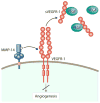
Cleavage of VEGFR-1 by MMP-14 into soluble (s)VEGFR-1. with the findings of previous studies, which showed that soluble VEGFR-1, generated by alternative splicing, can bind free VEGF to inhibit VEGF-induced proliferation in endothelial cells and trap free VEGF to ultimately preserve avascularity in the cornea .
Figure 6
Figure 6
As a result of corneal epithelial and stromal injury, bFGF mediates fibroblast activation, whereas stromal fibroblast MMP-14 initiates enzymatic activity. bFGF-mediated fibroblasts and stromal fibroblasts show upregulation of VEGF, and MMP-14 also mediates the degradation of ECM. Both upregulation of VEGF and ECM degradation enhance vascular endothelial cell proliferation, migration, and tube formation.
Figure 7
Figure 7
In the absence of mobilizing stimuli, such as G-CSF, proteolytic activities of MMP-14, -2, and -9 are relatively low due to inhibition by RECK. Functional membranal CD44 contributes to progenitor cell adhesion to the basement membrane components (retention). G-CSF signaling induces PI3k-mediated Akt phosphorylation, increasing MMP-14 and decreasing RECK expression. The opposed changes in MMP-14 and RECK levels result in MMP-14–mediated CD44 proteolysis as well as MMP-2 and MMP-9 secretion and activation. Collectively, these changes reduce progenitor cell retention and facilitate their egress and mobilization. [Adapted from Vagima et al. .]
Figure 8
Figure 8
Collagen upregulates MMP-14 through activation of the TGF-β/TβRI/Smad3/Snail pathway. MMP-14 can also be induced in the collagen microenvironment through theβ1-integrin/Src/Egr1 signaling pathway. [Adapted from Shields et al. with permission from Portland Press Limited.]

References

    1. Adamis AP, Meklir B, Joyce NC. In situ injury-induced release of basic-fibroblast growth factor from corneal epithelial cells. Am J Pathol. 1991;139:961–967. - PMC - PubMed
    1. Ager EI, Kozin SV, Kirkpatrick ND, et al. Blockade of MMP14 activity in murine breast carcinomas: implications for macrophages, vessels, and radiotherapy. J Natl Cancer Inst. 2015;107 - PMC - PubMed
    1. Al-Raawi D, Abu-El-Zahab H, El-Shinawi M, Mohamed MM. Membrane type-1 matrix metalloproteinase (MT1-MMP) correlates with the expression and activation of matrix metalloproteinase-2 (MMP-2) in inflammatory breast cancer. Int J Clin Exp Med. 2011;4:265–275. - PMC - PubMed
    1. Albuquerque RJ, Hayashi T, Cho WG, et al. Alternatively spliced vascular endothelial growth factor receptor-2 is an essential endogenous inhibitor of lymphatic vessel growth. Nat Med. 2009;15:1023–1030. - PMC - PubMed
    1. Alfranca A, Lopez-Oliva JM, Genis L, et al. PGE2 induces angiogenesis via MT1-MMP-mediated activation of the TGFbeta/Alk5 signaling pathway. Blood. 2008;112:1120–1128. - PubMed

Publication types

Substances