. 2016 Mar;25(3):597-604.
doi: 10.1002/pro.2853. Epub 2015 Dec 21.

NMR characterization of a 72 kDa transcription factor using differential isotopic labeling

Affiliations

NMR characterization of a 72 kDa transcription factor using differential isotopic labeling

Sulakshana P Mukherjee et al. Protein Sci. 2016 Mar.

Abstract

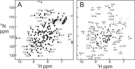
NF-κB is a major transcription factor that mediates a number of cellular signaling pathways. Crystal structure analysis gives an incomplete picture of the behavior of the protein, particularly in the free state; free monomers or dimers of NF-κB have never been crystallized. NMR analysis gives insights into the structure and dynamics of the protein in solution, but a necessary first step is the assignment of resonances. The size of the heterodimer of the Rel homology regions of the NF-κB monomers p65 and p50 (72 kDa) prohibits the straightforward use of triple-resonance spectroscopy to obtain the assignments. However, the dynamic nature of the free heterodimer, in particular the independence of the DNA-binding and dimerization domains of each monomer, allows the assignments made on differentially labeled smaller domains to be mapped successfully onto the spectrum of the larger full-length RHR. Problematic areas such as the p65 nuclear localization sequence, which is disordered in the free protein, can be approached by residue-specific labeling and comparison with previously-published spectra of a short peptide with the same sequence. Overall, this NMR analysis of NF-κB has given valuable insights into the highly dynamic nature of the free state, which is likely to play an important role in the functional cycle of NF-κB in the cell.

Keywords: IκBα; NF-κB; NMR; dynamics; structure.


Figures

Figure 1
Figure 1
A: Schematic representation of domain organization of p65 and p50. The residue numbering corresponds to mouse NF‐κB. RHR: Rel homology region; DBD: DNA‐binding domain; dd: dimerization domain; TAD: C‐terminal trans‐activation domain; NLS: nuclear localization sequence. B: Backbone representations of X‐ray crystal structures of the p65/p50 RHR heterodimer in complex with (left, PDB 1VKX) the κB DNA sequence and (middle, PDB 1NFI; right, PDB 1IKN) IκBα. Domains of p65 are colored the same as in part A. The domains of p50 (DBD and dd on the left, dd alone on the middle and right structures) are colored gray, signifying that these domains are deuterated and hence invisible in all of the NMR spectra reported here. The two strands of the duplex κB DNA sequence are shown in magenta and gold, and the 6 ankyrin repeats of IκBα are shown in gold.
Figure 2
Figure 2
1H–15N correlation spectra of domains of p65. A: 600 MHz HSQC spectrum of p65DBD (residues 19–191) B: 900 MHz TROSY spectrum of p65dd (residues 190–321) in complex with perdeuterated p50dd (residues 245–350). Selected assignments, obtained from 3D triple‐resonance spectra acquired with 15N, 13C‐labeled p65, are mapped onto the spectra.
Figure 3
Figure 3
A: Portion of an 800 MHz 1H–15N TROSY spectrum of 15N‐labeled, perdeuterated p65RHR (residues 19–325) in complex with perdeuterated p50RHR (residues 39–363) (black) superimposed on the 600 MHz HSQC spectrum of p65DBD (residues 19–191) (red) and the 900 MHz TROSY spectrum of p65dd (residues 190–321) in complex with deuterated p50dd (residues 245–350) (blue). Select resonances from the two domains are labeled in the corresponding colors. The complete spectra are shown in Supporting Information Figure S1. B: 600 MHz HSQC spectrum of p65DBD (black) superimposed on the HSQC spectrum of p65 DBD specifically labeled with 15N‐tyrosine (red). The tyrosine residues are labeled; the red cross peaks correspond to the tyrosines assigned in the spectra of the uniformly 15N‐labeled protein. C: 900 MHz TROSY spectrum of p65dd (residues 190–321) in complex with deuterated p50dd (residues 245–350) (black) superimposed on the HSQC spectrum of p65dd specifically labeled with 15N‐tyrosine and 15N‐methionine in complex with deuterated p50dd (residues 245–350) (red). The tyrosine and methionine residues are labeled; the cross peaks of Y227, M270, M284, and Y288 correspond to those assigned in the spectra of the uniformly‐labeled protein. The assignments of Y257, Y306, and M313 were made using information from the spectrum of the specifically‐labeled protein.
Figure 4
Figure 4
A: 900 MHz TROSY spectrum of p65dd in complex with perdeuterated p50dd (black) superimposed on the spectrum of p65dd in complex with perdeuterated p50dd and perdeuterated IκBα (residues 67–287) (red). Significantly shifted resonances are labeled in green, and those with lesser shifts in blue. Residues for which the shifted resonances in the IκBα complex were identified from those of the NLS peptide complex15 are labeled in magenta. Residues for which resonances could be identified in the free p65dd/p50dd spectrum but which did not show readily‐identifiable resonances in the IκBα‐bound spectrum are labeled in black. B: X‐ray crystal structure of the complex of p65RHR/p50dd with IκBα (1NFI)7 (the p65DBD has been omitted for clarity). The backbone of IκBα is shown in gold, the backbone of p65dd in blue and the backbone of p50dd in gray. Residues with large chemical shift changes upon addition of IκBα, as shown in blue and green in part A, are mapped onto the structure in red. Residues in the NLS, for which large chemical shift changes were inferred from the positions of the assigned cross peaks in the spectrum of the NLS peptide alone15 are shown in magenta.

References

    1. Hoffmann A, Natoli G, Ghosh G (2006) Transcriptional regulation via the NF‐kB signaling module. Oncogene 25:6706–6716. - PubMed
    1. Beg AA, Baldwin AS, Jr (1993) The IkB proteins: multifunctional regulators of Rel/NF‐kB transcription factors. Genes Dev 7:2064–2070. - PubMed
    1. Beg AA, Finco TS, Nantermet PV, Baldwin AS, Jr (1993) Tumor necrosis factor and interleukin‐1 lead to phosphorylation and loss of IkBa: a mechanism for NF‐kB activation. Mol Cell Biol 13:3301–3310. - PMC - PubMed
    1. Brown K, Park S, Kanno T, Franzoso G, Siebenlist U (1993) Mutual regulation of the transcriptional activator NF‐kappa B and its inhibitor, I kappa B‐alpha. Proc Natl Acad Sci USA 90:2532–2536. - PMC - PubMed
    1. Chen FE, Huang DB, Chen YQ, Ghosh G (1998) Crystal structure of p50/p65 heterodimer of transcription factor NF‐kB bound to DNA. Nature 391:410–413. - PubMed

Publication types

MeSH terms

LinkOut - more resources