. 2016 Feb 1;25(3):558-70.
doi: 10.1093/hmg/ddv496. Epub 2015 Dec 8.

The molecular basis of variable phenotypic severity among common missense mutations causing Rett syndrome

Affiliations

The molecular basis of variable phenotypic severity among common missense mutations causing Rett syndrome

Kyla Brown et al. Hum Mol Genet. .

Abstract

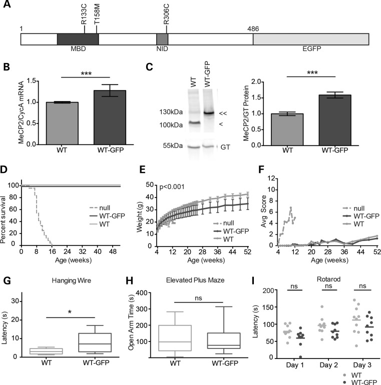
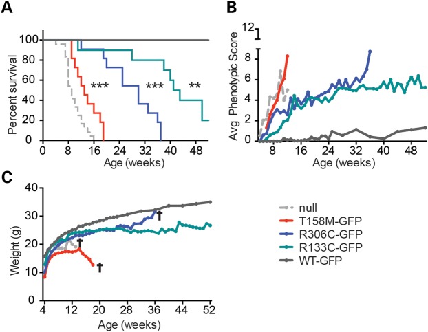
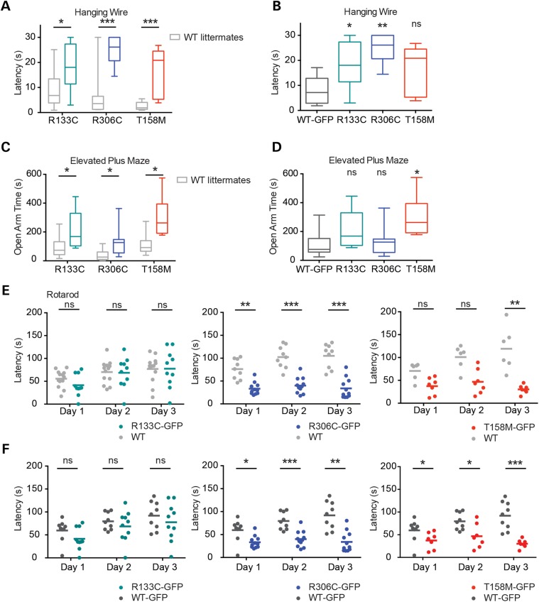
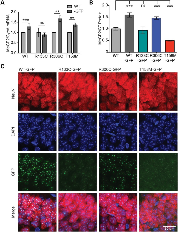
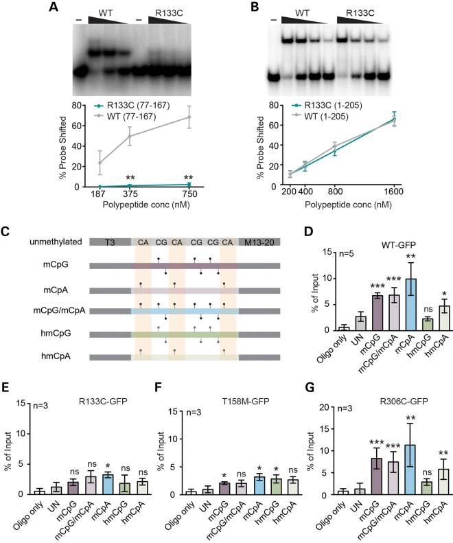
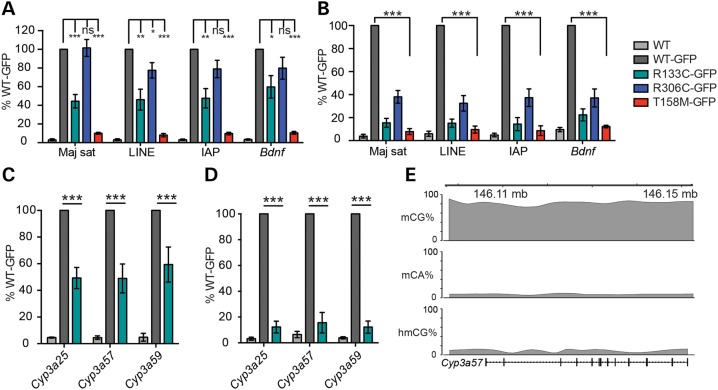
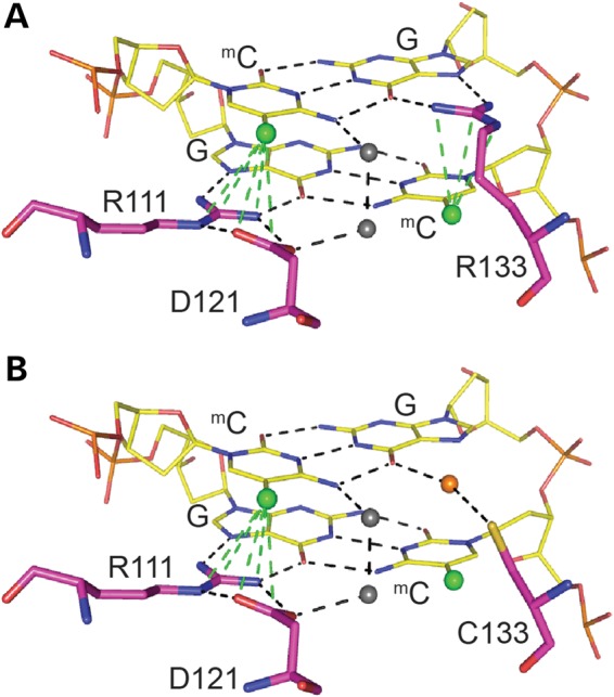
Rett syndrome is caused by mutations in the X-linked MECP2 gene, which encodes a chromosomal protein that binds to methylated DNA. Mouse models mirror the human disorder and therefore allow investigation of phenotypes at a molecular level. We describe an Mecp2 allelic series representing the three most common missense Rett syndrome (RTT) mutations, including first reports of Mecp2[R133C] and Mecp2[T158M] knock-in mice, in addition to Mecp2[R306C] mutant mice. Together these three alleles comprise ∼25% of all RTT mutations in humans, but they vary significantly in average severity. This spectrum is mimicked in the mouse models; R133C being least severe, T158M most severe and R306C of intermediate severity. Both R133C and T158M mutations cause compound phenotypes at the molecular level, combining compromised DNA binding with reduced stability, the destabilizing effect of T158M being more severe. Our findings contradict the hypothesis that the R133C mutation exclusively abolishes binding to hydroxymethylated DNA, as interactions with DNA containing methyl-CG, methyl-CA and hydroxymethyl-CA are all reduced in vivo. We find that MeCP2[T158M] is significantly less stable than MeCP2[R133C], which may account for the divergent clinical impact of the mutations. Overall, this allelic series recapitulates human RTT severity, reveals compound molecular aetiologies and provides a valuable resource in the search for personalized therapeutic interventions.


Figures

Figure 1.
Figure 1.
WT-GFP mice show minimal overt phenotype. (A) Schematic representation of MeCP2 with an EGFP tag. Missense mutations analysed in this study (T158M, R133C and R306C) are shown in relation to the MBD and NID. (B) Levels (mean ± SD) of Mecp2 transcripts in WT-GFP mouse brain (n = 9) compared with WT littermates (n = 9), expressed relative to Cyclophilin A transcript (CycA). (C) Representative western blot and quantification comparing MeCP2 protein abundance in WT-GFP mouse brain (double arrow-head, n = 6) versus WT littermates (single arrow-head, n = 6). Gamma tubulin (GT) served as an internal control. Mean ± SEM plotted. (D) The Kaplan–Meyer plots showing survival of WT-GFP mice (n = 8) compared with their WT littermates (n = 8) and Mecp2-null mice [Ref. (18)] (n = 24). (E) Growth curve showing average weight of WT-GFP mice (n = 8) compared with their WT littermates (n = 8). Using repeated measures ANOVA, the difference was consistent and significant (P < 0.001) over time, but at any single time point, the difference was not significant. (F) Phenotypic scoring (see the text) of WT-GFP or WT mice. For comparison, Mecp2-null scoring is shown. (GI) WT-GFP mice (n = 9) and their WT littermates (n = 10) were compared using three behavioural tests: (G) the hanging-wire test, (H) the elevated plus maze, and (I) the accelerating rotarod showing individual mean latencies (dots) and cohort mean latency (line) for each of three days of trials. Statistical analysis took all trials into account. Statistical tests were unpaired two-tailed t-test (B, C and E) and the Kolmogorov–Smirnov test (G–I). All behavioural paradigms were conducted on animals aged 8–10 weeks, and biochemical analyses were conducted using tissues from adults aged 6–12 weeks. Asterisks denote the following P values: *P < 0.05, **P < 0.01 and ***P < 0.001.
Figure 2.
Figure 2.
The Mecp2-GFP allelic series mimics the clinical severity of equivalent human mutations. (A) The Kaplan–Meyer plots showing reduced survival of T158M-GFP (red), R306C-GFP (blue) and R133C-GFP (green) mutants in comparison with WT-GFP (dark grey) and Mecp2-null (light grey) mice (18). Statistical significance is denoted as follows: *P < 0.05, **P < 0.01 and ***P < 0.001 (Mantel-Cox test) and calculated in comparison with the WT-GFP mice. (B) Graph showing average phenotypic score for each cohort over time. (C) Graph showing average weight over time. Cohorts comprised WT-GFP (n = 8), R133C-GFP (n = 10), R306C-GFP (n = 11), T158M-GFP (n = 11) and Mecp2-null mice (n = 12–24). A cross indicates that all mice in the cohort had been culled by this time point due to severity of the RTT-like phenotype.
Figure 3.
Figure 3.
Behavioural analysis of the Mecp2-GFP allelic series indicates that R133C-GFP mice are less severely affected than R306C-GFP and T158M-GFP. Mutant males and WT male littermates underwent behavioural analysis as in Figure 1. (A) Hanging-wire test, (C) elevated plus maze and (E) accelerating rotarod. R133C-GFP (n = 10, green) plus WT littermates (n = 13, grey); R306C-GFP (n = 10–11, blue) plus WT littermates (n = 9, grey); T158M-GFP (n = 7, red) plus WT littermates (n = 6, grey). B, D and F show comparisons between mutant males and WT-GFP males in the same series of tests. (E and F) Graphs showing mean time to descent for individuals (dots) and cohorts (lines) for each trial day. Statistical analysis took all trials into account, and significance is denoted as follows: *P < 0.05, **P < 0.01 and ***P < 0.001 (Kolmogorov–Smirnov test). All behavioural paradigms were conducted on animals aged 8–10 weeks.
Figure 4.
Figure 4.
R133C-GFP and T158M-GFP are less abundant than WT-GFP and have an abnormal pattern of sub-nuclear localization. (A) Graph showing level of Mecp2 transcript normalized to Cyclophilin A in mutant male mouse brains relative to WT littermates (mean ± SD). WT-GFP, n = 9; R133C-GFP, n = 4; R306C-GFP, n = 3; T158M-GFP, n = 3. (B) Quantification of western blots showing levels of WT-GFP, R306C-GFP, R133C-GFP and T158M-GFP protein in male mouse brain, relative to WT (mean ± SEM). GT served as an internal control; WT, n = 6; WT-GFP, n = 6; R133C-GFP, n = 3; R306C-GFP, n = 4; T158M-GFP, n = 6. (C) Representative images of the CA1 region of the hippocampus in adult male mice from the allelic series. Slices were imaged using the same confocal settings. Immunofluorescence was performed with DAPI (blue) and anti-NeuN (red). MeCP2 was imaged by virtue of its EGFP tag. R133C-GFP and T158M-GFP show mixed punctate and diffuse sub-nuclear localization. Scale bar = 20 μm. Statistical significance is denoted as follows: *P < 0.05, **P < 0.01 and ***P < 0.001 (two-tailed unpaired t-test). All biochemical analyses were conducted using tissues from adults aged 6–12 weeks.
Figure 5.
Figure 5.
Defective binding of MeCP2[R133C] and T158M to modified DNA in vitro and in vivo. Representative gels and quantification of EMSAs measuring MeCP2 peptide binding to mCG probes using (A) aa77-167 or (B) aa1-205 fragments of MeCP2 with (green) or without (grey) the R133C mutation. Mean percentage of probe shifted ± SEM is plotted from three replicated experiments. Triangle represents decreasing peptide concentration; –, no peptide. Statistical significance is denoted as: *P < 0.05, **P < 0.01 and ***P < 0.001 (unpaired two-tailed t-test). (C) ChIP oligonucleotide duplex probes containing symmetrical mCG or hmCG, or asymmetrical mCA or hmCA sites or both mCA and mCG sites together. Control DNA lacks all modifications. (DG) Results of five independent transient transfection experiments followed by ChIP of MeCP2-associated probe oligonucleotide fragments. Cells transiently expressed WT-GFP, R133C-GFP, T158M-GFP and R306C-GFP, respectively. Mean % input ± SD plotted (n ≥ 3).
Figure 6.
Figure 6.
Chromatin immunoprecipitation reveals abnormal binding of R133C-GFP to mCG-rich repetitive sequences and genes in mouse brain. (A) ChIP-qPCR of WT-MeCP2 (lacking a GFP tag) and knocked-in WT-GFP, R133C-GFP, R306C-GFP and T158M-GFP in the adult male mouse brain. Primers amplify, respectively, major satellite, LINE and IAP transposable elements and the Bdnf gene locus. The antibody was against GFP and therefore does not precipitate untagged WT MeCP2 (n ≥ 3). (B) Similar assay to (A) but with a lower and therefore more stringent cross-linking temperature (n = 4). (C) Column plot showing binding of R133C-GFP at the mCG-rich cytochrome p450 locus by GFP ChIP (n = 3). (D) The same assay as (C) but using a more stringent cross-linking temperature. Plots are expressed as % WT-GFP value. Error bars represent ± SEM, and statistical significance is denoted as: *P < 0.05, **P < 0.01 and ***P < 0.001 (unpaired, two-tailed t-test). Each replicate experiment used tissue from a separate animal. (E) Schematic of the Cyp3a57 locus depicting cytosine methylation context. All biochemical analyses were conducted using tissues from adults aged 6–12 weeks.
Figure 7.
Figure 7.
Predictive modelling of MeCP2 MBD binding to methylated DNA reveals a loss of specificity with the R133C mutation. (A) Predicted model of MBD binding to a methylated CpG pair. Arginine 111 and 133 make contact with the guanines and form hydrogen bonds with the methylcytosines of the CpG pair. Arginine 111 forms a hydrogen bond ‘clip’ with aspartic acid 121. (B) Predicted model of MBD[R133C] binding to a methylated CpG pair. The cysteine 133 interaction with guanine is now dependent on water molecules, and there is no significant interaction with the methylcytosine. (A and B) Hydrogen bonds are indicated by dotted lines, methyl groups by green balls, and water molecules by grey and orange balls.

References

    1. Amir R.E., Van den Veyver I.B., Wan M., Tran C.Q., Francke U., Zoghbi H.Y. (1999) Rett syndrome is caused by mutations in X-linked MECP2, encoding methyl-CpG-binding protein 2. Nature Genet., 23, 185–188. - PubMed
    1. Meins M., Lehmann J., Gerresheim F., Herchenbach J., Hagedorn M., Hameister K., Epplen J.T. (2005) Submicroscopic duplication in Xq28 causes increased expression of the MECP2 gene in a boy with severe mental retardation and features of Rett syndrome. J Med Genet., 42, e12. - PMC - PubMed
    1. Guy J., Hendrich B., Holmes M., Martin J.E., Bird A. (2001) A mouse Mecp2-null mutation causes neurological symptoms that mimic Rett syndrome. Nat Genet, 27, 322–326. - PubMed
    1. Chen R.Z., Akbarian S., Tudor M., Jaenisch R. (2001) Deficiency of methyl-CpG binding protein-2 in CNS neurons results in a Rett-like phenotype in mice. Nat Genet, 27, 327–331. - PubMed
    1. Brendel C., Belakhov V., Werner H., Wegener E., Gartner J., Nudelman I., Baasov T., Huppke P. (2011) Readthrough of nonsense mutations in Rett syndrome: evaluation of novel aminoglycosides and generation of a new mouse model. J Mol Med (Berl), 89, 389–398. - PMC - PubMed

Publication types

MeSH terms