. 2016 Aug;27(8):2346-56.
doi: 10.1681/ASN.2015050515. Epub 2015 Dec 8.

Macula Densa Nitric Oxide Synthase 1β Protects against Salt-Sensitive Hypertension

Affiliations

Macula Densa Nitric Oxide Synthase 1β Protects against Salt-Sensitive Hypertension

Yan Lu et al. J Am Soc Nephrol. 2016 Aug.

Abstract

Nitric oxide (NO) is an important negative modulator of tubuloglomerular feedback responsiveness. We recently found that macula densa expresses α-, β-, and γ-splice variants of neuronal nitric oxide synthase 1 (NOS1), and NOS1β expression in the macula densa increases on a high-salt diet. This study tested whether upregulation of NOS1β expression in the macula densa affects sodium excretion and salt-sensitive hypertension by decreasing tubuloglomerular feedback responsiveness. Expression levels of NOS1β mRNA and protein were 30- and five-fold higher, respectively, than those of NOS1α in the renal cortex of C57BL/6 mice. Furthermore, macula densa NO production was similar in the isolated perfused juxtaglomerular apparatus of wild-type (WT) and nitric oxide synthase 1α-knockout (NOS1αKO) mice. Compared with control mice, mice with macula densa-specific knockout of all nitric oxide synthase 1 isoforms (MD-NOS1KO) had a significantly enhanced tubuloglomerular feedback response and after acute volume expansion, significantly reduced GFR, urine flow, and sodium excretion. Mean arterial pressure increased significantly in MD-NOS1KO mice (P<0.01) but not NOS1flox/flox mice fed a high-salt diet. After infusion of angiotensin II, mean arterial pressure increased by 61.6 mmHg in MD-NOS1KO mice versus 32.0 mmHg in WT mice (P<0.01) fed a high-salt diet. These results indicate that NOS1β is a primary NOS1 isoform expressed in the macula densa and regulates the tubuloglomerular feedback response, the natriuretic response to acute volume expansion, and the development of salt-sensitive hypertension. These findings show a novel mechanism for salt sensitivity of BP and the significance of tubuloglomerular feedback response in long-term control of sodium excretion and BP.

Keywords: NOS1; hypertension; macula densa; salt-sensitive; tubuloglomerular feedback.


Figures

Figure 1.
Figure 1.
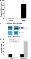
Expression of NOS1 splice variants in the macula densa of C57BL/6 mice. The macula densa cells were isolated with LCM, and mRNA was measured with real-time PCR. (A) NOS1β level was about 32-fold higher than NOS1α (n=7). Protein levels of NOS1 splice variants were measured with Western blot. (B and C) In the brain, NOS1β is <5% of NOS1α; however, in renal cortex, NOS1β is more than fivefold higher than NOS1α (n=5). *P<0.01 versus NOS1α.
Figure 2.
Figure 2.
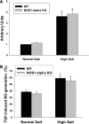
Function of NOS1 splice variants in the macula densa. The NOS1β mRNA was measured with real-time PCR and compared between NOS1αKO and C57BL/6 WT mice. (A) There were no differences in NOS1β mRNA levels in the macula densa from mice fed both normal- (0.4% NaCl) and high-salt diets (4% NaCl; n=7). (B) NO generation by the macula densa was measured in the isolated perfused JGA with DAF-FM DA. There were no differences in NO generation by the macula densa from mice fed both normal- and high-salt diets (n=6). *P<0.01 versus normal-salt diet. TGF, tubuloglomerular feedback.
Figure 3.
Figure 3.
Generation of NKCC2-Cre mouse strain. To characterize NKCC2-Cre mice that we developed, we generated NKCC2-Cre-Rosa mice by crossing with 29-Gt-Rosa-26Sortm2Sho/J mice. Kidney slices were labeled with a primary antibody against GFP (to identify Cre) or NKCC2 (to identify TAL and macula densa) and fluorescent secondary antibodies. (Aa) and (Ab) show typical transition positions in medulla from TALs that express NKCC2 (marked with yellow arrows) to thin ascending limbs that do not express NKCC2 (marked with blue arrows). (Aa) Was probed with an NKCC2 antibody. (Ab) Was probed with a GFP antibody. (Ac) Shows GFP staining in macula densa and TAL in renal cortex. (B) Immunofluorescence staining against GFP in isolated perfused JGA of NKCC2-Cre-Rosa mice. GFP was strongly expressed in the macula densa. MD, macula densa.
Figure 4.
Figure 4.
Development of tissue–specific NOS1KO mice. MD-NOS1KO mice were generated by crossing NKCC2-Cre mice with NOS1tm2Plhflox mice. (Aa) NOS1 was strongly stained in a kidney slice of a littermate of a C57BL/6 WT mouse with a C–terminal primary antibody, whereas (Ab) there was no staining of NOS1 in the macula densa in a kidney slice of an MD-NOS1KO mouse. (B) NO generation by the macula densa was absent at both 10 and 80 mM NaCl in the MD-NOS1KO mice. *P<0.01 versus 10 mM NaCl (n=5); **P<0.01 versus WT (n=5). (C) No apparent abnormalities in size and structure of the JGA from a KO mouse. Left and right panels show structure and constriction of an Af-Art, respectively, while NaCl concentration in tubular perfusate increased from 10 to 80 mM; the center panel shows an intact macula densa plaque of the same perfused JGA at a different focus level. (D) Tubuloglomerular feedback response was enhanced both (Da) in vitro measured in isolated perfused JGA (n=7) and (Db) in vivo measured with micropuncture (n=6). *P<0.01 versus WT. G, glomerulus; MD, macula densa; TGF, tubuloglomerular feedback.
Figure 5.
Figure 5.
Renal clearance function in response to acute volume expansion. Basal GFR and sodium excretion rate were measured in MD-NOS1KO mice and NOS1flox/flox mice as controls. Renal clearance function was measured during 0–60 and 60–90 minutes after acute volume expansion by infusion of 3% body wt of 0.9% NaCl saline. (A) GFR, (B) urinary flow, (C) Na excretion, and (D) K excretion rate were measured. Numbers in parentheses indicate numbers of mice studied per group. *P<0.01 versus control; #P<0.05 versus NOS1flox/flox mice. VE, volume expansion.
Figure 6.
Figure 6.
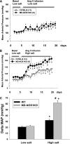
BP response to a high-salt diet. The mice were fed a low-salt diet (0.1% NaCl) for 10 days and then, switched to a high-salt diet (4% NaCl) for 2 weeks. MAP was measured by telemetry in MD-NOS1KO and NOS1flox/flox mice. Numbers in parentheses indicate numbers of mice studied per group. *P<0.01 versus low-salt control; #P<0.01 versus NOS1flox/flox.
Figure 7.
Figure 7.
BP response to subpressor AngII infusion plus a high-salt diet. The mice were fed a low-salt diet (0.1% NaCl), and basal MAP was measured in MD-NOS1KO and littermates of C57BL/6 WT mice. Then, a subpressor AngII (600 ng/min per kilogram) was infused. (A) One group of mice was continually fed a low-salt diet, whereas (B) another group of mice was switched to a high-salt diet (4% NaCl) and (C) comparison of the delta change of MAP in these two groups of mice. Numbers in parentheses indicate numbers of mice studied per group. *P<0.05 versus low salt; #P<0.01 versus WT.

Comment in

References

    1. Goormaghtigh N: Les segments neuro-myo-artériels juxtaglomérulaires du rein. Arch Biol (Liege) 43: 575–591, 1932
    1. Zimmerman KW: Ueber den bau des glomerulus der saeugerniere. Z Microskop Anat Forsch 32: 176–287, 1933
    1. Thurau K, Schnermann J: The sodium concentration in the macula densa cells as a regulating factor for glomerular filtration (micropuncture experiments). Klin Wochenschr 43: 410–413, 1965 - PubMed
    1. Liu R, Pittner J, Persson AE: Changes of cell volume and nitric oxide concentration in macula densa cells caused by changes in luminal NaCl concentration. J Am Soc Nephrol 13: 2688–2696, 2002 - PubMed
    1. Schnermann J, Briggs JP: The macula densa is worth its salt. J Clin Invest 104: 1007–1009, 1999 - PMC - PubMed

Publication types

MeSH terms

Substances