. 2016 Sep;9(5):1278-87.
doi: 10.1038/mi.2015.129. Epub 2015 Dec 9.

Aspirin-triggered resolvin D1 is produced during self-resolving gram-negative bacterial pneumonia and regulates host immune responses for the resolution of lung inflammation

Affiliations

Aspirin-triggered resolvin D1 is produced during self-resolving gram-negative bacterial pneumonia and regulates host immune responses for the resolution of lung inflammation

R E Abdulnour et al. Mucosal Immunol. 2016 Sep.

Abstract

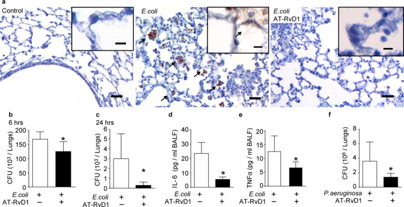
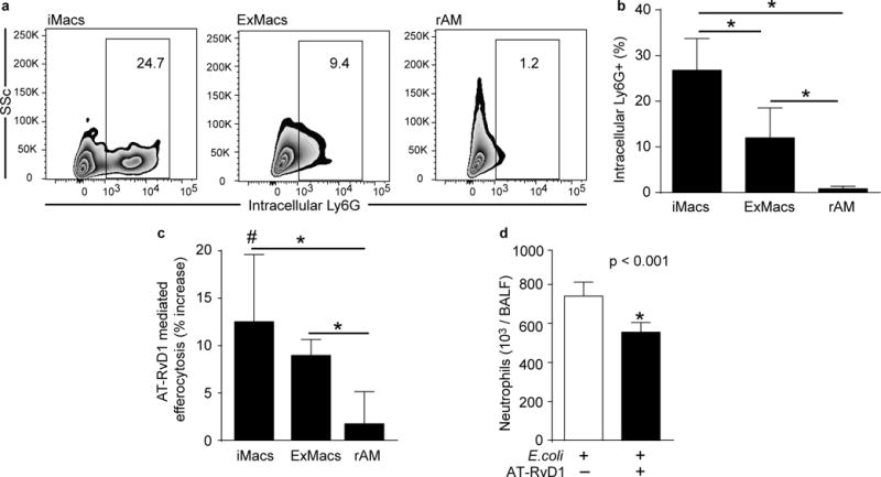
Bacterial pneumonia is a leading cause of morbidity and mortality worldwide. Host responses to contain infection and mitigate pathogen-mediated lung inflammation are critical for pneumonia resolution. Aspirin-triggered resolvin D1 (AT-RvD1; 7S,8R,17R-trihydroxy-4Z,9E,11E,13Z,15E,19Z-docosahexaenoic acid) is a lipid mediator (LM) that displays organ-protective actions in sterile lung inflammation, and regulates pathogen-initiated cellular responses. Here, in a self-resolving murine model of Escherichia coli pneumonia, LM metabololipidomics performed on lungs obtained at baseline, 24, and 72 h after infection uncovered temporal regulation of endogenous AT-RvD1 production. Early treatment with exogenous AT-RvD1 (1 h post infection) enhanced clearance of E. coli and Pseudomonas aeruginosa in vivo, and lung macrophage phagocytosis of fluorescent bacterial particles ex vivo. Characterization of macrophage subsets in the alveolar compartment during pneumonia identified efferocytosis by infiltrating macrophages (CD11b(Hi) CD11c(Low)) and exudative macrophages (CD11b(Hi) CD11c(Hi)). AT-RvD1 increased efferocytosis by these cells ex vivo, and accelerated neutrophil clearance during pneumonia in vivo. These anti-bacterial and pro-resolving actions of AT-RvD1 were additive to antibiotic therapy. Taken together, these findings suggest that the pro-resolving actions of AT-RvD1 during pneumonia represent a novel host-directed therapeutic strategy to complement the current antibiotic-centered approach for combatting infections.


Conflict of interest statement

declaration: C.N.S. is an inventor on patents [resolvins] assigned to BWH and licensed to Resolvyx Pharmaceuticals. C.N.S. was scientific founder of Resolvyx Pharmaceuticals and owns founder stock in the company. C.N.S.’ interests were reviewed and are managed by the Brigham and Women’s Hospital and Partners HealthCare in accordance with their conflict of interest policies. B.D.L. is an inventor on patents [resolvins] assigned to BWH and licensed to Resolvyx Pharmaceuticals. B.D.L.’s interests were reviewed and are managed by the Brigham and Women’s Hospital and Partners HealthCare in accordance with their conflict of interest policies. The remaining authors declare that they have no competing interests or other interests that might be perceived to influence the results and/or discussion reported in this manuscript.

Figures

Figure 1
Figure 1. Endogenous AT-RvD1 is produced during bacterial pneumonia
(a) Time course of murine lung CFU, and BAL neutrophils and macrophages, at indicated time points after infection (E. coli, 106 CFU, IB). Results expressed as mean ± SD, n > 5 mice in two independent experiments, * P < 0.05 vs 0 and 72 hrs. (b–f) Lipids were extracted from murine lungs 0, 24, and 72 hrs after infection and LM-SPM profiles were obtained using LC-MS/MS metabololipidomics as in Methods. (b) Two-dimensional score plot and (c) loading plot of a principal component analysis of murine LM-SPM signature profiles. The white ellipse in the score plot denotes 95% confidence regions. (d) Representative multiple reaction monitoring (MRM) chromatogram (m/z = 375>141) and (e) MS/MS spectrum, obtained from infected murine lung, used for the identification of AT-RvD1 (7S,8R,17R-trihydroxy-docosa-4Z,9E,11E,13Z,15E,19Z-hexaenoic acid). (f) AT-RvD1 levels in murine lungs during pneumonia (n = 4 per time point, mean ± SD, * P < 0.05 vs 0 hrs).
Figure 2
Figure 2. AT-RvD1 enhances bacterial clearance
(a) Representative lung sections from control mice, and 24 hrs after infection and treatment with AT-RvD1 or vehicle stained with an anti-E. coli antibody. Arrowheads indicate E. coli, bar = 50 μm, inset bar = 10 μm. Lung E. coli CFU counts (b) 6 hrs and (c) 24 hrs after infection (106 CFU, IB) in mice treated with AT-RvD1 or vehicle. Levels of (d) IL-6 and (e) TNF-α 6 hrs after infection determined by bead-based immunoassay. (f) Lung P. aeruginosa CFU counts 6 hrs after infection (106 CFU, IB) in mice treated with AT-RvD1 or vehicle. In all experiments, AT-RvD1 (100 ng, IV) or vehicle (< 0.1% EtOH in saline vol/vo, IV) were given 1 hr after E. coli. Results are expressed as mean ± SD n = 4–7 per group. * P < 0.05
Figure 3
Figure 3. AT-RvD1 increases bacterial phagocytosis by CD11c+ lung cells
Ultra-thin perfused lung sections were obtained from naïve mice, stained with anti-CD11c, and treated with AT-RvD1 (10 nM, 45 minutes, 37°C) or vehicle. Sections were then co-incubated with E. coli particles labelled with a pH-sensitive probe (100 μg/ml, 60 minutes, RT) and imaged using a fluorescence microscope. Particle dose was chosen to give minimal phagocytosis in vehicle treated sections. Representative images of (a) control and (b) AT-RvD1 treated lung sections. (c) Proportion of CD11c+ lung cells participating in phagocytosis of E. coli particles. * P < 0.05. (d) Lipocalin 2 levels in BAL fluid 6 hrs after E. coli infection determined by ELISA. Results are expressed as mean ± SD n = > 8 sections per group in two independent experiments. * P < 0.05 vs vehicle treated un-infected mice, # P < 0.05 vs vehicle treated infected mice.
Figure 4
Figure 4. Macrophage subsets during E. coli pneumonia
BAL was obtained from mice at determined time-points after E. coli infection (106 CFU, IB) for macrophage subset enumeration by flow cytometry. (a) Representative flow cytometry dot plots from BAL gated on macrophages (SScHi CD45+ F4/80+). (b) Identification of macrophage subsets based on CD11b and CD11c surface expression as infiltrating (iMacs, CD11cLow CD11bHi), exudative (ExMacs, CD11cHi CD11bHi), and resident Alveolar Macrophages (rAM, CD11cHi CD11b). Time course of BAL macrophage subset (c) fractions and (d) counts. Results are expressed as mean ± SD. n = 10 mice per group from 2 independent experiments, * P < 0.05
Figure 5
Figure 5. Recruited macrophages participate in efferocytosis of neutrophils during resolution of pathogen-mediated inflammation
(a) Representative dot plots and (b) bar-graph of intracellular Ly6G staining gated on macrophage subsets in BAL obtained 48 hrs after infection. Results are expressed as mean ± SD. n = 4 sections per group in two independent experiments. (c) Percent increase in efferocytosis of apoptotic neutrophils by macrophage subsets after treatment with AT-RvD1 compared to vehicle (10nM, 30 min, 37°C). Macrophage and apoptotic neutrophil preparations are detailed in Methods. Results are expressed as mean ± SD. n = > 10 per group. ♯ P < 0.05 vs vehicle treated (d) Neutrophil count in BAL obtained from mice 48 hrs after high-dose infection (E. coli, 2×10⁶ CFU, IB) and treatment with AT-RvD1 (100ng, IB) 24 and 36 hrs after E. coli. Results are expressed as mean ± SD. n = 10 mice per group from 2 independent experiments. In all figures, * P < 0.05
Figure 6
Figure 6. AT-RvD1 enhances bacterial clearance in combination with antibiotics
(a) Lung CFU counts and (b) BAL fluid LPS concentrations (Endotoxin Units/ml BAL fluid) 6 hrs after E. coli infection, and (c) BALF neutrophils and (d) BALF macrophage to neutrophil ratio (a resolution index) 48 hrs after infection with E. coli (106 CFU, IB) in mice treated with AT-RvD1 (100 ng, IV, 1 hr after infection) or vehicle (< 0.1% EtOH in saline vol/vol), with or without ciprofloxacin (0.2 mg/kg, IP, 1 hr after infection). Results are expressed as mean ± SD. n = 5–7 per group. * P < 0.05 (e) Representative hematoxylin and eosin stains of murine lungs 48 hrs after infection. n = 3 per group.

References

    1. Serhan CN. Resolution phase of inflammation: novel endogenous anti-inflammatory and proresolving lipid mediators and pathways. Annual review of immunology. 2007;25:101–137. - PubMed
    1. Matthay MA, Ware LB, Zimmerman GA. The acute respiratory distress syndrome. J Clin Invest. 2012;122(8):2731–2740. - PMC - PubMed
    1. Serhan CN, Hong S, Gronert K, Colgan SP, Devchand PR, Mirick G, et al. Resolvins: A Family of Bioactive Products of Omega-3 Fatty Acid Transformation Circuits Initiated by Aspirin-Treatment that Counter Proinflammation Signals. J Exp Med. 2002;196(8):1025–1037. - PMC - PubMed
    1. Sun YP, Oh SF, Uddin J, Yang R, Gotlinger K, Campbell E, et al. Resolvin D1 and its aspirin-triggered 17R epimer. Stereochemical assignments, anti-inflammatory properties, and enzymatic inactivation. J Biol Chem. 2007;282(13):9323–9334. - PubMed
    1. Serhan CN, Petasis NA. Resolvins and protectins in inflammation resolution. Chemical reviews. 2011;111(10):5922–5943. - PMC - PubMed

MeSH terms