Review
. 2016 Jul;283(14):2577-98.
doi: 10.1111/febs.13616. Epub 2015 Dec 31.

Protease signaling in animal and plant-regulated cell death

Affiliations
Review

Protease signaling in animal and plant-regulated cell death

Guy S Salvesen et al. FEBS J. 2016 Jul.

Abstract

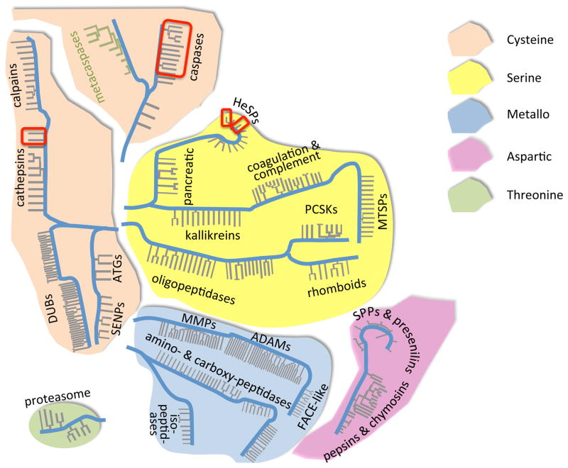
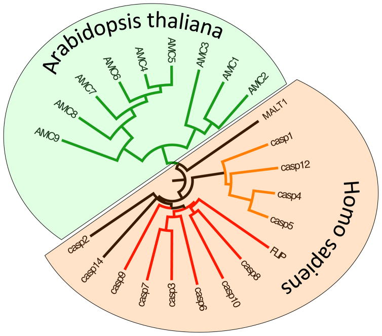
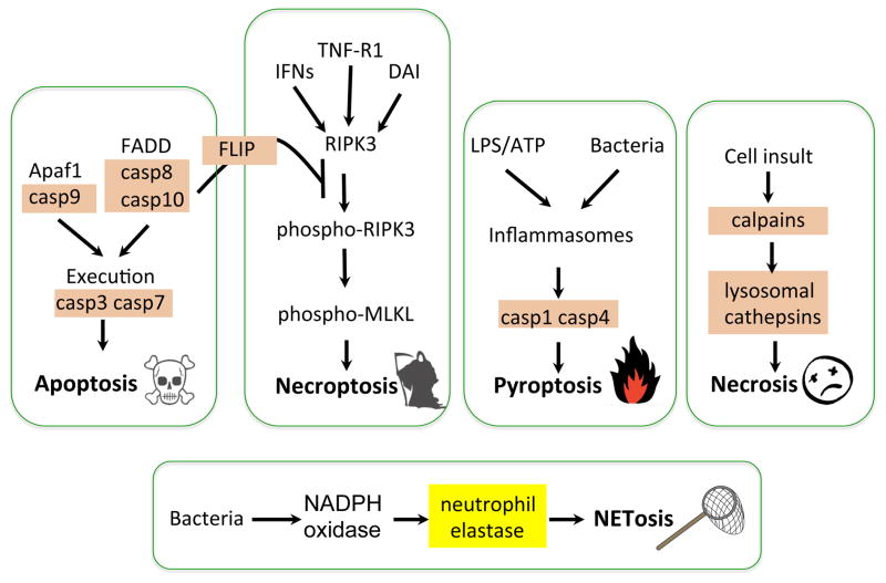
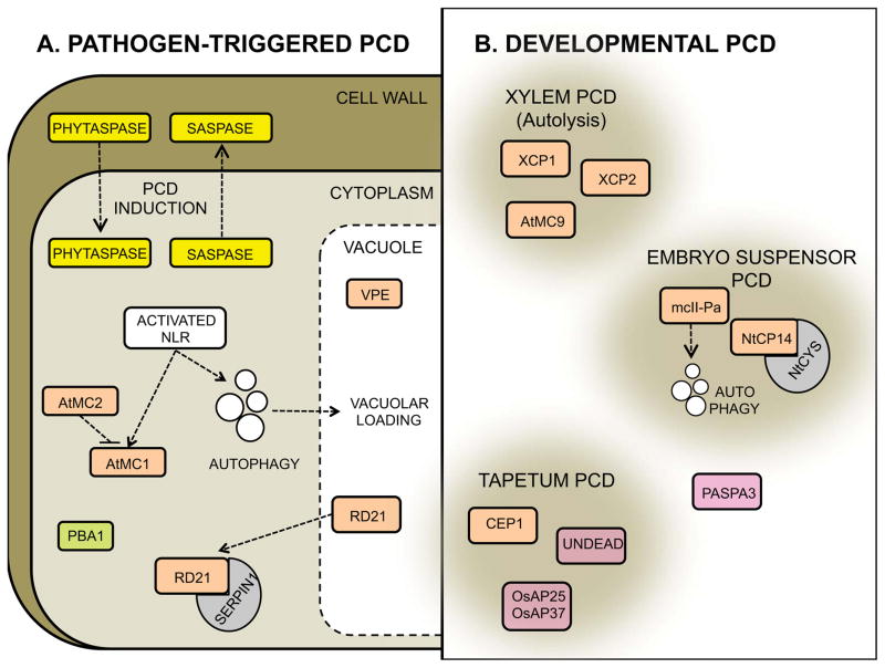
This review aims to highlight the proteases required for regulated cell death mechanisms in animals and plants. The aim is to be incisive, and not inclusive of all the animal proteases that have been implicated in various publications. The review also aims to focus on instances when several publications from disparate groups have demonstrated the involvement of an animal protease, and also when there is substantial biochemical, mechanistic and genetic evidence. In doing so, the literature can be culled to a handful of proteases, covering most of the known regulated cell death mechanisms: apoptosis, regulated necrosis, necroptosis, pyroptosis and NETosis in animals. In plants, the literature is younger and not as extensive as for mammals, although the molecular drivers of vacuolar death, necrosis and the hypersensitive response in plants are becoming clearer. Each of these death mechanisms has at least one proteolytic component that plays a major role in controlling the pathway, and sometimes they combine in networks to regulate cell death/survival decision nodes. Some similarities are found among animal and plant cell death proteases but, overall, the pathways that they govern are kingdom-specific with very little overlap.

Keywords: NETosis; apoptosis; caspase; cathepsin; metacaspase; necroptosis; necrosis; peptidase; proteolysis; pyroptosis.


Figures

Fig. 1
Fig. 1
Human Protease Landscape. Protease catalytic types come from unrelated evolutionary origins. The intent of this map is to show the diversity of human proteases, based on phylogenetic relationships of most human protease catalytic domains collected from the MEROPS database. Family relationships are gathered by the grey lines, and evolutionary divergence by the blue mainlines (not to an evolutionary scale). Within a catalytic class some families and clans have no common origin – shown by disconnected blue mainlines. The thickness and spacing of the grey lines does not signify anything, and is just a device to cluster some protease families with a large number of members. A. thaliana metacaspases (green) are overlaid on this human map to display their relation to caspases. Red boxes highlight some of the human proteases considered in this review as playing roles in regulated cell death.
Fig. 2
Fig. 2
Vertebrate and Plant clan CD Proteases. In addition to the proteases identified here, plants and mammals contain two clan CD members in common: legumain and separase (not shown). Members specific for each kingdom are human caspase family, which can be divided into apoptotic caspases (red) and inflammatory caspases (orange). We have not included caspase-2 in the apoptotic caspases, as some would do, simply because the evidence for its role is so bewildering with many conflicting reports [–185]. Caspase-14 is not an apoptotic caspase, and the closest relative to the caspases, the paracaspase MALT1 is a survival protease that operated via the NFkB pathway [186]. Two types of metacaspases can be distinguished in A. thaliana: Type I (AMC1–3) and Type II (AMC 4–9).
Fig. 3
Fig. 3
Protease Zymogen Activation Mechanisms. Schematic of most common activation mechanisms with examples covered in the review, plus other classic examples in italics. The cartoon explains the mechanism of zymogen stabilization, and the processes required to activate each example.
Fig. 4
Fig. 4
Regulated Cell Death Pathways. The schematics illustrate the involvement of cysteine proteases in most regulated cell death pathways, with the exception of NETosis, which requires the serine protease neutrophil elastase. Different sets of caspases are important for either apoptotic or pyroptotic cell death. In addition to its role in initiating the extrinsic pathway of apoptosis, caspase-8 regulates necroptosis by forming a complex with the pseudocaspase FLIP. Although the story is still quite murky in humans, regulated necrosis has been shown in C. elegans to depend on calpains and lysosomal cathepsins.
Fig. 5
Fig. 5
Proteases identified during different types of plant cell death. A) Pathogen-triggered cell death. The plant subtilases phytaspases and saspases, with caspase-like activity are activated during pathogen-triggered cell death. Whereas phytaspases enter the cytosol from the apoplast upon induction, saspases relocalize from the cytosol to the apoplast. The Arabidopsis metacaspase 1 (AtMC1) acts as a positive regulator of the hypersensitive response triggered upon NLR activation. AtMC2 negatively regulates this process. Autophagy acts additively to AtMC1, contributing to HR. The vacuolar VPE and the proteasome subunit PBA1 display caspase-1 and -3 activities, respectively during pathogen-triggered cell death. RD21 is a positive regulator of necrotroph-triggered cell death. The protease inhibitor SERPIN1 binds and blocks RD21 activity in the cytoplasm, which might ensure timely activation of the protease. B) A few representative types of developmental cell death are displayed with the associated proteases identified to date. The cysteine proteases XCP1, XCP2 and AtMC9 have been shown to jointly regulate post-mortem cell clearance during xylem formation. Elimination of the embryo suspensor has been shown to involve the metacaspase mcII-Pa which operates upstream of autophagy to orchestrate this type of cell death. The positive regulator of suspensor cell death NtCP14 is negatively regulated by the cystatin NtCYS, to avoid uncontrolled activation of the protease. Finally, the cysteine protease CEP1 and the aspartic proteases UNDEAD and OsAP25 and OsAP37, shown to positively regulate tapetum cell death during pollen development are highlighted. PASPA3 is included in the scheme as it is common to several types of developmental cell death. Serine proteases are displayed in yellow, cysteine proteases in orange, aspartic proteases in pink and threonine proteases in green in accordance with Fig 1.

References

    1. Kroemer G, Galluzzi L, Vandenabeele P, Abrams J, Alnemri ES, Baehrecke EH, Blagosklonny MV, El-Deiry WS, Golstein P, Green DR, Hengartner M, Knight RA, Kumar S, Lipton SA, Malorni W, Nunez G, Peter ME, Tschopp J, Yuan J, Piacentini M, Zhivotovsky B, Melino G. Classification of cell death: recommendations of the Nomenclature Committee on Cell Death 2009. Cell Death Differ. 2009;16:3–11. - PMC - PubMed
    1. Galluzzi, Bravo-San Pedro L, Vitale JM, Aaronson I, Abrams SA, Adam JM, Alnemri D, Altucci ES, Andrews L, Annicchiarico-Petruzzelli D, Baehrecke M, Bazan EH, Bertrand NG, Bianchi MJ, Blagosklonny K, Blomgren MV, Borner K, Bredesen C, Brenner DE, Campanella C, Candi M, Cecconi E, Chan F, Chandel FK, Cheng NS, Chipuk EH, Cidlowski JE, Ciechanover JA, Dawson A, Dawson TM, De Laurenzi VL, De Maria V, Debatin R, Di Daniele KM, Dixit N, Dynlacht VM, El-Deiry BD, Fimia WS, Flavell GM, Fulda RA, Garrido S, Gougeon C, Green ML, Gronemeyer DR, Hajnoczky H, Hardwick G, Hengartner JM, Ichijo MO, Joseph H, Jost B, Kaufmann PJ, Kepp T, Klionsky O, Knight DJ, Kumar RA, Lemasters S, Levine JJ, Linkermann B, Lipton A, Lockshin SA, Lopez-Otin RA, Lugli C, Madeo E, Malorni F, Marine W, Martin JC, Martinou SJ, Medema JC, Meier JP, Melino P, Mizushima S, Moll N, Munoz-Pinedo U, Nunez C, Oberst G, Panaretakis A, Penninger T, Peter JM, Piacentini ME, Pinton M, Prehn P, Puthalakath JH, Rabinovich H, Ravichandran GA, Rizzuto KS, Rodrigues R, Rubinsztein CM, Rudel DC, Shi T, Simon Y, Stockwell HU, Szabadkai BR, Tait G, Tang SW, Tavernarakis HL, Tsujimoto N, Vanden Berghe Y, Vandenabeele T, Villunger P, Wagner AEF, et al. Essential versus accessory aspects of cell death: recommendations of the NCCD 2015. Cell Death Differ. 2015;22:58–73. - PMC - PubMed
    1. Buhl D, Snyder LE, Schwartz PR, Edrich J. HNCO in the Galactic Centre. Nature. 1973;243:513–514.
    1. Drag M, Salvesen GS. Emerging principles in protease-based drug discovery. Nat Rev Drug Discov. 2010;9:690–701. - PMC - PubMed
    1. Polgar L. The catalytic triad of serine peptidases. Cell Mol Life Sci. 2005;62:2161–72. - PMC - PubMed