

Decreased expression of RASSF1A tumor suppressor gene is associated with worse prognosis in clear cell renal cell carcinoma
- PMID: 26648328
- PMCID: PMC4734610
- DOI: 10.3892/ijo.2015.3251
Decreased expression of RASSF1A tumor suppressor gene is associated with worse prognosis in clear cell renal cell carcinoma
Abstract
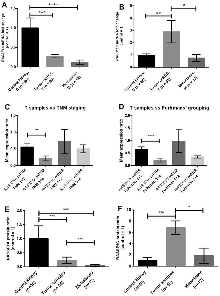
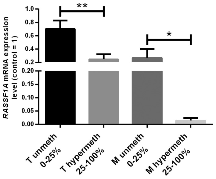
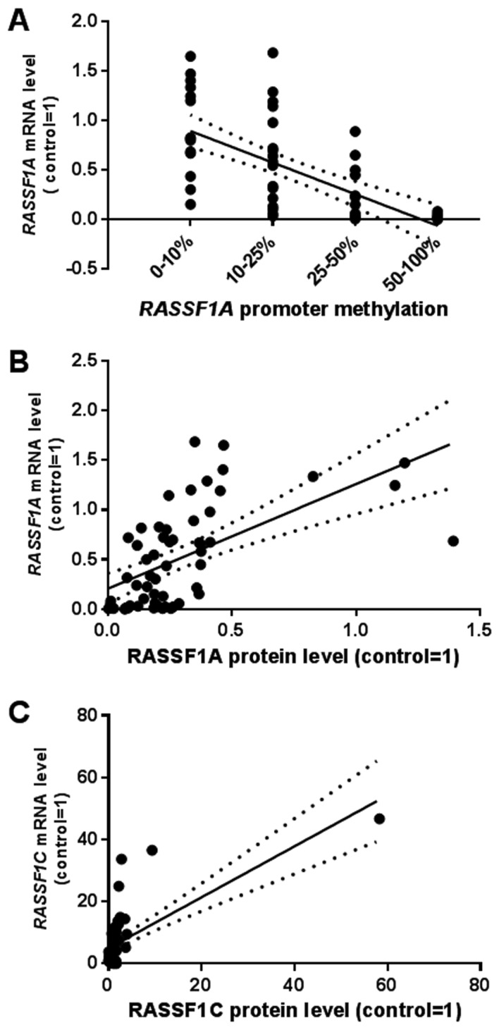
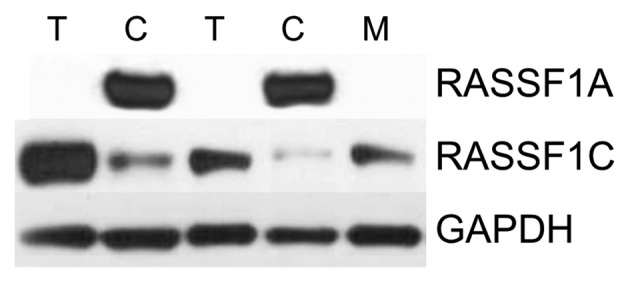
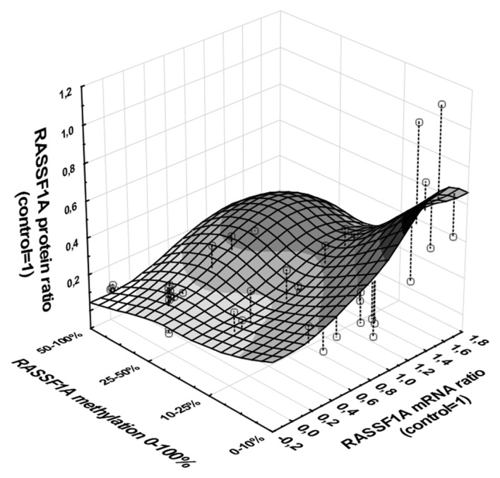
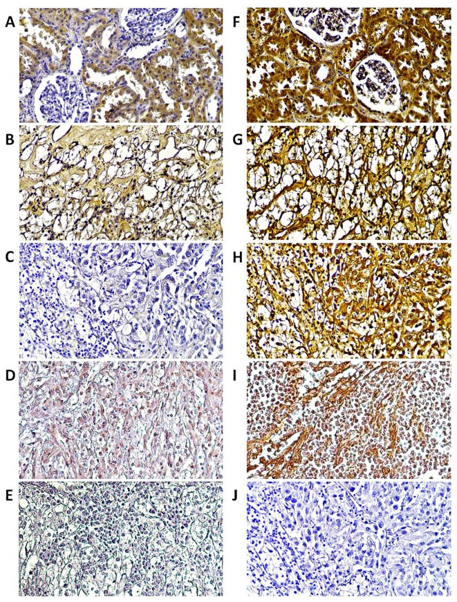
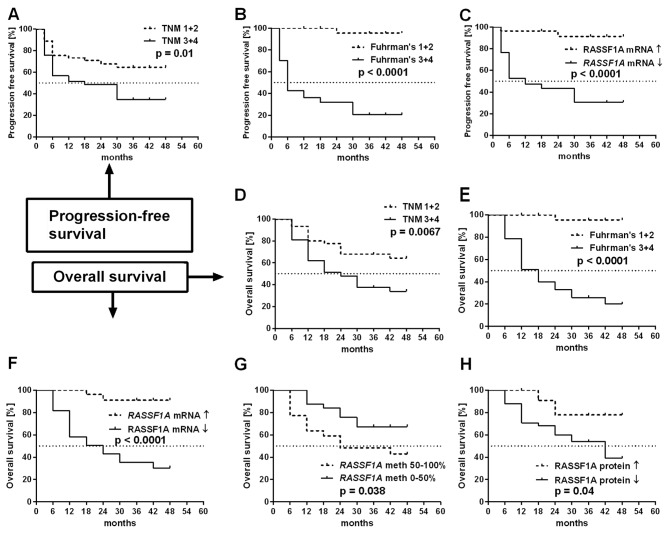
Clear-cell renal cell carcinoma (ccRCC) is the most common subtype of RCC (70-80%) and is associated with poor prognosis in 40% of cases mainly due to metastasis in the course of the disease. RASSF1, with its isoforms RASSF1A and RASSF1C, is a tumor suppressor gene which has not been fully analyzed in ccRCC yet. The epigenetic downregulation of RASSF1A is commonly associated with promoter hypermethylation. The aim of the present study was to compare the ccRCC outcomes with the expression of RASSF1A and RASSF1C. Tissues were obtained from 86 ccRCC patients. RASSF1A and RASSF1C mRNA levels were assessed in tumor and matched normal kidney tissue, and in 12 samples of local metastases by quantitative PCR (qPCR). RASSF1A and RASSF1C proteins levels were semi-quantified in 58 samples by western blot analysis and their tissue localization was assessed by immunohistochemistry. Hypermethylation of RASSF1A promoter was measured by high-resolution-melting methylation-specific qPCR. RASSF1A mRNA levels were 4 and 5 times lower in 66% of tumor and 75% metastasized samples. RASSF1A hypermethylation was found in 40% of analyzed T cases. RASSF1A protein expression was 5 or 20 times decreased in 70% tumor and 75% metastatic samples, respectively. RASSF1A hypermethylation, mRNA and protein levels were associated with TNM progression and higher Fuhrman's grading. Decreased RASSF1A expression, hypermethylation, TNM and Fuhrman's grading were associated with poorer overall survival (OS). Cox hazard ratio (HR) analysis revealed predictor role of RASSF1A mRNA levels on OS and progression-free survival (PFS) in relation to Fuhrman's grading (OS HR=2.25, PFS HR=2.93). RASSF1C levels were increased in ccRCC; no correlations with clinicopathological variables were found. We conclude that RASSF1C gene is not involved in ccRCC progression and we propose that the measurements of RASSF1A mRNA levels in paired tumor-normal kidney tissue could serve as a new prognostic factor in ccRCC.
Figures
Similar articles
-
Overexpression of the YAP1 oncogene in clear cell renal cell carcinoma is associated with poor outcome.Oncol Rep. 2017 Jul;38(1):427-439. doi: 10.3892/or.2017.5642. Epub 2017 May 15. Oncol Rep. 2017. PMID: 28504812
-
RASSF1A promoter methylation and expression analysis in normal and neoplastic kidney indicates a role in early tumorigenesis.Mol Cancer. 2007 Jul 16;6:49. doi: 10.1186/1476-4598-6-49. Mol Cancer. 2007. PMID: 17634119 Free PMC article.
-
Frequent epigenetic suppression of tumor suppressor gene glutathione peroxidase 3 by promoter hypermethylation and its clinical implication in clear cell renal cell carcinoma.Int J Mol Sci. 2015 May 11;16(5):10636-49. doi: 10.3390/ijms160510636. Int J Mol Sci. 2015. PMID: 25970749 Free PMC article.
-
The tumor suppressor RASSF1A in human carcinogenesis: an update.Histol Histopathol. 2005 Apr;20(2):645-63. doi: 10.14670/HH-20.645. Histol Histopathol. 2005. PMID: 15736067 Review.
-
Development of a prognostic risk model for clear cell renal cell carcinoma by systematic evaluation of DNA methylation markers.Clin Epigenetics. 2021 May 4;13(1):103. doi: 10.1186/s13148-021-01084-8. Clin Epigenetics. 2021. PMID: 33947447 Free PMC article.
Cited by
-
FBP1 /miR-24-1/enhancer axis activation blocks renal cell carcinoma progression via Warburg effect.Front Oncol. 2022 Aug 1;12:928373. doi: 10.3389/fonc.2022.928373. eCollection 2022. Front Oncol. 2022. PMID: 35978816 Free PMC article.
-
Sodium Butyrate Enhances Curcuminoids Permeability through the Blood-Brain Barrier, Restores Wnt/β-Catenin Pathway Antagonists Gene Expression and Reduces the Viability of Glioblastoma Cells.Int J Mol Sci. 2021 Oct 19;22(20):11285. doi: 10.3390/ijms222011285. Int J Mol Sci. 2021. PMID: 34681943 Free PMC article.
-
Dioscin inhibits SCC15 cell proliferation via the RASSF1A/MST2/YAP axis.Mol Med Rep. 2021 Jun;23(6):414. doi: 10.3892/mmr.2021.12053. Epub 2021 Mar 31. Mol Med Rep. 2021. PMID: 33786612 Free PMC article.
-
MiR-216a exerts tumor-suppressing functions in renal cell carcinoma by targeting TLR4.Am J Cancer Res. 2018 Mar 1;8(3):476-488. eCollection 2018. Am J Cancer Res. 2018. PMID: 29637002 Free PMC article.
-
High mucin 5AC expression predicts adverse postoperative recurrence and survival of patients with clear-cell renal cell carcinoma.Oncotarget. 2017 Mar 4;8(35):59777-59790. doi: 10.18632/oncotarget.15894. eCollection 2017 Aug 29. Oncotarget. 2017. PMID: 28938681 Free PMC article.
References
-
- Massari F, Bria E, Maines F, Milella M, Giannarelli D, Cognetti F, Pappagallo G, Tortora G, Porta C. Adjuvant treatment for resected renal cell carcinoma: Are all strategies equally negative? Potential implications for trial design with targeted agents. Clin Genitourin Cancer. 2013;11:471–476. doi: 10.1016/j.clgc.2013.04.018. - DOI - PubMed
-
- Keizman D, Rouvinov K, Sella A, Gottfried M, Maimon N, Kim JJ, Eisenberger MA, Sinibaldi V, Peer A, Carducci MA, et al. Is there a ‘trial effect’ on outcome of patients with meta-static renal cell carcinoma treated with sunitinib? Cancer Res Treat. 2015 Mar 5; doi: 10.4143/crt.2014.289. (Epub ahead of print) - DOI - PMC - PubMed
Publication types
MeSH terms
Substances
LinkOut - more resources
Full Text Sources
Other Literature Sources
