. 2016 Feb;13(2):1083-90.
doi: 10.3892/mmr.2015.4650. Epub 2015 Dec 7.

N-methyl-N-nitro-N'-nitrosoguanidine induces the expression of CCR2 in human gastric epithelial cells promoting CCL2-mediated migration

Affiliations

N-methyl-N-nitro-N'-nitrosoguanidine induces the expression of CCR2 in human gastric epithelial cells promoting CCL2-mediated migration

Jie Cai et al. Mol Med Rep. 2016 Feb.

Abstract

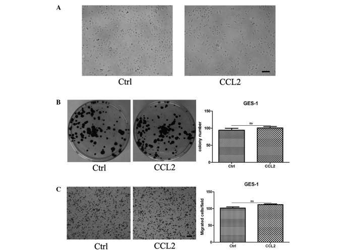
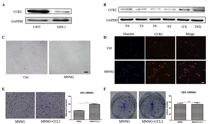
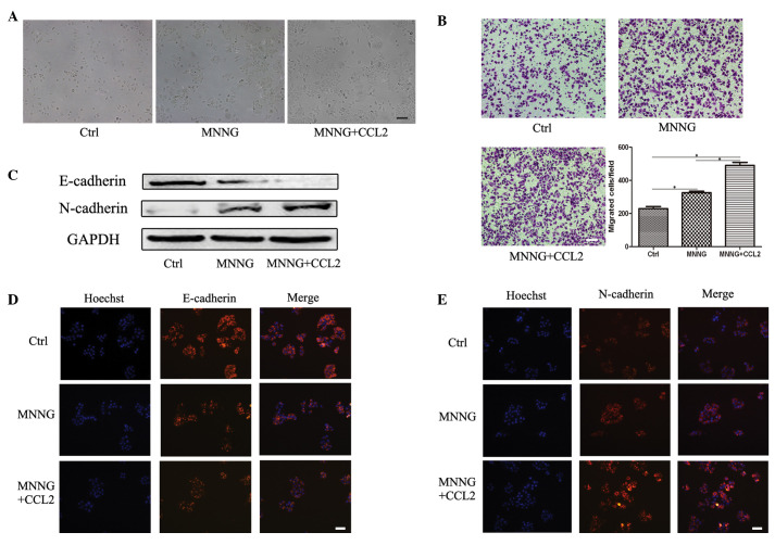
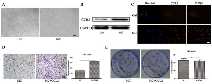
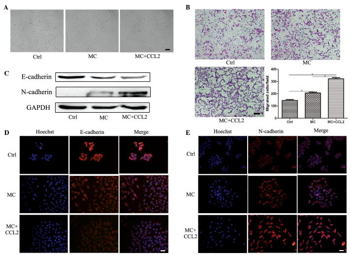
Chronic inflammation has a decisive role in tumorigenesis, particularly in gastric carcinogenesis. The CC chemokine ligand 2 (CCL2), an important inflammatory cytokine, is involved in the initiation, development and progression of various types of cancer. However, the role of CCL2 in gastric cancer remains to be elucidated. The present study demonstrated that recombinant CCL2 stimulation caused no effect on the morphology, proliferation and migration of human GES-1 gastric mucosa epithelial cells, in which the protein expression of CC-chemokine receptor 2 (CCR2) was markedly low. However, the expression of CCR2 was significantly upregulated in the GES-1 cells following pretreatment with the chemical carcinogen, N-methyl-N-nitro-N'-nitrosoguanidine (MNNG), for 12 h or transformed with MNNG (MC cells). The present study used CCL2 to stimulate MNNG pretreated GES-1 cells and MC cells, and demonstrated that CCL2 clearly promoted their migration and the epithelial-mesenchymal transition (EMT). However, no effect was observed on the proliferative ability of the cells. Taken together, these findings suggested that the CCL2/CCR2 chemokine signaling may regulate the EMT in gastric epithelial cells and resulted in gastric carcinogenesis in response to the intake of the carcinogen, MNNG.


Figures

Figure 1
Figure 1
CCL2 revealed no direct effect on the morphology, proliferation and migration of the GES-1 cells. Following persistent pretreatment with CCL2 for 9 days, the GES-1 cells were digested and collected for analysis. (A) Representative images of the cells (magnification, ×100; scale bar, 100 µm). (B) Representative images of the cell colony formation assay and the quantification of colony number. (C) Representative images of the Transwell migration assay (magnification, ×100; scale bar, 100 µm) and the quantification of the numbers of migrated cells. *P<0.05. Ctrl, GES-1 cells; CCL2, GES-1 cells treated with CC chemokine ligand 2 for 9 days; ns, non-significant.
Figure 2
Figure 2
Pretreatment with MNNG induces the expression of CCR2 in GES-1 cells. (A) Western blotting was performed to determine the protein expression of CCR2 in U937 and GES-1 cells. (B) Western blotting was performed to determine the protein expression of CCR2 in GES-1 cells treated with MNNG for 0, 2, 4, 8, 12 and 24 h. (C) Representative images of cells (magnification, ×100; scale bar, 100 µm). (D) Immunofluorescence staining of CCR2 was performed to determine the expression levels of CCR2 (magnification, ×200; scale bar, 50 µm). (E) A Transwell assay was performed to determine the migration capability of the cells. The cells were seeded onto the top of the upper chamber and the number of cells that migrated through the uncoated filter in response to CCL2 in the lower chamber was quantified (magnification, ×100; scale bar, 100 µm; *P<0.05). (F) A cell colony formation assay of the MNNG-pretreated GES-1 cells cultured alone or in the presence of CCL2 (*P<0.05). Ctrl, GES-1 cells; CCL2, CC chemokine ligand 2; ns, non-significant; MNNG, cells treated for 12 h with N-methyl-N-nitro-N′-nitrosoguanidine.
Figure 3
Figure 3
CCL2 promotes the EMT in MNNG-pretreated GES-1 cells. The GES-1 cells were pretreated with MNNG and subsequently induced with CCL2 for 9 days. (A) Representative images of the cells (magnification, ×100; scale bar, 100 µm). (B) A Transwell migration assay was performed to assess the migration capacity of the cells (magnification, ×100; scale bar, 100 µm; *P<0.05). (C) Western blotting was performed to determine the protein expression levels of E-cadherin and N-cadherin. Immunofluorescence staining was performed to determine the protein expression levels of (D) E-cadherin and (E) N-cadherin (magnification, ×200; scale bar, 50 µm). Ctrl, GES-1 cells; MNNG, N-methyl-N-nitro-N′-nitrosoguanidine-pretreated GES-1 cells; CCL2, CC chemokine ligand 2; MNNG+CCL2, MNNG-pretreated GES-1 cells induced with CCL2 for 9 days.
Figure 4
Figure 4
CCR2 is upregulated in the MNNG transformed GES-1 cell line. (A) Representative images of the GES-1 and MC cells (magnification, ×100; scale bar, 100 µm). (B) Western blotting was performed to determine the protein expression of CCR2 in the GES-1 and MC cells. (C) Immunofluorescence staining of CCR2 was performed to assess the expression of CCR2 in the cells (magnification, ×200; scale bar, 50 µm). (D) A Transwell assay was performed to determine the migration ability of the MC cells. The MC cells were seeded onto the top of upper chamber and the number of cells that migrated through the uncoated filter in response to CCL2 in the lower chamber was quantified (magnification, ×100; scale bar, 100 µm; *P<0.05). (E) A cell colony formation assay of the MC cells cultured alone or in the presence of CCL2 was performed (*P<0.05). Ctrl, GES-1 cells; MC, MC cells; CCR2, CC chemokine ligand 2.
Figure 5
Figure 5
CCL2 promotes the EMT in the MNNG transformed GES-1 cell line. The MNNG transformed GES-1 cells (MC cells) were stimulated with CCL2 for 9 days. (A) Representative images of the cells (magnification, ×100; scale bar, 100 µm). (B) A Transwell migration assay was performed to determine the migration ability of the cell line (magnification, ×100; scale bar, 100 µm; *P<0.05). (C) Western blotting was performed to determine the protein expression levels of E-cadherin and N-cadherin. Immunofluorescence staining was performed to asses the protein expression of (D) E-cadherin and (E) N-cadherin (magnification, ×200; scale bar, 50 µm). Ctrl, GES-1 cells; MC, MC cells; CCL2, CC chemokine ligand 2; MC + CCL2, MC cells were stimulated with CCL2 for 9 days.

References

    1. Hussain SP, Harris CC. Inflammation and cancer: An ancient link with novel potentials. Int J Cancer. 2007;121:2373–2380. doi: 10.1002/ijc.23173. - DOI - PubMed
    1. Landskron G, De la Fuente M, Thuwajit P, Thuwajit C, Hermoso MA. Chronic inflammation and cytokines in the tumor microenvironment. J Immunol Res. 2014;2014(149185) doi: 10.1155/2014/149185. - DOI - PMC - PubMed
    1. Grivennikov SI, Greten FR, Karin M. Immunity, inflammation and cancer. Cell. 2010;140:883–899. doi: 10.1016/j.cell.2010.01.025. - DOI - PMC - PubMed
    1. Schetter AJ, Heegaard NH, Harris CC. Inflammation and cancer: Interweaving microRNA, free radical, cytokine and p53 pathways. Carcinogenesis. 2010;31:37–49. doi: 10.1093/carcin/bgp272. - DOI - PMC - PubMed
    1. Schauer IG, Zhang J, Xing Z, Guo X, Mercado-Uribe I, Sood AK, Huang P, Liu J. Interleukin-1β promotes ovarian tumorigenesis through a p53/NF-κB-mediated inflammatory response in stromal fibroblasts. Neoplasia. 2013;15:409–420. doi: 10.1593/neo.121228. - DOI - PMC - PubMed

Publication types

MeSH terms