. 2015 Dec 9:5:17946.
doi: 10.1038/srep17946.

Aflibercept Traps Galectin-1, an Angiogenic Factor Associated with Diabetic Retinopathy

Affiliations

Aflibercept Traps Galectin-1, an Angiogenic Factor Associated with Diabetic Retinopathy

Atsuhiro Kanda et al. Sci Rep. .

Abstract

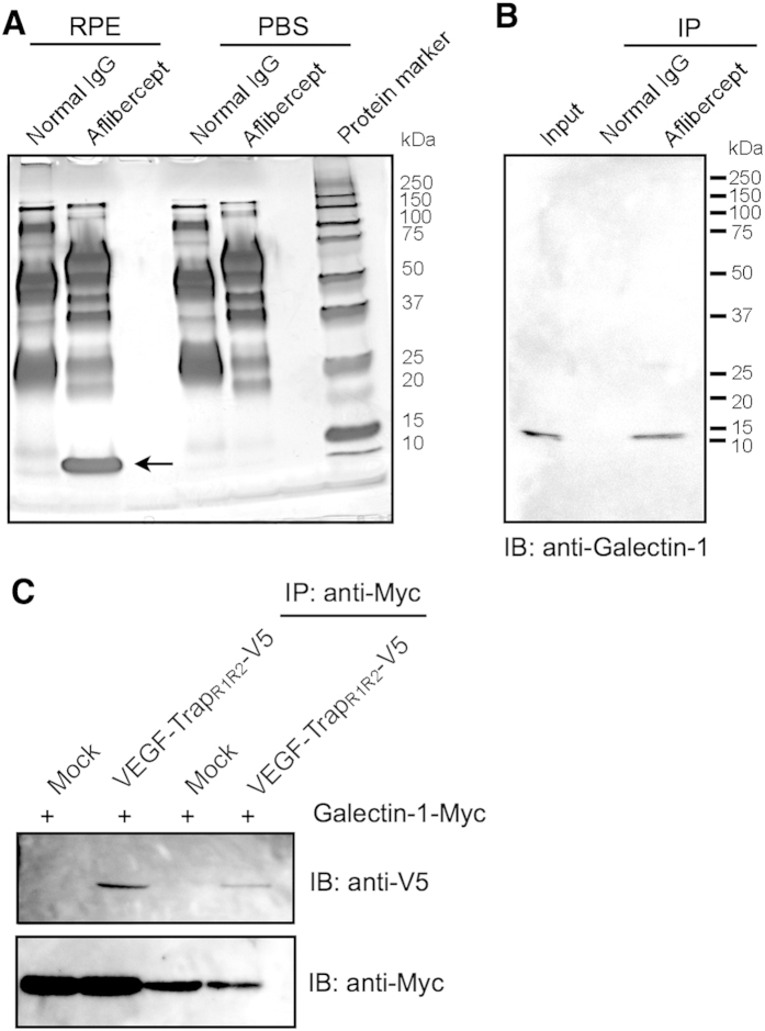
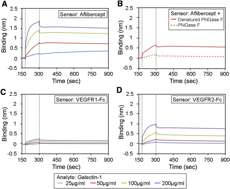
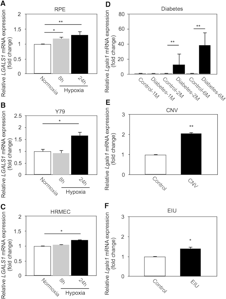
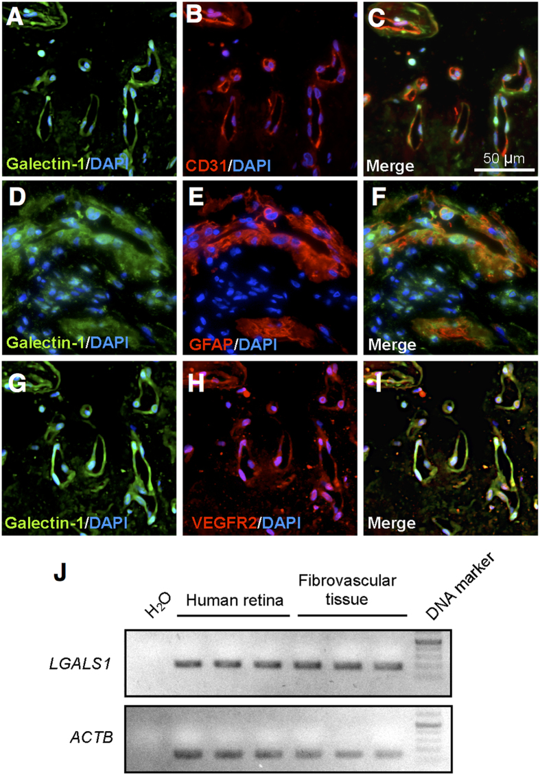
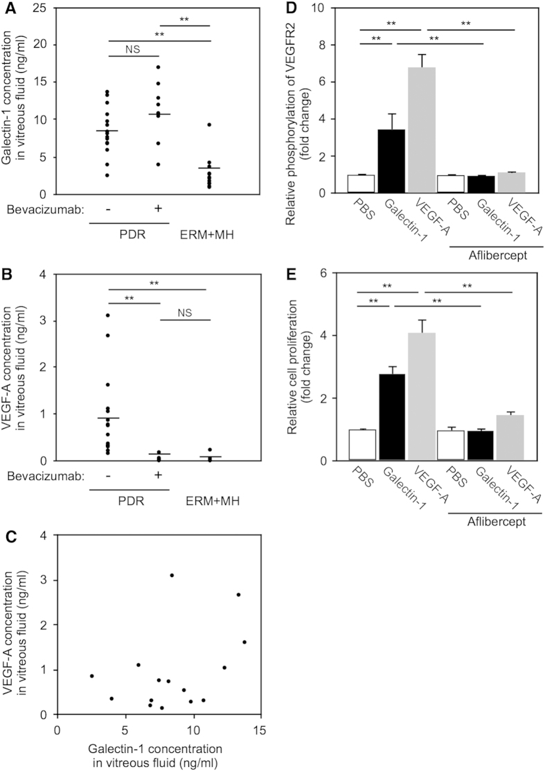
Vascular endothelial growth factor (VEGF)-A-driven angiogenesis contributes to various disorders including cancer and proliferative diabetic retinopathy (PDR). Among several VEGF-A blockers clinically used is aflibercept, a chimeric VEGFR1/VEGFR2-based decoy receptor fused to the Fc fragment of IgG1 (i.e., VEGFR1/VEGFR2-Fc). Here, we revealed a novel anti-angiogenic function for aflibercept beyond its antagonism against VEGF family members. Immunoprecipitation and mass spectrometry analyses identified galectin-1 as an aflibercept-interacting protein. Biolayer interferometry revealed aflibercept binding to galectin-1 with higher affinity than VEGFR1-Fc and VEGFR2-Fc, which was abolished by deglycosylation of aflibercept with peptide:N-glycosidase F. Retinal LGALS1/Galectin-1 mRNA expression was enhanced in vitro by hypoxic stimulation and in vivo by induction of diseases including diabetes. Galectin-1 immunoreactivity co-localized with VEGFR2 in neovascular tissues surgically excised from human eyes with PDR. Compared with non-diabetic controls, intravitreal galectin-1 protein levels were elevated in PDR eyes, showing no correlation with increased VEGF-A levels. Preoperative injection of bevacizumab, a monoclonal antibody to VEGF-A, reduced the VEGF-A, but not galectin-1, levels. Galectin-1 application to human retinal microvascular endothelial cells up-regulated VEGFR2 phosphorylation, which was eliminated by aflibercept. Our present findings demonstrated the neutralizing efficacy of aflibercept against galectin-1, an angiogenic factor associated with PDR independently of VEGF-A.


Figures

Figure 1
Figure 1. Molecular binding of aflibercept with galectin-1.
(A) Human RPE cell extracts were applied with aflibercept- or normal IgG-immobilized protein G beads. The eluted proteins were separated by SDS-PAGE and visualized with silver staining. A single protein band around 14 kDa was detected (arrow). (B) Co-IP of human RPE cell extracts using aflibercept was performed, followed by SDS-PAGE and immunoblot (IB) analyses for galectin-1. (C) Co-transfection and Co-IP. V5-tagged VEGF-TrapR1R2 expression plasmids were co-transfected into HEK293T cells with Myc-tagged Galectin-1/LGASL1 expression constructs. IP was performed with anti-Myc antibody followed by detection with anti-V5 antibody.
Figure 2
Figure 2. Binding affinity of aflibercept with galectin-1.
(A) Kinetic analysis of galectin-1 binding with aflibercept using the BLItz biolayer interferometry system. Sensorgrams obtained using aflibercept-loaded biosensors incubated with different concentrations of galectin-1 as analytes. Dotted lines indicate the start of the binding (left) and the dissociation (right) phase. (B) Sensorgrams obtained using biosensors loaded with aflibercept with (dotted line) or without (solid line) deglycosylation (+ PNGase F or Denatured PNGase F, respectively) and incubated with galectin-1 at the concentration of 50 μg/ml. (C,D) Sensorgrams obtained using biosensors loaded with VEGFR1-Fc (C) or VEGFR2-Fc (D) and incubated with different concentrations of galectin-1.
Figure 3
Figure 3. Induction of retinal Galectin-1/LGALS1 mRNA in vitro and in vivo.
(AC) LGALS1 mRNA expression in hypoxia (1% O2)-stimulated cell culture using RPE (A), Y79 (B) and HRMEC (C). (D–F) Lgals1 mRNA expression in retinal tissues from disease model mice with streptozotocin-induced diabetes (D), CNV (E) and EIU (F). *p < 0.05, **p < 0.01 (n = 6 per group).
Figure 4
Figure 4. Localization of galectin-1 in human PDR fibrovascular tissues.
(AC) Double labeling of galectin-1 (green), CD31 (red) and DAPI (blue) in PDR fibrovascular tissues. (DF) Double labeling of galectin-1 (green), GFAP (red) and DAPI (blue) in PDR fibrovascular tissues. (G–I) Double labeling of galectin-1 (green), VEGFR2 (red) and DAPI (blue) in PDR fibrovascular tissues. Scale bar = 30 μm. (J) End-point PCR analysis. Gene expression of LGALS1 in healthy human retinas and PDR fibrovascular tissues.
Figure 5
Figure 5. Elevation of galectin-1 protein levels in human PDR vitreous fluids and suppression of galectin-1-mediated VEGFR2 activation by aflibercept in HRMEC.
(A) Protein levels of galectin-1 in PDR eyes with (n = 8) or without (n = 15) bevacizumab pretreatment and in control (ERM + MH) eyes (n = 12). Black symbols indicate individual samples in each group with a bar showing the average. **p < 0.01. (B) Protein levels of VEGF-A in PDR eyes with or without bevacizumab pretreatment and in control eyes. (C) No correlation (p = 0.137, r2 = 0.150) between galectin-1 and VEGF-A in PDR eyes without bevacizumab pretreatment. (D) VEGFR2 phosphorylation induced by galectin-1 as well as VEGF-A, both of which were significantly suppressed by aflibercept. Data are shown as relative values compared to PBS (n = 5 per group). (E) HRMEC proliferation induced by galectin-1 and VEGF-A, both of which were significantly suppressed by aflibercept. Data are shown as relative values compared to PBS (n = 8 per group).

References

    1. Folkman J. Tumor angiogenesis: therapeutic implications. N Engl J Med 285, 1182–1186 (1971). - PubMed
    1. Hicklin D. J. & Ellis L. M. Role of the vascular endothelial growth factor pathway in tumor growth and angiogenesis. J Clin Oncol 23, 1011–1027 (2005). - PubMed
    1. Hoeben A. et al. Vascular endothelial growth factor and angiogenesis. Pharmacol Rev 56, 549–580 (2004). - PubMed
    1. Adamis A. P. et al. Increased vascular endothelial growth factor levels in the vitreous of eyes with proliferative diabetic retinopathy. Am J Ophthalmol 118, 445–450 (1994). - PubMed
    1. Ferrara N. Vascular endothelial growth factor and age-related macular degeneration: from basic science to therapy. Nat Med 16, 1107–1111 (2010). - PubMed

Publication types

MeSH terms

Substances