. 2015 Dec 1:6:355.
doi: 10.3389/fphys.2015.00355. eCollection 2015.

Structure and Function of SLC4 Family [Formula: see text] Transporters

Affiliations

Structure and Function of SLC4 Family [Formula: see text] Transporters

Ying Liu et al. Front Physiol. .

Abstract

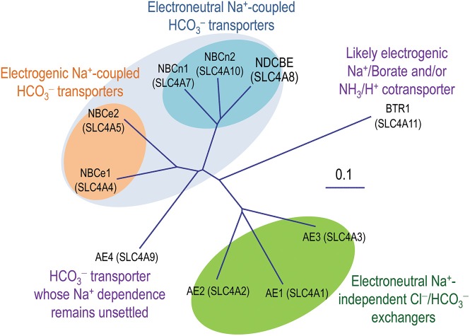
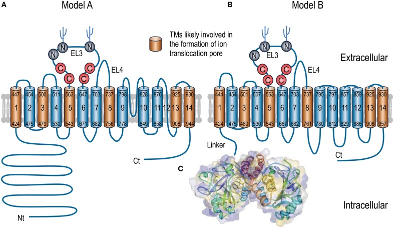
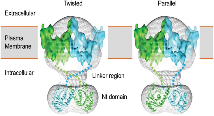
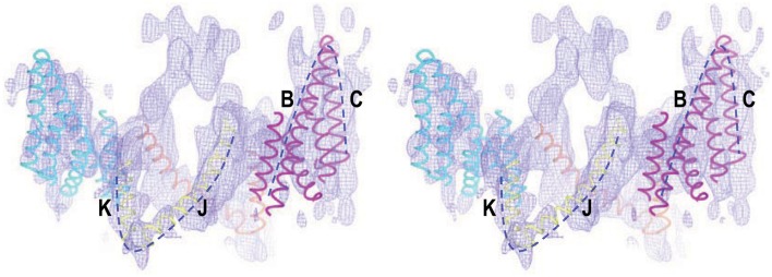
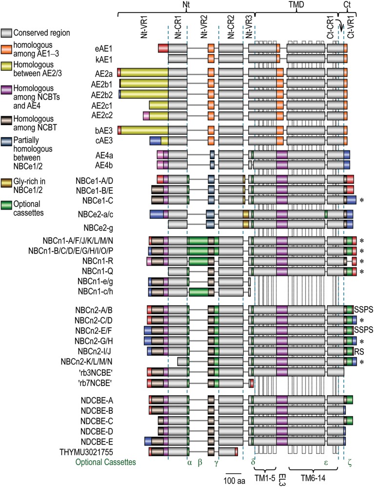
The solute carrier SLC4 family consists of 10 members, nine of which are [Formula: see text] transporters, including three Na(+)-independent Cl(-)/[Formula: see text] exchangers AE1, AE2, and AE3, five Na(+)-coupled [Formula: see text] transporters NBCe1, NBCe2, NBCn1, NBCn2, and NDCBE, as well as "AE4" whose Na(+)-dependence remains controversial. The SLC4 [Formula: see text] transporters play critical roles in pH regulation and transepithelial movement of electrolytes with a broad range of demonstrated physiological relevances. Dysfunctions of these transporters are associated with a series of human diseases. During the past decades, tremendous amount of effort has been undertaken to investigate the topological organization of the SLC4 transporters in the plasma membrane. Based upon the proposed topology models, mutational and functional studies have identified important structural elements likely involved in the ion translocation by the SLC4 transporters. In the present article, we review the advances during the past decades in understanding the structure and function of the SLC4 transporters.

Keywords: N-glycosylation; acid-base balance; alternative splicing; bicarbonate transporter; cysteine scanning mutagenesis; metabolic acidosis; solute carrier; topology structure.


Figures

Figure 1
Figure 1
Phylogenetic tree of SLC4 family. A multiple sequence alignment was performed with human sequences of eAE1 (NP_000333.1), AE2a (NP_003031.3), bAE3 (NP_005061.2), AE4a (NM_031467), NBCe1-A (AAC51645.1), NBCe2-c (AAK97072.1), NBCn1-E (ACH61961), NBCn2-A (NP_071341.2), NDCBE-A (AAY79176), and BTR1 (NP_114423) with the online software Clustal Omega from the European Bioinformatics Institute. The unrooted phylogenetic tree was generated by using Treview based on the sequence alignment (Page, 1996).
Figure 2
Figure 2
Putative models of topological organization of SLC4 transporters (A,B) and 3D crystal structure of Nt domain of AE1 (C). For the convenience to compare the sequence assignments, both topological models A and B use human NBCe1-A for illustration. The numbers (according to accession #AAC51645.1) at both ends of each putative TM indicate the proposed initial and last residues of the TM in each model. Experimentally, model A is largely based on the studies with AE1 by N-glycosylation scanning mutagenesis (Popov et al., 1997, 1999) and cysteine scanning mutagenesis (Tang et al., ; Zhu et al., 2003). The front half of TMD (TM1-TM9) of model B is basically consistent with model A except for some minor deviations in the boundaries of TMs. The assignment of the Ct half from EL5-TM14 in model B is based upon the cysteine-scanning mutagenesis study on NBCe1 (Zhu et al., 2010a,b) and the structural modelings with AE1 (Barneaud-Rocca et al., ; Bonar et al., 2013). Panel C shows the 3D structure of the dimer of the Nt domain of human AE1. The model was created based on the crystal structure of the Nt of human AE1 (PDB ID #4KY9) using Protein Workshop 4.2.0 from RCSB Protein Data Bank (Moreland et al., 2005). The hydrophobicity surface (blue least, red most) was generated by using a Euclidean Distance Transform (Xu and Zhang, 2009).
Figure 3
Figure 3
3D structure model of human AE1 dimers obtained by cryo-EM. Shown here are two potential organizations of the AE1 dimers: twisted (left) and parallel (right). The two molecules of AE1 are shown in different color. The figures are modified from Jiang et al. (2013).
Figure 4
Figure 4
Stereo view of cryo-EM structure of the TMD of human AE1 fitted with structural elements of chloride channel ClC. The dotted lines show the two V-shaped structures in the TMD of AE1 that fit well with helices B + C and J + K of ClC, respectively. The figure was modified from Yamaguchi et al. (2010b) with permission.
Figure 5
Figure 5
Alignment of expression variants of SLC4 family HCO3- transporters. The diagram was based upon a sequence alignment with Clustal Omega from the European Bioinformatics Institute Details. Accession numbers for the SLC4 variants are available in Supplemental Table S1. The colors denoting homology among different SLC4 members are shown in the legend. The extreme Nt and Ct ends of different SLC4 members with identical color pattern do not denote homology unless specified elsewhere in the main text. The dotted vertical boxes in TMD indicate the putative transmembrane helices shown in model A in Figure 2. *Proteins containing PDZ-binding motif at the Ct end.

References

    1. Abramson J., Wright E. M. (2009). Structure and function of Na+-symporters with inverted repeats. Curr. Opin. Struct. Biol. 19, 425–432. 10.1016/j.sbi.2009.06.002 - DOI - PMC - PubMed
    1. Abuladze N., Song M., Pushkin A., Newman D., Lee I., Nicholas S., et al. . (2000). Structural organization of the human NBC1 gene: kNBC1 is transcribed from an alternative promoter in intron 3. Gene 251, 109–122. 10.1016/S0378-1119(00)00204-3 - DOI - PubMed
    1. Ahmed S., Thomas G., Ghoussaini M., Healey C. S., Humphreys M. K., Platte R., et al. . (2009). Newly discovered breast cancer susceptibility loci on 3p24 and 17q23.2. Nat. Genet. 41, 585–590. 10.1038/ng.354 - DOI - PMC - PubMed
    1. Alper S. L. (2009). Molecular physiology and genetics of Na+-independent SLC4 anion exchangers. J. Exp. Biol. 212, 1672–1683. 10.1242/jeb.029454 - DOI - PMC - PubMed
    1. Alrefai W. A., Tyagi S., Nazir T. M., Barakat J., Anwar S. S., Hadjiagapiou C., et al. . (2001). Human intestinal anion exchanger isoforms: expression, distribution, and membrane localization. Biochim. Biophys. Acta 1511, 17–27. 10.1016/S0005-2736(00)00366-7 - DOI - PubMed

LinkOut - more resources