

doi: 10.1155/2015/342312.
Epub 2015 Nov 15.
An Unusual Case of Laryngeal Paraganglioma in a Patient with Carotid Body Paraganglioma: Multimodality Imaging Findings
Affiliations
- PMID: 26649218
- PMCID: PMC4662993
- DOI: 10.1155/2015/342312
Item in Clipboard
An Unusual Case of Laryngeal Paraganglioma in a Patient with Carotid Body Paraganglioma: Multimodality Imaging Findings
Case Rep Radiol.
2015.
Abstract
Multiple paragangliomas of the head and neck are rare conditions. Carotid paragangliomas are most common multiple paragangliomas. Laryngeal paragangliomas are very rare neuroendocrine tumors and usually are seen as symptomatic solitary lesions. We present multimodality imaging findings of incidentally detected laryngeal paraganglioma in a woman with synchronous carotid body paraganglioma and positive family history. To the best of our knowledge, this is the first case of laryngeal and carotid body paragangliomas in a patient with positive family history. Radiologists should keep in mind that paragangliomas may occur in various locations as multiple tumors.
Figures
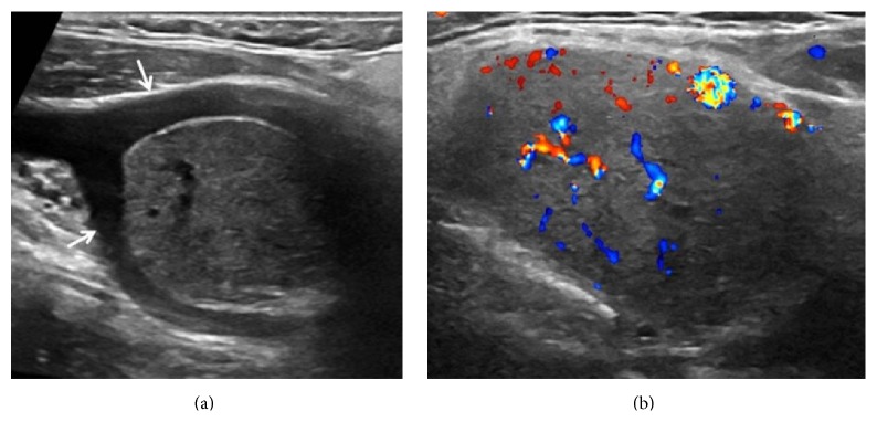
Gray-scale ultrasound (a) shows well defined, solid mass that causes splaying of the internal and external carotid arteries (arrows). High vascularity of lesions is seen in Doppler ultrasound image (b).
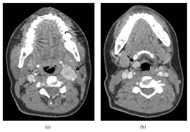
Axial contrast enhanced computed tomography demonstrates vascular mass (arrow) at left carotid bifurcation (a). Right supraglottic, well defined laryngeal mass (arrow) is seen in lower level contrast enhanced computed tomography image (b).
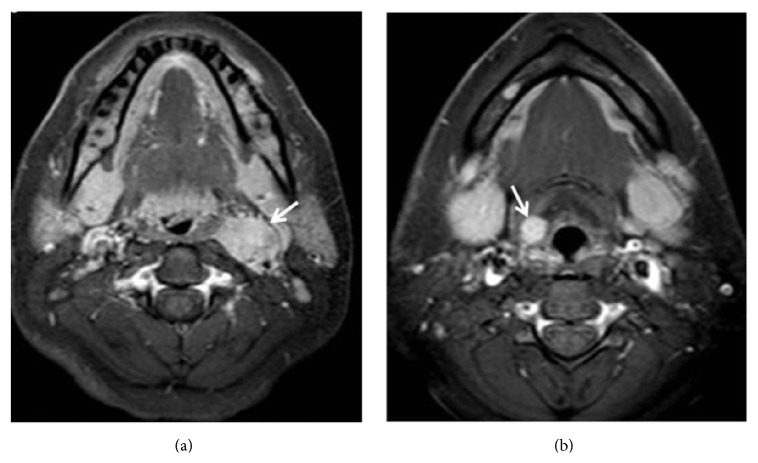
Enhanced lesions (arrows) are seen at left carotid bifurcation (a) and right supraglottic larynx (b) in axial contrast enhanced T1 weighted turbo spin echo spectral fat saturation inversion recovery image (T1 TSE SPIR).
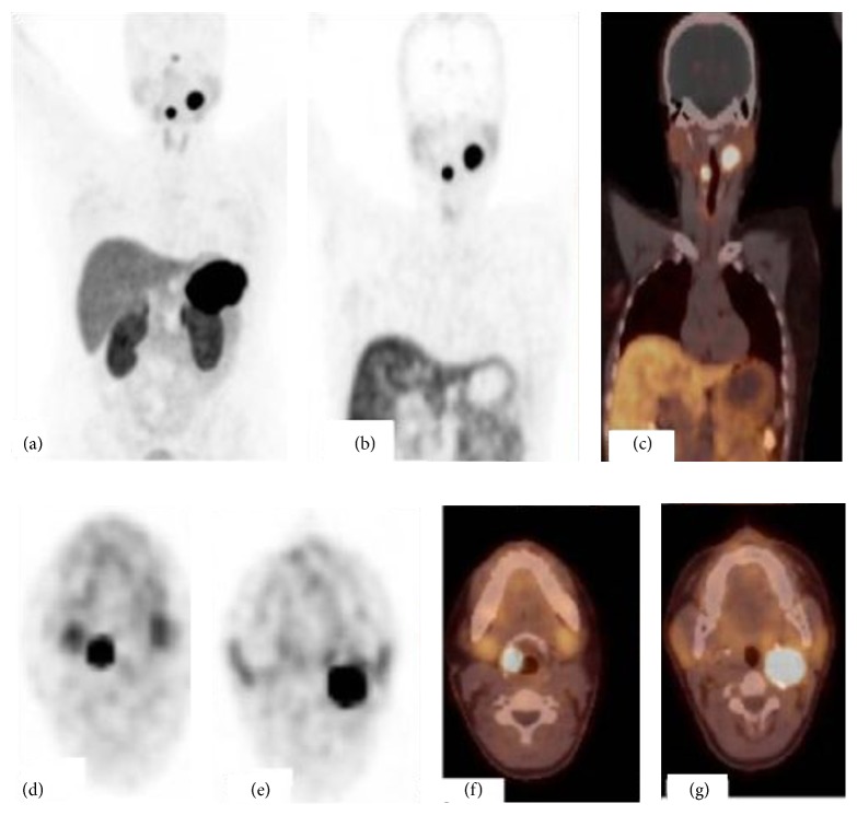
68Ga-DOTANOC PET/CT; maximum intensity projection (MIP) image (a), coronal PET and PET/CT fusion images (b, c), and axial PET and PET/CT fusion images (d, e, f, and g) show an intense uptake by the right laryngeal paraganglioma and left carotid body paraganglioma.
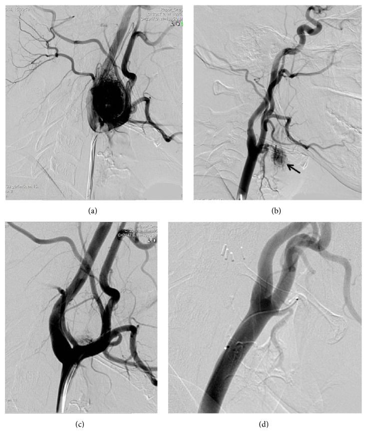
A lateral cervical angiogram from the left common carotid artery demonstrates the intense blush at the carotid bifurcation corresponding to the carotid body paraganglioma (a). Right common carotid artery lateral cervical angiogram shows significant tumor vascularity (arrow) at the larynx (b). Principal arterial feeder to the tumor from superior thyroid artery. Significant reduction is seen in tumor vascularity after embolization of the ascending pharyngeal and proximal occipital artery with polyvinyl alcohol particles measuring 255 to 350 (c). Superselective catheterization of the superior thyroid artery after embolization shows complete devascularization (d).
References
-
- Bikhazi P. H., Roeder E., Attaie A., Lalwani A. K. Familial paragangliomas: the emerging impact of molecular genetics on evaluation and management. American Journal of Otology. 1999;20(5):639–643. - PubMed
LinkOut - more resources
Full Text Sources
Other Literature Sources
