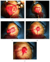
. 2015:2015:698385.
doi: 10.1155/2015/698385. Epub 2015 Nov 16.

One-Stage Reconstruction of Scalp after Full-Thickness Oncologic Defects Using a Dermal Regeneration Template (Integra)

Affiliations

One-Stage Reconstruction of Scalp after Full-Thickness Oncologic Defects Using a Dermal Regeneration Template (Integra)

Barbara De Angelis et al. Biomed Res Int. 2015.

Abstract

The use of Dermal Regeneration Template (DRT) can be a valid alternative for scalp reconstruction, especially in elderly patients where a rapid procedure with an acceptable aesthetic and reliable functional outcome is required. We reviewed the surgical outcome of 20 patients, 14 (70%) males and 6 (30%) females, who underwent application of DRT for scalp reconstruction for small defects (group A: mean defect size of 12.51 cm(2)) and for large defects (group B: mean defect size of 28.7 cm(2)) after wide excision of scalp neoplasm (basal cell carcinoma and squamous cell carcinoma). In group A, the excisions were performed to the galeal layer avoiding pericranium, and in group B the excisions were performed including pericranium layer with subsequent coverage of the exposed bone with local pericranial flap. In both the groups (A and B) after the excision of the tumor, the wound bed was covered with Dermal Regeneration Template. In 3 weeks we observed the complete healing of the wound bed by secondary intention with acceptable cosmetic results and stable scars. Scalp reconstruction using a DRT is a valid coverage technique for minor and major scalp defects and it can be conducted with good results in elderly patients with multiple comorbidities.


Figures
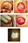
Figure 1
Figure 1
Patient of group A: (a) excision of the lesion including the galeal layer, avoiding the pericranium, (b) detail of loss of substance after the excision, (c) remodelling of a single piece of Integra Double Layer (DL) (5 cm × 5 cm), (d) Integra after remodelling which has then been shaped to size, and (e) moulage.
Figure 2
Figure 2
Patient of group B: (a) excision of the lesion including the pericranium, (b) harvesting of local pericranial flap, (c) phase of pericranial flap rotation, (d) local flap fixed, and (e) coverage with Integra Double Layer which has then been shaped to size.
Figure 3
Figure 3
(a) Integra Double Layer after sixteen days and (b) removal of the silicone layer of Integra.
Figure 4
Figure 4
Patient of group A: reepithelization time.
Figure 5
Figure 5
Patient of group B: reepithelization time.
Figure 6
Figure 6
Patient number 1 of group A: (a) preoperative appearance with lesion, (b) postoperative appearance after 25 days, and (c) postoperative appearance after 35 days.
Figure 7
Figure 7
Patient of group B: (a) preoperative appearance with big lesion, (b) detail of the lesion, (c) postoperative appearance in frontal projection after 45 days, and (d) postoperative appearance in back projection after 45 days.
Figure 8
Figure 8
Patient number 2 of group B: (a) preoperative appearance with lesion; 85-year-old patient, affected by chronic lymphocytic leukemia, renal failure, and arterial hypertension; defect size of 65.94 cm2; (b) postoperative appearance after reconstruction with pericranial local flap and Integra. After 20 days from surgery the patient had a local recurrence promptly excised and covered with DRT. Initial lesion healed and area of the recurrence not yet healed; (c) detail of lesion.
Figure 9
Figure 9
Histological analysis: (a) switching between skin covered by skin and ulcerated area, (b) side with the overlying skin epidermidis, (c) ulcerated area with underlying lattice partially amorphous material, and (d) higher magnification image 1 particularly of amorphous material.

References

    1. Snow S. N., Stiff M. A., Bullen R., Mohs F. E., Chao W.-H. Second-intention healing of exposed facial-scalp bone after Mohs surgery for skin cancer: review of ninety-one cases. Journal of the American Academy of Dermatology. 1994;31(3 I):450–454. doi: 10.1016/s0190-9622(94)70209-8. - DOI - PubMed
    1. Cruz A. P., Campbell R. M., Perlis C. S., Dufresne R. G., Jr. Double purse-string closure for scalp and extremity wounds. Dermatologic Surgery. 2007;33(3):369–373. doi: 10.1111/j.1524-4725.2007.33076.x. - DOI - PubMed
    1. Onishi K., Maruyama Y., Hayashi A., Inami K. Repair of scalp defect using a superficial temporal fascia pedicle VY advancement scalp flap. British Journal of Plastic Surgery. 2005;58(5):676–680. doi: 10.1016/j.bjps.2005.01.009. - DOI - PubMed
    1. Murtagh J. The rotation flap. Australian Family Physician. 2001;30(10, article 973) - PubMed
    1. Shin T. M., Bordeaux J. S. Repair of a large, full-thickness scalp defect with exposed bone using a thin transposition flap. Dermatologic Surgery. 2013;39(4):646–648. doi: 10.1111/dsu.12053. - DOI - PubMed