. 2016 Jun 1;193(11):1281-91.
doi: 10.1164/rccm.201507-1499OC.

IFN-γ-Producing T-Helper 17.1 Cells Are Increased in Sarcoidosis and Are More Prevalent than T-Helper Type 1 Cells

Affiliations

IFN-γ-Producing T-Helper 17.1 Cells Are Increased in Sarcoidosis and Are More Prevalent than T-Helper Type 1 Cells

Joris Ramstein et al. Am J Respir Crit Care Med. .

Abstract

Rationale: Pulmonary sarcoidosis is classically defined by T-helper (Th) cell type 1 inflammation (e.g., IFN-γ production by CD4(+) effector T cells). Recently, IL-17A-secreting cells have been found in lung lavage, invoking Th17 immunity in sarcoidosis. Studies also identified IL-17A-secreting cells that expressed IFN-γ, but their abundance as a percentage of total CD4(+) cells was either low or undetermined.

Objectives: Based on evidence that Th17 cells can be polarized to Th17.1 cells to produce only IFN-γ, our goal was to determine whether Th17.1 cells are a prominent source of IFN-γ in sarcoidosis.

Methods: We developed a single-cell approach to define and isolate major Th-cell subsets using combinations of chemokine receptors and fluorescence-activated cell sorting. We subsequently confirmed the accuracy of subset enrichment by measuring cytokine production.

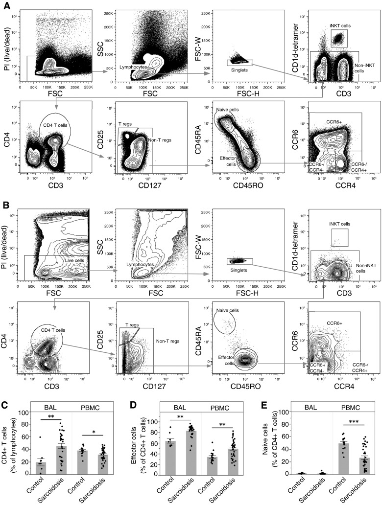
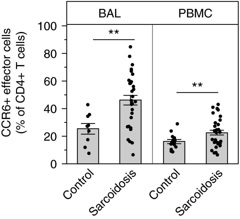
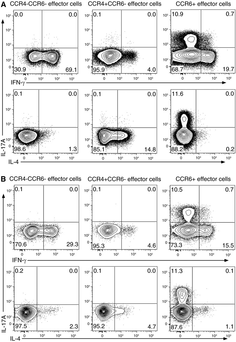
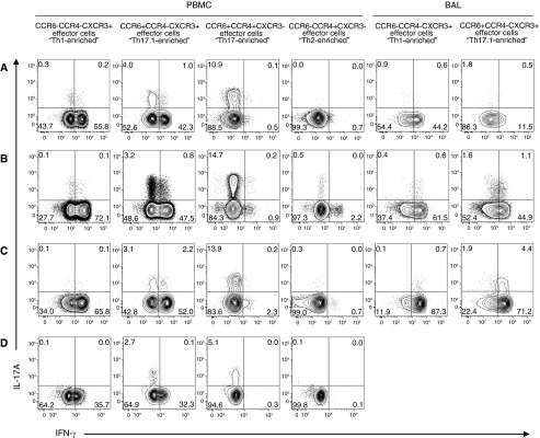
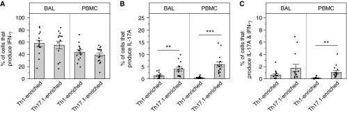
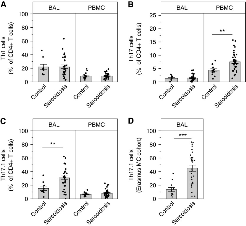
Measurements and main results: Discrimination between Th17 and Th17.1 cells revealed very high percentages of Th17.1 cells in lung lavage in sarcoidosis compared with controls in two separate cohorts. No differences in Th17 or Th1 lavage cells were found compared with controls. Lung lavage Th17.1-cell percentages were also higher than Th1-cell percentages, and approximately 60% of Th17.1-enriched cells produced only IFN-γ.

Conclusions: Combined use of surface markers and functional assays to study CD4(+) T cells in sarcoidosis revealed a marked expansion of Th17.1 cells that only produce IFN-γ. These results suggest that Th17.1 cells could be misclassified as Th1 cells and may be the predominant producer of IFN-γ in pulmonary sarcoidosis, challenging the Th1 paradigm of pathogenesis.

Keywords: chemokine receptor; inflammation; lymphocyte.


Figures

Figure 1.
Figure 1.
Flow cytometry sorting strategy and comparative analysis of CD4+ T-cell subsets. Peripheral blood mononuclear cells (PBMCs) and bronchoalveolar lavage (BAL) cells were stained and analyzed by an LSRII Fortessa cytometer (BD Biosciences). (A and B) Representative sample is shown for blood (A) and BAL cells (B) from a patient with sarcoidosis. Lymphocytes were analyzed using the following series of subgates: live cells, lymphocytes, singlets, non–invariant natural killer (iNKT), CD4 T cells (CD4+CD3+), nonregulatory T cells (CD4+CD127+CD25 T cells), memory effector cells (CD45RA/CD45RO+ CD4 T cells), and three subpopulations of memory effector cells using CCR4 and CCR6. We excluded iNKT cells (CD1d tetramer-PBS57), regulatory T cells (CD4+CD3+CD127CD25+), and naive T cells (CD4+CD3+CD45RA+CD45RO) from the CD4+ population. (C) CD4+ T cells as a percentage of lymphocytes are significantly increased in the lungs of patients with sarcoidosis (**P = 0.0019), and decreased in the blood (*P = 0.012). (D) Effector cells as a percentage of CD4+ T cells are significantly increased in both the lungs (**P = 0.0015) and the blood (**P = 0.0004) of patients with sarcoidosis. (E) There are essentially no naive cells in the lungs, and a significant decrease of naive cells as a percentage of CD4+ T cells in the blood of patients with sarcoidosis (***P < 0.0001). Data are expressed as mean (histogram bars) ± SEM. Overlaid dots represent individual patient values. BAL, n = 32 subjects with sarcoidosis and 9 control subjects; PBMC, n = 35 subjects with sarcoidosis and 18 control subjects. FSC = forward scatter; PI = propidium iodide; SSC = side scatter; T regs = T regulatory cells.
Figure 2.
Figure 2.
Significant increases in CCR6+ effector cells, consistent with the T-helper 17 lineage, in sarcoidosis. Bronchoalveolar lavage (BAL) cells and peripheral blood mononuclear cells (PBMCs) were stained and analyzed by flow cytometry as described in Methods. As a percentage of CD4+ T cells, CCR6+ effector cells are significantly increased in both the lungs (**P = 0.0006) and the blood (**P = 0.0051) of patients with sarcoidosis compared with control subjects. Data are shown as mean (histogram bars) ± SEM. Overlaid dots represent individual patient values. BAL, n = 32 subjects with sarcoidosis and 9 control subjects; PBMC, n = 35 subjects with sarcoidosis and 18 control subjects.
Figure 3.
Figure 3.
Isolation of discrete T-helper (Th)-cell populations from peripheral blood mononuclear cells (PBMCs) using FACS sorting confirmed accurate separation and revealed that CCR6+ cells demonstrated different patterns of cytokine secretion. FACS-sorted cells were stimulated and stained with fluorescent antibodies, as described in Methods. Shown is a representative sarcoidosis (A) and healthy control (B) sample of FACS-sorted PBMC effector cell subsets demonstrating predicted patterns of cytokine secretion for Th1 (CCR6CCR4) and Th2 (CCR4+CCR6) cells. FACS-sorted CCR6+ effector cells show increased frequencies of cells expressing IFN-γ or IL-17 and a low frequency of cells expressing both cytokines. FACS = fluorescence-activated cell sorter.
Figure 4.
Figure 4.
T-helper (Th) 17.1 cells showed coproduction of IFN-γ and IL-17A to a small extent but significant production of IFN-γ, which was not found for Th17 cells. Shown are representative dot plot images displaying IFN-γ versus IL-17A production from FACS-sorted effector cell subsets from subjects with sarcoidosis (AC) and a healthy control subject (D). After FACS sorting by surface markers, the memory effector cell subsets were stimulated, fixed, permeabilized, stained intracellularly for cytokines, and analyzed by flow cytometry. These methods revealed marked differences in IL-17 and IFN-γ cytokine production between Th17- and Th17.1-enriched cells. In the bronchoalveolar lavage (BAL) samples, too few events fell within the CCR4+ gate to quantify CCR6+CCR4+CXCR3 “Th17-enriched” cells and CCR6CCR4+CXCR3 “Th2-enriched.” Functional stimulation assays were not available from healthy control BAL samples owing to a paucity of available cells. FACS = fluorescence-activated cell sorter.
Figure 5.
Figure 5.
T-helper (Th) 17.1-enriched cells were equipotent in their ability to produce IFN-γ compared with Th1-enriched cells. Bronchoalveolar lavage (BAL) and peripheral blood mononuclear cells (PBMCs) cells were FACS sorted and subsequently stimulated and stained for intracellular cytokines, as described in Methods. (AC) Functional data collected from flow cytometry on patients with sarcoidosis were compiled and displayed. (A) Percentage of cells producing IFN-γ only. (B) Percentage of cells producing IL-17A only (BAL, **P = 0.0056; PBMC, ***P < 0.0001). (C) Percentage of polyfunctional cells producing both IL-17A and IFN-γ (PBMC, **P = 0.0024). Data are expressed as mean (histogram bars) ± SEM. Overlaid dots represent individual patient values. Sarcoidosis BAL, n = 14, and PBMC, n = 15. FACS = fluorescence-activated cell sorter.
Figure 6.
Figure 6.
T-helper (Th) 17.1 cells from sarcoidosis bronchoalveolar lavage (BAL) fluid were significantly increased in two separate sarcoidosis cohorts and present in higher percentages compared with Th1 cells. BAL cells and peripheral blood mononuclear cells (PBMCs) were stained, FACS sorted, and the acquired data were analyzed to include all three chemokine receptors to define Th1 and Th17.1 cells, as described in Methods, Table 1, and Figure E1. (A) There were no significant differences in the percentages of Th1 cells (CCR6CCR4CXCR3+ effector cells) in the blood or lungs in subjects with sarcoidosis compared with healthy control subjects. (B) The percentages of Th17 cells (defined CCR6+CCR4+CXCR3 effector cells) are significantly increased in the blood of patients with sarcoidosis (**P = 0.0002), but not in the lungs, compared with healthy control subjects. (C) The percentages of Th17.1 cells (defined CCR6+CCR4CXCR3+) were significantly increased in the lungs of patients with sarcoidosis (**P = 0.0016), but not in the blood. (D) The percentages of Th17.1 cells were markedly increased in the BAL of patients with sarcoidosis from the Erasmus MC cohort compared with similarly aged disease control subjects (***P ≤ 0.0001). Data are expressed as mean (histogram bars) ± SEM. Overlaid dots represent individual patient values. U.S. cohort: BAL, n = 32 subjects with sarcoidosis and 9 control subjects; PBMC, n = 35 subjects with sarcoidosis and 18 control. Erasmus MC cohort: BAL, n = 30 subjects with sarcoidosis and 12 control subjects. FACS = fluorescence-activated cell sorter.

Comment in

  • Sarcoidosis and T-Helper Cells. Th1, Th17, or Th17.1?
    Georas SN, Chapman TJ, Crouser ED. Georas SN, et al. Am J Respir Crit Care Med. 2016 Jun 1;193(11):1198-200. doi: 10.1164/rccm.201512-2419ED. Am J Respir Crit Care Med. 2016. PMID: 27248588 Free PMC article. No abstract available.

References

    1. Chen ES, Moller DR. Sarcoidosis—scientific progress and clinical challenges. Nat Rev Rheumatol. 2011;7:457–467. - PubMed
    1. Hunninghake GW, Crystal RG. Pulmonary sarcoidosis: a disorder mediated by excess helper T-lymphocyte activity at sites of disease activity. N Engl J Med. 1981;305:429–434. - PubMed
    1. Robinson BW, McLemore TL, Crystal RG. Gamma interferon is spontaneously released by alveolar macrophages and lung T lymphocytes in patients with pulmonary sarcoidosis. J Clin Invest. 1985;75:1488–1495. - PMC - PubMed
    1. Agostini C, Meneghin A, Semenzato G. T-lymphocytes and cytokines in sarcoidosis. Curr Opin Pulm Med. 2002;8:435–440. - PubMed
    1. Prasse A, Georges CG, Biller H, Hamm H, Matthys H, Luttmann W, Virchow JC., Jr Th1 cytokine pattern in sarcoidosis is expressed by bronchoalveolar CD4+ and CD8+ T cells. Clin Exp Immunol. 2000;122:241–248. - PMC - PubMed

Publication types

Substances