. 2015 Dec 9;10(12):e0144174.
doi: 10.1371/journal.pone.0144174. eCollection 2015.

Newly Characterized Region of CP190 Associates with Microtubules and Mediates Proper Spindle Morphology in Drosophila Stem Cells

Affiliations

Newly Characterized Region of CP190 Associates with Microtubules and Mediates Proper Spindle Morphology in Drosophila Stem Cells

Karen M Plevock et al. PLoS One. .

Abstract

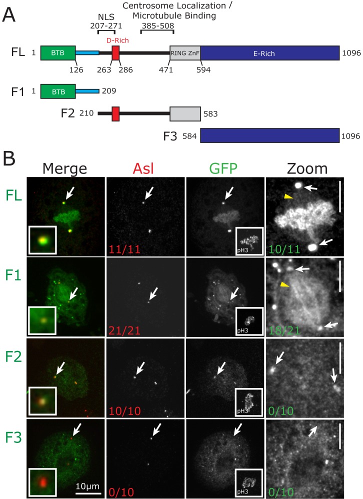
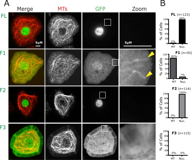
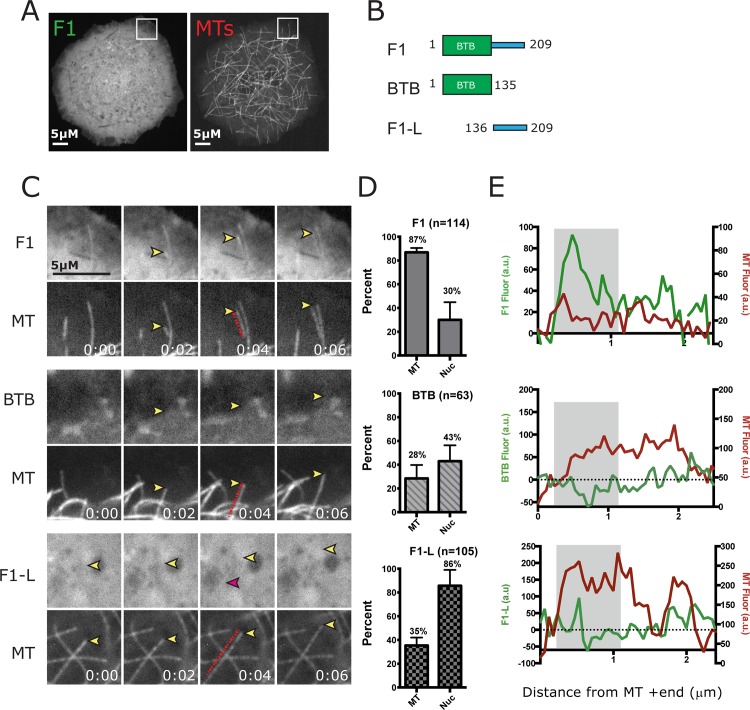
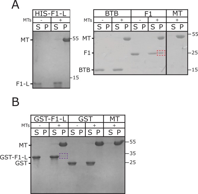
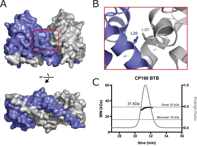
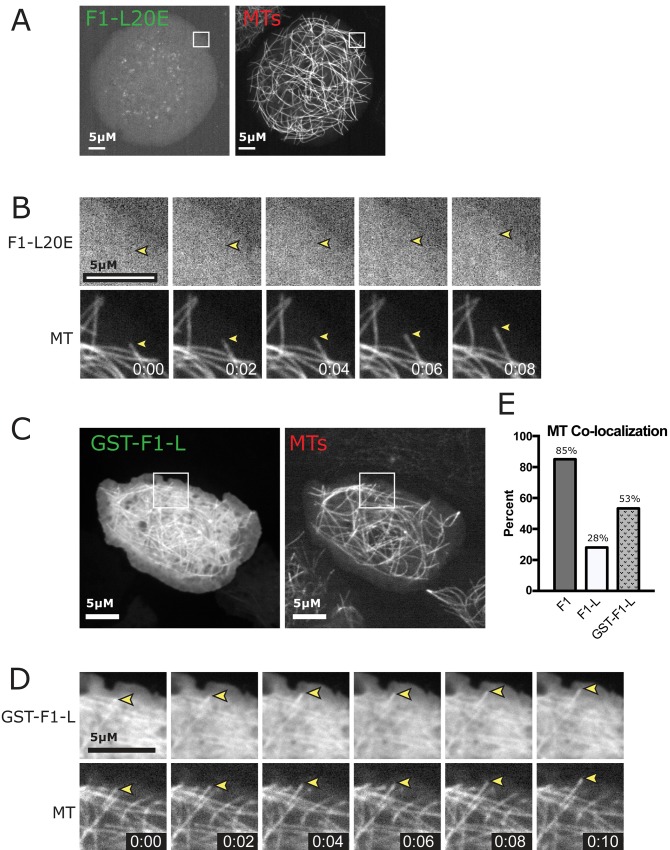
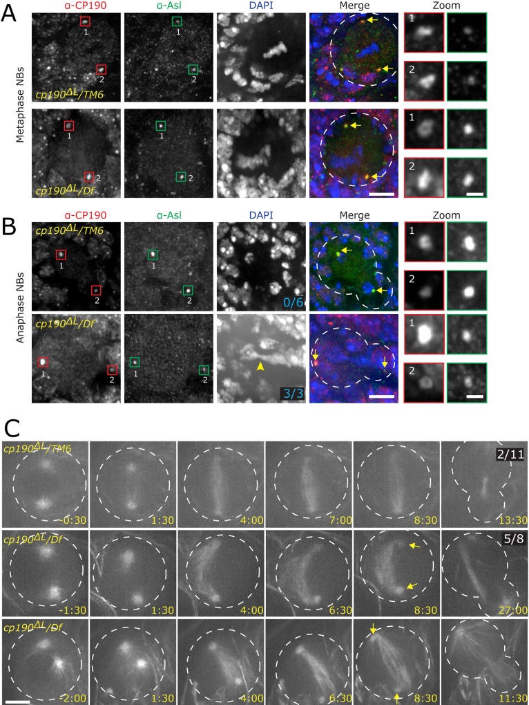
CP190 is a large, multi-domain protein, first identified as a centrosome protein with oscillatory localization over the course of the cell cycle. During interphase it has a well-established role within the nucleus as a chromatin insulator. Upon nuclear envelope breakdown, there is a striking redistribution of CP190 to centrosomes and the mitotic spindle, in addition to the population at chromosomes. Here, we investigate CP190 in detail by performing domain analysis in cultured Drosophila S2 cells combined with protein structure determination by X-ray crystallography, in vitro biochemical characterization, and in vivo fixed and live imaging of cp190 mutant flies. Our analysis of CP190 identifies a novel N-terminal centrosome and microtubule (MT) targeting region, sufficient for spindle localization. This region consists of a highly conserved BTB domain and a linker region that serves as the MT binding domain. We present the 2.5 Å resolution structure of the CP190 N-terminal 126 amino acids, which adopts a canonical BTB domain fold and exists as a stable dimer in solution. The ability of the linker region to robustly localize to MTs requires BTB domain-mediated dimerization. Deletion of the linker region using CRISPR significantly alters spindle morphology and leads to DNA segregation errors in the developing Drosophila brain neuroblasts. Collectively, we highlight a multivalent MT-binding architecture in CP190, which confers distinct subcellular cytoskeletal localization and function during mitosis.


Conflict of interest statement

Competing Interests: The authors have declared that no competing interests exist.

Figures

Fig 1
Fig 1. CP190 localization to the mitotic spindle is driven by its N-terminal region.
A, Domain structure of CP190. Shown is the BTB domain (green), linker (blue), D-rich domain (red), Zinc finger domain (grey), and the E-rich domain (blue). Indicated above the graphic representation are the previously identified nuclear localization signal (NLS) and the centrosome localization/MT binding domain [11]. Below the full length (FL) CP190 is a schematic of fragments used for this study (F1, F2, and F3). B, Drosophila S2 cells transfected with the indicated GFP-CP190 constructs (green). Shown are mitotic cells fixed and stained for Asterless (Asl, red) to mark centrosomes and pH3 (mitotic specific histone marker, inset in the GFP column). White arrows designate the centrosome. Red numbers on Asl column indicate the fraction of mitotic cells that exhibit GFP localization to centrosomes. Zoom of GFP channel (right column) is contrast enhanced to emphasize GFP signal on the mitotic spindle (yellow arrowheads). Green numbers indicate the fraction of mitotic cells with GFP signal at the spindle. Scale bars (B) = 10 μm, (zoom) = 5 μm.
Fig 2
Fig 2. Interphase cells serve as an excellent model to study CP190 MT association.
A, Drosophila S2 cells expressing GFP-CP190 constructs and TagRFP-Tubulin. Box in GFP channel is zoomed to highlight GFP localization to MTs (right column). F1 localizes to centrosomes and is unique in its localization to MTs during interphase, similar to what we document in mitosis (Fig 1B ). B, Quantification of the percent of cells in which CP190 constructs co-localize with MTs or show nuclear (Nuc) localization. FL and F2 are localized to the nucleus during interphase. F3 is cytoplasmic during interphase. F1 localizes to MTs robustly during interphase (see S1 Fig). Scale bars = 5 μm.
Fig 3
Fig 3. CP190-F1 is enriched at the plus ends of growing MTs.
A, Images of S2 cells expressing GFP-F1 and TagRFP-Tubulin. Box indicates inset for zoom in C. B, Schematic of the CP190-F1 sub-fragments F1, BTB and Linker domain (F1-L) analyzed for MT association. C, Live-cell imaging of F1 reveals enrichment at the plus end of MTs. Yellow arrowhead indicates a growing MT plus end. D, Graphs indicate percent of cells with MT and Nuclear localization. E, Graphs shown to the far right are line scans along the MTs indicated in frames 0:04 (red dashed line) in C. X-axes are arbitrary fluorescence units (a.u.) and y-axis is distance in microns from the MT +tip end (coordinate x = 0). F1 linescan indicates its enrichment at the MT +ends (grey area on graph). In contrast, the BTB domain and F1-L show very weak localization to some MTs (pink arrowhead, grey box on graph). Scale bars = 5μm. Time = min:s.
Fig 4
Fig 4. CP190-F1 binds directly to MTs.
MT co-sedimentation assay with purified components. A, Coomassie stained gel shows CP190-Linker (F1-L), CP190-BTB domain and CP190-F1 fragment incubated without (-) and with (+) MTs followed by high-speed centrifugation. Supernatant (S) and pellet (P) fractions are run separately. F1 co-sediments with MTs (red dotted box), while neither the F1-L linker nor the BTB domain show MT-binding. B, Coomassie stained gel shows the F1-L linker artificially dimerized as a GST fusion (GST-F1-L) and GST alone incubated without (-) and with (+) MTs followed by high-speed centrifugation. Supernatant (S) and pellet (P) fractions are run separately. Although we were not able to fully recapitulate F1 MT binding, dimerized linker (GST-F1-L) is able to weakly associate with MTs (purple dotted box).
Fig 5
Fig 5. The CP190 BTB domain is highly conserved across species.
CP190 F1 region sequence alignment across six species shows a high level of conservation within the CP190 BTB domain. Residue numbers correspond to Drosophila melanogaster CP190. Conservation is mapped on the alignment. Residues with 100% identity are mapped in green and residues with 100% similarity are mapped in yellow. Below the CP190 sequences (last row) are displayed residues that are highly conserved among other Drosophila melanogaster BTB domain containing proteins (not CP190) and are likely involved in the BTB domain fold. Residues from this set that are divergent in CP190 are indicated above the Drosophila CP190 sequence by a red rectangle. This alignment suggests that there are residues important for the BTB domain structural fold and others that are specifically unique to CP190. Secondary structure is mapped on the alignment. Arrows indicate β-sheets and rectangles indicate α-helices. Red letters in the linker region highlight basic (R and K) residues. The SGLP motif is underlined. Solvent accessible surface area (SASA) of a theoretical monomer as well as the buried surface area (BSA) of the homodimer is plotted above the alignment. B, The BTB domain adopts a dimeric fold and makes extensive structural contacts with its dimeric mate. One BTB domain is shown in color with secondary structure elements colored as in Figure 5A. The dimeric mate is colored grey. The β1 and β6’ strands from dimeric mates form an antiparallel, two-stranded β-sheet. C, The BTB domain shown in surface representation, rotated 90° about the x-axis relative to the orientation shown in B. Conservation is colored on the structure (above) following the scheme in A. Electrostatics are indicated on the structure below.
Fig 6
Fig 6. The BTB domain exists as a dimer and is critical for F1 MT binding.
A, Space-filling model showing homodimeric mates in the crystal lattice with one molecule colored purple, the other grey. The β1 strands wrap along the length of the opposing molecule. B, Zoom of the dimer interface shows the hydrophobic leucine (L20) residue that was mutated to a charged glutamic acid residue (L20E). This mutation destabilizes the BTB domain at the dimer interface. C, CP190’s BTB domain exists as a stable dimer in solution as shown by SEC-MALS. The x-axis is the time of the run in minutes, the left y-axis is the MW (black: kDa), and the right y-axis is the Raleigh ratio (grey). The predicted CP190 BTB domain monomer and dimer molecular weights are indicated by dotted lines (16 and 32 kDa respectively). The experimentally determined mass of the eluted CP190 BTB domain is plotted as a black line and corresponds to a homodimer.
Fig 7
Fig 7. Artificially dimerized F1-L localizes to MT.
A, S2 cell expressing the F1-L20E dimerization interface mutant and TagRFP-Tubulin. F1-L20E is unable to localize to MTs in vivo. White box indicates zoom for B. B, Time-lapse from boxed region showing that F1-L20E does not localize to MTs (yellow arrowhead). C, S2 cells expressing GST-F1-L and TagRFP-Tubulin. White box indicates zoom for D. D, Time-lapse images from boxed region in C showing robust localization of GST-F1-L to the MT lattice. Scale bars = 5 μm. E, Percent of cells showing MT-Colocalization of F1, F1-L and GST-F1-L. Localization is partially rescued with artificially dimerized F1-L. Time is indicated in min:s.
Fig 8
Fig 8. CP190-L is important for spindle formation in developing brain.
Drosophila NBs were fixed and stained as indicated. A. Metaphase NBs are shown with CP190 in red, Asl to mark the centrosome in green, and DAPI in blue. WT control (cp190 ΔL /TM6) is in the top row and mutant cp190 ΔL / Df p11 in the bottom row. Boxed regions indicating centrosomes are magnified and shown on the far right panels (numbers next to centrosome indicate which zoomed centrosome is displayed). Yellow arrows point out centrosomes in the merged channel. White dotted line indicates NB outline. B.Fixed anaphase NBs. Labeling is the same as in A. Note a lagging chromosome in cp190 ΔL / Df p11 (yellow arrowhead), which is never seen in WT (frequency indicated in DAPI channel). Scale bar for A and B = 5μm, zoom = 1μm. C, Live imaging of MTs in WT (cp190 ΔL /TM6) and cp190 ΔL / Df p11mutant NBs, note bent spindle (frequency indicated in right most image) and detached centrosome (yellow arrows) in mutant cell. Scale bar = 5μm. Panels in C from top to bottom correspond to S1 Movie, S2 Movie and S3 Movie, respectively.

Similar articles

Cited by

References

    1. Mitchison T, Kirschner M (1984) Dynamic instability of microtubule growth. Nature 312: 237–242. - PubMed
    1. Weisenbe Rc (1972) Microtubule Formation in-Vitro in Solutions Containing Low Calcium Concentrations. Science 177: 1104–&. - PubMed
    1. Dictenberg JB, Zimmerman W, Sparks CA, Young A, Vidair C, et al. (1998) Pericentrin and gamma-tubulin form a protein complex and are organized into a novel lattice at the centrosome. J Cell Biol 141: 163–174. - PMC - PubMed
    1. Moritz M, Braunfeld MB, Sedat JW, Alberts B, Agard DA (1995) Microtubule nucleation by gamma-tubulin-containing rings in the centrosome. Nature 378: 638–640. - PubMed
    1. Goshima G, Mayer M, Zhang N, Stuurman N, Vale RD (2008) Augmin: a protein complex required for centrosome-independent microtubule generation within the spindle. J Cell Biol 181: 421–429. 10.1083/jcb.200711053 - DOI - PMC - PubMed

Publication types

LinkOut - more resources