

Complex Evolutionary and Genetic Patterns Characterize the Loss of Scleral Ossification in the Blind Cavefish Astyanax mexicanus
- PMID: 26649887
- PMCID: PMC4674125
- DOI: 10.1371/journal.pone.0142208
Complex Evolutionary and Genetic Patterns Characterize the Loss of Scleral Ossification in the Blind Cavefish Astyanax mexicanus
Abstract
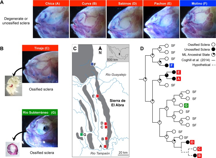
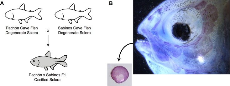
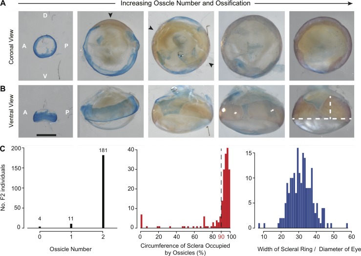
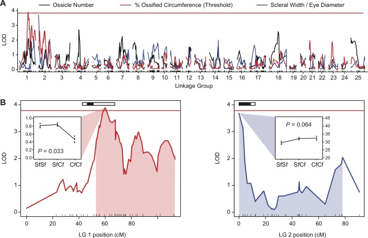
The sclera is the tough outer covering of the eye that provides structural support and helps maintain intraocular pressure. In some fishes, reptiles, and birds, the sclera is reinforced with an additional ring of hyaline cartilage or bone that forms from scleral ossicles. Currently, the evolutionary and genetic basis of scleral ossification is poorly understood, especially in teleost fishes. We assessed scleral ossification among several groups of the Mexican tetra (Astyanax mexicanus), which exhibit both an eyed and eyeless morph. Although eyed Astyanax surface fish have bony sclera similar to other teleosts, the ossicles of blind Astyanax cavefish generally do not form. We first sampled cavefish from multiple independent populations and used ancestral character state reconstructions to determine how many times scleral ossification has been lost. We then confirmed these results by assessing complementation of scleral ossification among the F1 hybrid progeny of two cavefish populations. Finally, we quantified the number of scleral ossicles present among the F2 hybrid progeny of a cross between surface fish and cavefish, and used this information to identify quantitative trait loci (QTL) responsible for this trait. Our results indicate that the loss of scleral ossification is common-but not ubiquitous-among Astyanax cavefish, and that this trait has been convergently lost at least three times. The presence of wild-type, ossified sclera among the F1 hybrid progeny of a cross between different cavefish populations confirms the convergent evolution of this trait. However, a strongly skewed distribution of scleral ossicles found among surface fish x cavefish F2 hybrids suggests that scleral ossification is a threshold trait with a complex genetic basis. Quantitative genetic mapping identified a single QTL for scleral ossification on Astyanax linkage group 1. We estimate that the threshold for this trait is likely determined by at least three genetic factors which may control the severity and onset of lens degeneration in cavefishes. We conclude that complex evolutionary and genetic patterns underlie the loss of scleral ossification in Astyanax cavefish.
Conflict of interest statement
Figures
Similar articles
-
Dual roles of the retinal pigment epithelium and lens in cavefish eye degeneration.J Exp Zool B Mol Dev Evol. 2020 Nov;334(7-8):438-449. doi: 10.1002/jez.b.22923. Epub 2020 Jan 12. J Exp Zool B Mol Dev Evol. 2020. PMID: 31930686 Free PMC article.
-
Two - three loci control scleral ossicle formation via epistasis in the cavefish Astyanax mexicanus.PLoS One. 2017 Feb 9;12(2):e0171061. doi: 10.1371/journal.pone.0171061. eCollection 2017. PLoS One. 2017. PMID: 28182695 Free PMC article.
-
Quantitative genetic analysis of retinal degeneration in the blind cavefish Astyanax mexicanus.PLoS One. 2013;8(2):e57281. doi: 10.1371/journal.pone.0057281. Epub 2013 Feb 20. PLoS One. 2013. PMID: 23437360 Free PMC article.
-
Evolutionary Genetics of the Cavefish Astyanax mexicanus.Adv Genet. 2016;95:117-59. doi: 10.1016/bs.adgen.2016.03.001. Epub 2016 Jun 13. Adv Genet. 2016. PMID: 27503356 Review.
-
The complex origin of Astyanax cavefish.BMC Evol Biol. 2012 Jun 30;12:105. doi: 10.1186/1471-2148-12-105. BMC Evol Biol. 2012. PMID: 22747496 Free PMC article. Review.
Cited by
-
The elusive scleral cartilages: Comparative anatomy and development in teleosts and avians.Anat Rec (Hoboken). 2025 Jul;308(7):1838-1850. doi: 10.1002/ar.25345. Epub 2023 Nov 9. Anat Rec (Hoboken). 2025. PMID: 37943147 Free PMC article. Review.
-
Dual roles of the retinal pigment epithelium and lens in cavefish eye degeneration.J Exp Zool B Mol Dev Evol. 2020 Nov;334(7-8):438-449. doi: 10.1002/jez.b.22923. Epub 2020 Jan 12. J Exp Zool B Mol Dev Evol. 2020. PMID: 31930686 Free PMC article.
-
Neural Crest Transplantation Reveals Key Roles in the Evolution of Cavefish Development.Integr Comp Biol. 2018 Sep 1;58(3):411-420. doi: 10.1093/icb/icy006. Integr Comp Biol. 2018. PMID: 29718239 Free PMC article.
-
Behavioural changes controlled by catecholaminergic systems explain recurrent loss of pigmentation in cavefish.Proc Biol Sci. 2018 May 16;285(1878):20180243. doi: 10.1098/rspb.2018.0243. Proc Biol Sci. 2018. PMID: 29720416 Free PMC article.
-
Quantification and comparison of teleost scleral cartilage development and growth.J Anat. 2022 Oct;241(4):1014-1025. doi: 10.1111/joa.13727. Epub 2022 Jul 21. J Anat. 2022. PMID: 36574601 Free PMC article.
References
Publication types
MeSH terms
Grants and funding
LinkOut - more resources
Full Text Sources
Other Literature Sources
Miscellaneous
