. 2015 Dec 9;10(12):e0144510.
doi: 10.1371/journal.pone.0144510. eCollection 2015.

Biophysical Characterization of G-Quadruplex Recognition in the PITX1 mRNA by the Specificity Domain of the Helicase RHAU

Affiliations

Biophysical Characterization of G-Quadruplex Recognition in the PITX1 mRNA by the Specificity Domain of the Helicase RHAU

Emmanuel O Ariyo et al. PLoS One. .

Abstract

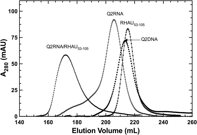
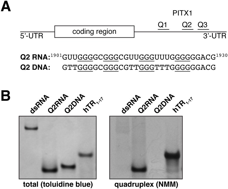
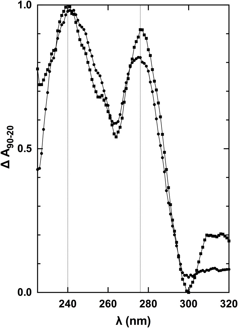
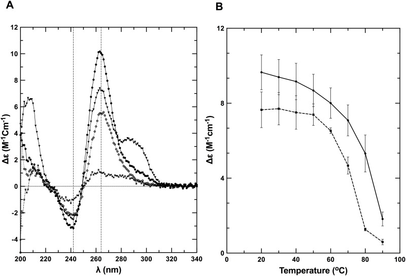
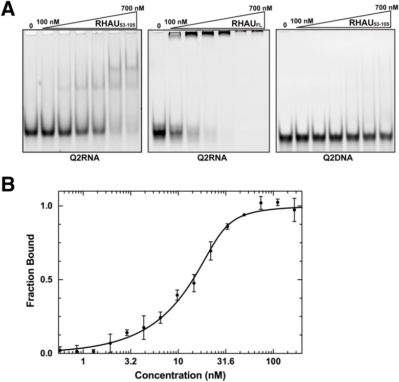
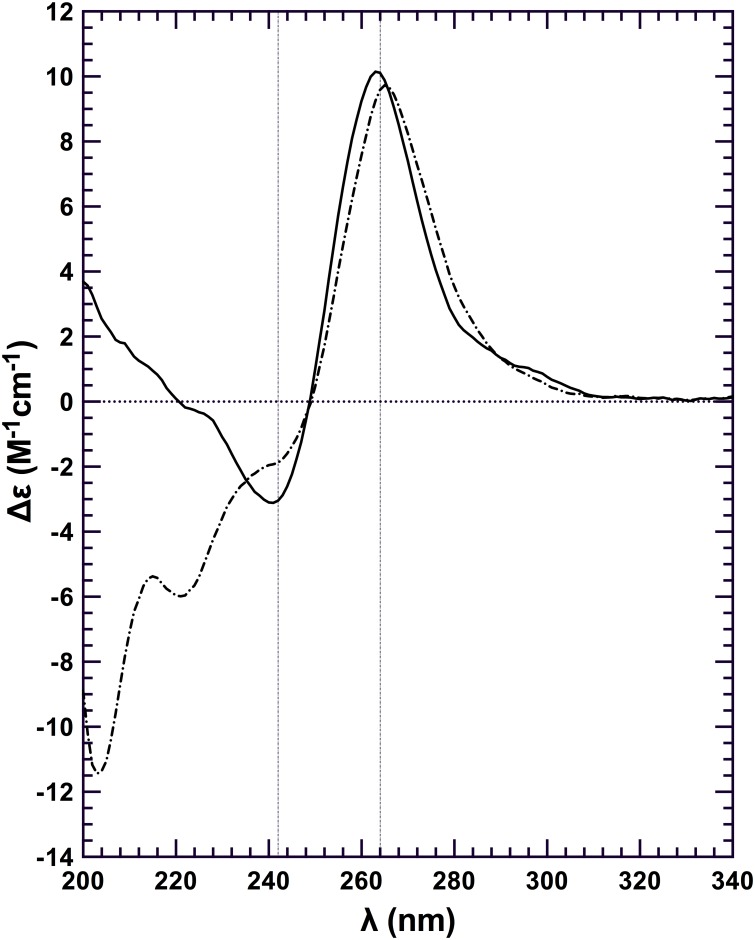
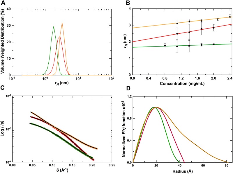
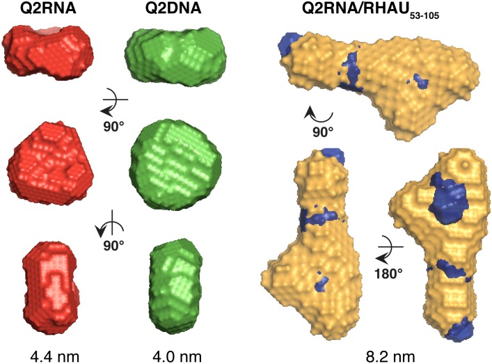
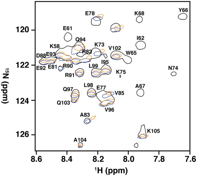
Nucleic acids rich in guanine are able to fold into unique structures known as G-quadruplexes. G-quadruplexes consist of four tracts of guanylates arranged in parallel or antiparallel strands that are aligned in stacked G-quartet planes. The structure is further stabilized by Hoogsteen hydrogen bonds and monovalent cations centered between the planes. RHAU (RNA helicase associated with AU-rich element) is a member of the ATP-dependent DExH/D family of RNA helicases and can bind and resolve G-quadruplexes. RHAU contains a core helicase domain with an N-terminal extension that enables recognition and full binding affinity to RNA and DNA G-quadruplexes. PITX1, a member of the bicoid class of homeobox proteins, is a transcriptional activator active during development of vertebrates, chiefly in the anterior pituitary gland and several other organs. We have previously demonstrated that RHAU regulates PITX1 levels through interaction with G-quadruplexes at the 3'-end of the PITX1 mRNA. To understand the structural basis of G-quadruplex recognition by RHAU, we characterize a purified minimal PITX1 G-quadruplex using a variety of biophysical techniques including electrophoretic mobility shift assays, UV-VIS spectroscopy, circular dichroism, dynamic light scattering, small angle X-ray scattering and nuclear magnetic resonance spectroscopy. Our biophysical analysis provides evidence that the RNA G-quadruplex, but not its DNA counterpart, can adopt a parallel orientation, and that only the RNA can interact with N-terminal domain of RHAU via the tetrad face of the G-quadruplex. This work extends our insight into how the N-terminal region of RHAU recognizes parallel G-quadruplexes.


Conflict of interest statement

Competing Interests: The authors have declared that no competing interests exist.

Figures

Fig 1
Fig 1. Purification of protein and nucleic acid components.
Elution profiles obtained on a HiLoad Superdex 75 26/60 column, with each species labeled.
Fig 2
Fig 2. Q2RNA, but not Q2DNA, stains with a parallel G4 dye.
(A) Schematic showing the G4 forming regions of PITX1 mRNA; the RNA and DNA equivalent sequences with the guanylate tracts underlined are also shown. 250 pmol of Q2RNA and Q2DNA were separated by native-Tris-borate EDTA (TBE) polyacrylamide gel electrophoresis, stained with (B) toluidine blue, and (C) G4-specific dye N-methyl mesoporphyrin IX alongside their positive (G4: hTR1-17) and negative (double stranded RNA) controls.
Fig 3
Fig 3. Normalized thermal difference spectra of Q2RNA and DNA counterpart.
TDS of Q2RNA (circles) and Q2DNA (squares) in 10 mM Tris, pH 7.5, 100 mM KCl, 1 mM EDTA. For details of analysis, see Materials and Methods.
Fig 4
Fig 4. Q2RNA and its DNA counterpart adopt G4 structures.
(A) Far-UV CD spectra of Q2RNA and Q2DNA obtained in 10 mM Tris, pH 7.5, 100 mM KCl, 1 mM EDTA buffer of Q2RNA at 20°C (closed circles) and at 80°C (open circles) as well as of Q2DNA at 20°C (closed triangles) and at 80°C (closed squares). (B) CD melting curves of Q2RNA (——) and Q2DNA (------) monitored by spectropolarimetry at 264 nm in the same buffer.
Fig 5
Fig 5. RHAU53-105 forms a complex with Q2RNA.
(A) Electrophoretic mobility shift assays (EMSA) were performed using a constant 150 nM concentration of Q2RNA or Q2DNA and a variable concentration from 0–700 nM of RHAU53-105 or full-length RHAU. The 12% native Tris borate-EDTA (TBE) polyacrylamide gels were stained with SYBR Gold for visualization. (B) Microscale thermophoresis measurements performed using 3’-FAM Q2RNA (25 nM) in complex with RHAU53-105 at several concentrations (0.6–250 nM). Reverse T-Jump signals from the traces were fit as described in the Materials & Methods.
Fig 6
Fig 6. RHAU53-105 binding is not sufficient for G4 unwinding.
Far-UV CD spectra of Q2RNA (——); and Q2RNA/RHAU53-105 complex (··—··) at 20 μM and 20°C with all the spectra normalized to the number of nucleotides. The G4 features of the RNA in the context of the complex are observed in the region unique to nucleic acids (~250–320 nm).
Fig 7
Fig 7. SAXS data collection and analysis.
(A) DLS profiles of Q2RNA, Q2DNA and Q2RNA/RHAU53-105 complex. (B) Concentration dependence of hydrodynamic radii measured by DLS of the molecules in (A). (C) Merged SAXS data of Q2RNA, Q2DNA and Q2RNA/RHAU53-105 complex. (D) The corresponding pair distance distribution functions. (Color code: red, Q2RNA; green, Q2DNA; golden, Q2RNA/RHAU53-105 complex).
Fig 8
Fig 8. SAXS envelopes of Q2RNA, Q2DNA and Q2RNA/RHAU53-105 complex.
Color code: Red, Q2RNA; green, Q2DNA; golden, Q2RNA/RHAU53-105 complex. D max values are shown beneath each species. The SAXS shape model of RHAU53-105 (blue) was superimposed on the complex.
Fig 9
Fig 9. Amino acids in and adjacent to the RSM mediate recognition of RNA G4.
15N-HSQC spectral overlay of RHAU53-105 free (black) and in complex with Q2RNA (orange) or hTR1-20 (blue) with a subset of assigned resonances labeled. Data for RHAU53-105 and RHAU53-105/hTR1-20 were previously obtained, and used for comparison (30).

References

    1. Hoogsteen K. Crystal and Molecular Structure of a Hydrogen-Bonded Complex between 1-Methylthymine and 9-Methyladenine. Acta Crystallogr. 1963;16(9):907 10.1107/S0365110x63002437 WOS:A19631326A00007. - DOI
    1. Burge S, Parkinson GN, Hazel P, Todd AK, Neidle S. Quadruplex DNA: sequence, topology and structure. Nucleic Acids Res. 2006;34(19):5402–15. 10.1093/nar/gkl655 - DOI - PMC - PubMed
    1. Bochman ML, Paeschke K, Zakian VA. DNA secondary structures: stability and function of G-quadruplex structures. Nat Rev Genet. 2012;13(11):770–80. 10.1038/nrg3296 - DOI - PMC - PubMed
    1. Sacca B, Lacroix L, Mergny JL. The effect of chemical modifications on the thermal stability of different G-quadruplex-forming oligonucleotides. Nucleic Acids Res. 2005;33(4):1182–92. 10.1093/nar/gki257 - DOI - PMC - PubMed
    1. Zhang AY, Bugaut A, Balasubramanian S. A sequence-independent analysis of the loop length dependence of intramolecular RNA G-quadruplex stability and topology. Biochemistry. 2011;50(33):7251–8. 10.1021/bi200805j - DOI - PMC - PubMed

Publication types