. 2015 Dec 9:4:e11012.
doi: 10.7554/eLife.11012.

Precise assembly of complex beta sheet topologies from de novo designed building blocks

Affiliations

Precise assembly of complex beta sheet topologies from de novo designed building blocks

Indigo Chris King et al. Elife. .

Abstract

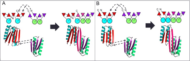
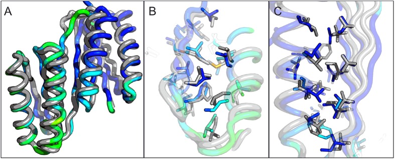
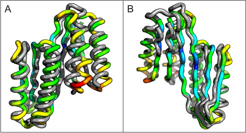
Design of complex alpha-beta protein topologies poses a challenge because of the large number of alternative packing arrangements. A similar challenge presumably limited the emergence of large and complex protein topologies in evolution. Here, we demonstrate that protein topologies with six and seven-stranded beta sheets can be designed by insertion of one de novo designed beta sheet containing protein into another such that the two beta sheets are merged to form a single extended sheet, followed by amino acid sequence optimization at the newly formed strand-strand, strand-helix, and helix-helix interfaces. Crystal structures of two such designs closely match the computational design models. Searches for similar structures in the SCOP protein domain database yield only weak matches with different beta sheet connectivities. A similar beta sheet fusion mechanism may have contributed to the emergence of complex beta sheets during natural protein evolution.

Keywords: E. coli; beta sheets; biophysics; computational biology; protein design; protein folding; structural biology; systems biology.


Conflict of interest statement

The authors declare that no competing interests exist.

Figures

Figure 1.
Figure 1.. Generation of protein domains with single extended beta sheets by inserting one beta sheet containing protein into another.
(A) Insertion of a ferrrodoxin domain (purple) into TOP7 (red). (B) Insertion of one ferrodoxin domain into another. In both cases, two beta strands from each partner (red and purple) are concatenated to form the central strand pair of the fusion protein (pink). DOI: http://dx.doi.org/10.7554/eLife.11012.003
Figure 2.
Figure 2.. Comparison of the crystal structure of ferredoxin-TOP7 fusion to design model.
(A) Backbone superposition of the crystal structure of ferredoxin-TOP7 (4KYZ, chain A) with the design model. The backbones of the two proteins are nearly identical. (B, C) The core sidechain packing in the ferrodoxin-TOP7 fusion is very similar in the crystal structure and design model both in the insert (B) and host (C) domains. The crystal structure is colored by B-factor and the design model is in gray. DOI: http://dx.doi.org/10.7554/eLife.11012.004
Figure 2—figure supplement 1.
Figure 2—figure supplement 1.. The circular dichroism spectrum of ferrodoxin-TOP7 has the shape expected for an alpha/beta protein.
DOI: http://dx.doi.org/10.7554/eLife.11012.005
Figure 3.
Figure 3.. Comparison of the crystal structure of the ferredoxin-ferredoxin fusion to the design model.
The crystal structure (5CW9) aligns well with the design model over both the helices (A) and the fused beta sheet (B). DOI: http://dx.doi.org/10.7554/eLife.11012.006
Figure 3—figure supplement 1.
Figure 3—figure supplement 1.. Circular dichroism spectra of ferrrodoxin-ferrodoxin at 25°C.
DOI: http://dx.doi.org/10.7554/eLife.11012.007
Figure 3—figure supplement 2.
Figure 3—figure supplement 2.. Ferredoxin-Ferredoxin 2Fo-Fc omit map superimposed with crystal structure shows core packing of host (A) and insert (B) domains.
DOI: http://dx.doi.org/10.7554/eLife.11012.008
Figure 4.
Figure 4.. Top two SCOP domain structural homologues for Fd-Top7 (A) and Fd-Fd (B) designed domain found in TM-align searches.
Ribbon diagrams are shown on left, the strand connectivity, at the right. The beta strand connectivity is quite different in the designs than in these closest structural matches. DOI: http://dx.doi.org/10.7554/eLife.11012.009
Figure 4—figure supplement 1.
Figure 4—figure supplement 1.. Parent domain PDB structures (2KL8, 1QYS) and daughter designed folds (5CW9,4KYZ) (pink) mapped into the α+β region of the SCOP domains network of Nepomnyachi et al. (A) and zoomed region (B) highlighting parent, designed, and first neighbor folds.
DOI: http://dx.doi.org/10.7554/eLife.11012.010
Figure 4—figure supplement 2.
Figure 4—figure supplement 2.. Neutral drift mutant models, relative changes to predicted free energy of folding in REU (Rosetta Energy Units), and multiple sequence alignment of parent and designed sequences, showing mutations in ferredoxin-top7 (A) and ferredoxin-ferredoxin (B).
DOI: http://dx.doi.org/10.7554/eLife.11012.011

References

    1. Aroul-Selvam R, Hubbard T, Sasidharan R. Domain insertions in protein structures. Journal of Molecular Biology. 2004;338:633–641. doi: 10.1016/j.jmb.2004.03.039. - DOI - PMC - PubMed
    1. Ay J, Gotz F, Borriss R, Heinemann U. Structure and function of the bacillus hybrid enzyme GluXyn-1: native-like jellyroll fold preserved after insertion of autonomous globular domain. Proceedings of the National Academy of Sciences of the United States of America. 1998;95:6613–6618. doi: 10.1073/pnas.95.12.6613. - DOI - PMC - PubMed
    1. Ben-Tal N, Kolodny R. Representation of the protein universe using classifications, maps, and networks. Israel Journal of Chemistry. 2014;54:1286–1292. doi: 10.1002/ijch.201400001. - DOI
    1. Berrondo M, Ostermeier M, Gray JJ. Structure prediction of domain insertion proteins from structures of individual domains. Structure. 2008;16:513–527. doi: 10.1016/j.str.2008.01.012. - DOI - PMC - PubMed
    1. Collinet B, Herve M, Pecorari F, Minard P, Eder O, Desmadril M. Functionally accepted insertions of proteins within protein domains. Journal of Biological Chemistry. 2000;275:17428–17433. doi: 10.1074/jbc.M000666200. - DOI - PubMed

Publication types

MeSH terms

Substances