. 2016 Mar;157(3):1122-34.
doi: 10.1210/en.2015-1480. Epub 2015 Dec 9.

Ferredoxin 1b (Fdx1b) Is the Essential Mitochondrial Redox Partner for Cortisol Biosynthesis in Zebrafish

Affiliations

Ferredoxin 1b (Fdx1b) Is the Essential Mitochondrial Redox Partner for Cortisol Biosynthesis in Zebrafish

Aliesha Griffin et al. Endocrinology. 2016 Mar.

Abstract

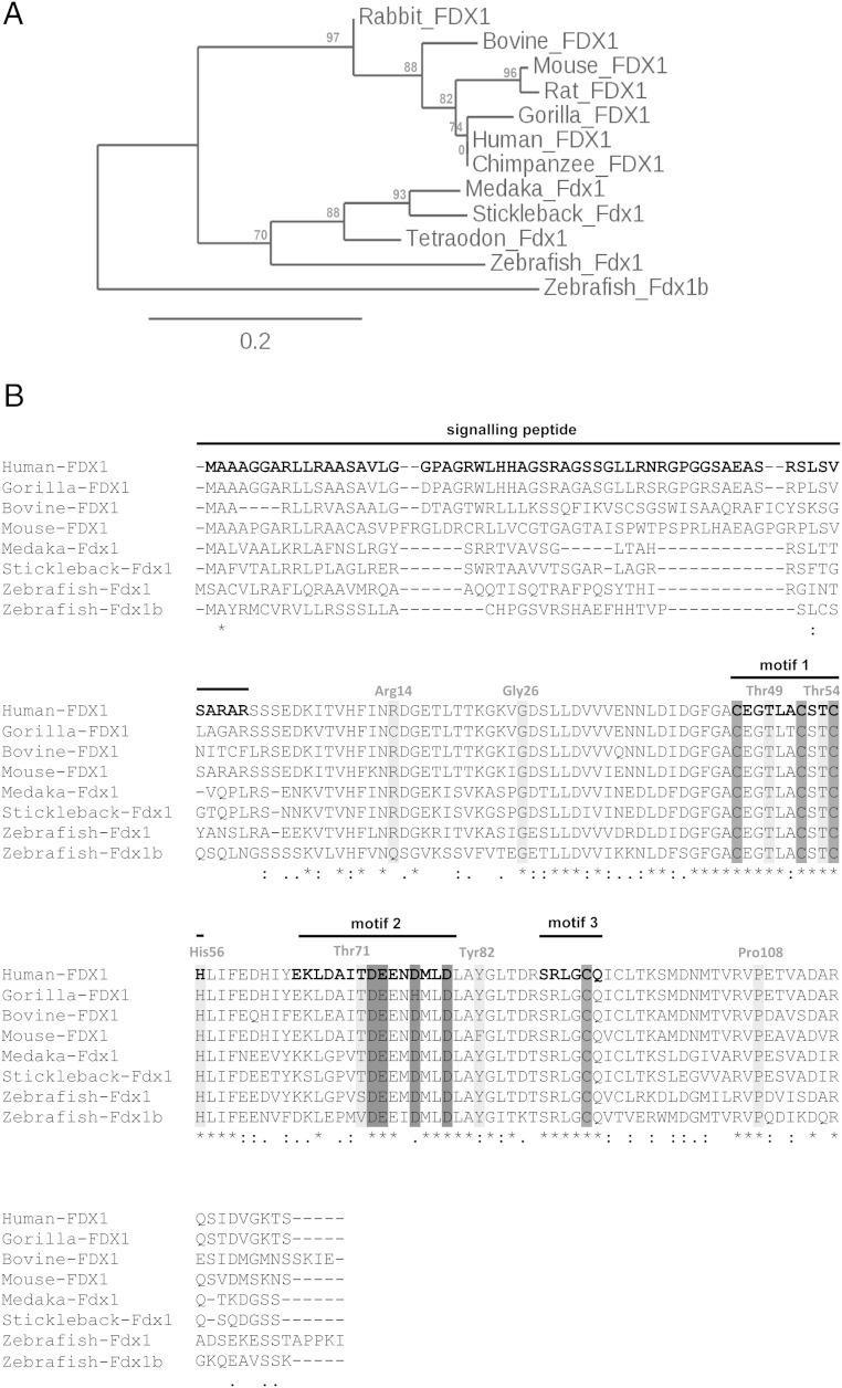
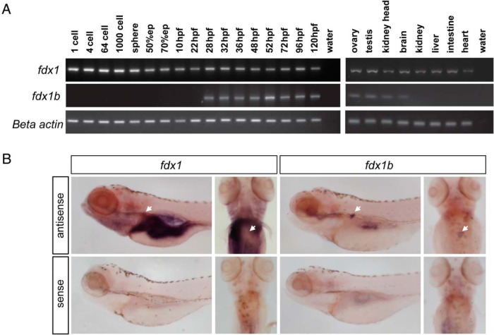
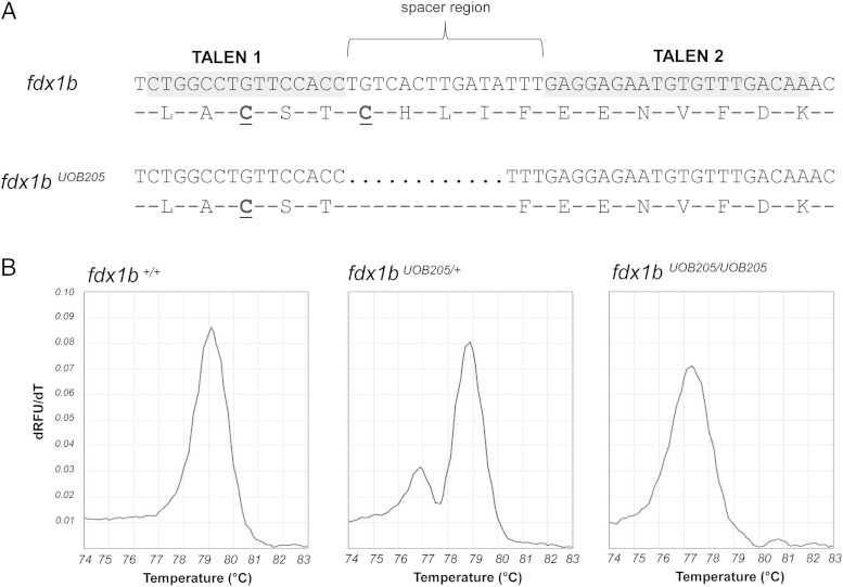
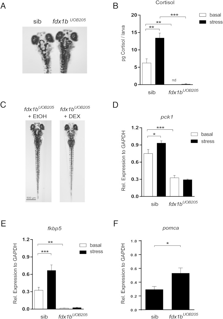
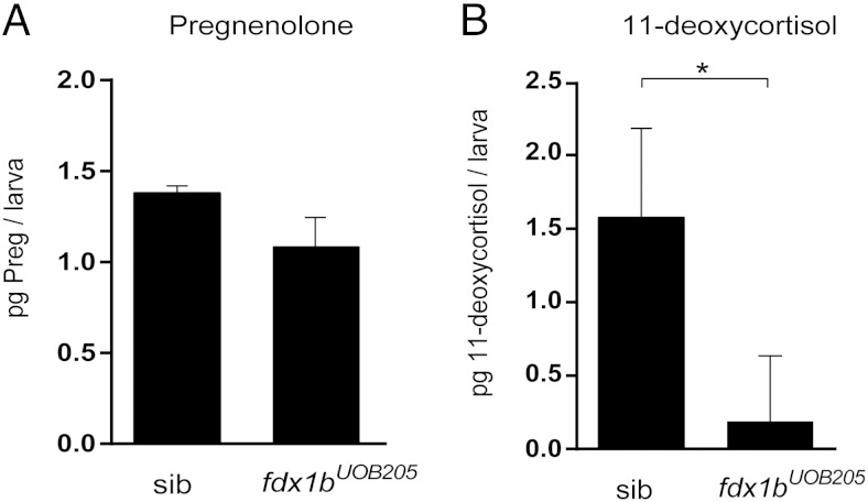
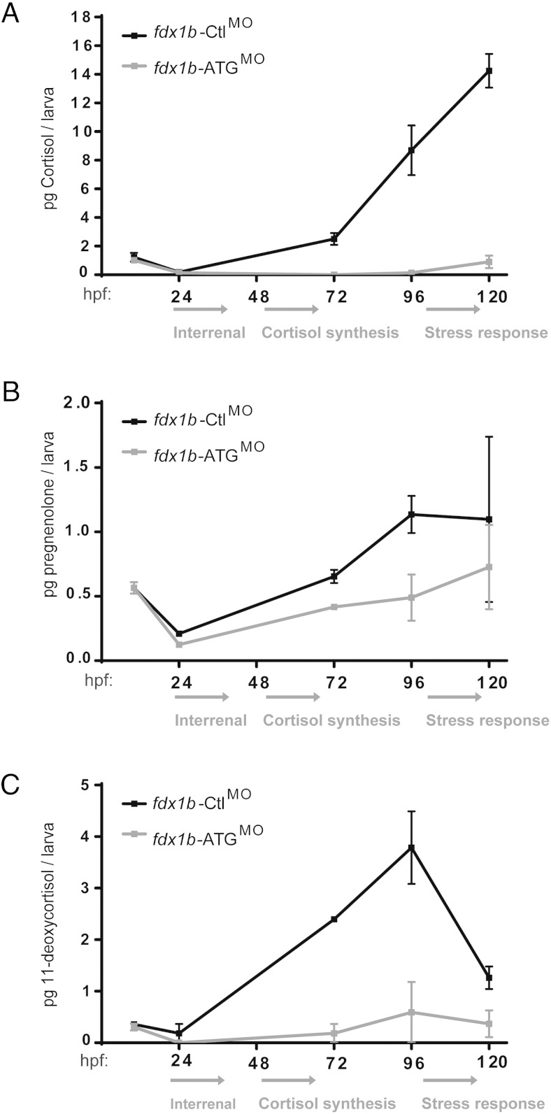
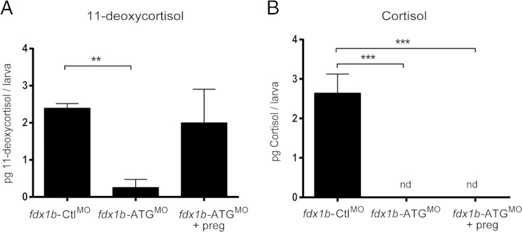
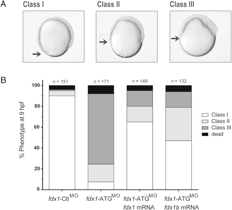
Mitochondrial cytochrome P450 (CYP) enzymes rely on electron transfer from the redox partner ferredoxin 1 (FDX1) for catalytic activity. Key steps in steroidogenesis require mitochondrial CYP enzymes and FDX1. Over 30 ferredoxin mutations have been explored in vitro; however, no spontaneously occurring mutations have been identified in humans leaving the impact of FDX1 on steroidogenesis in the whole organism largely unknown. Zebrafish are an important model to study human steroidogenesis, because they have similar steroid products and endocrine tissues. This study aimed to characterize the influence of ferredoxin on steroidogenic capacity in vivo by using zebrafish. Zebrafish have duplicate ferredoxin paralogs: fdx1 and fdx1b. Although fdx1 was observed throughout development and in most tissues, fdx1b was expressed after development of the zebrafish interrenal gland (counterpart to the mammalian adrenal gland). Additionally, fdx1b was restricted to adult steroidogenic tissues, such as the interrenal, gonads, and brain, suggesting that fdx1b was interacting with steroidogenic CYP enzymes. By using transcription activator-like effector nucleases, we generated fdx1b mutant zebrafish lines. Larvae with genetic disruption of fdx1b were morphologically inconspicuous. However, steroid hormone analysis by liquid chromatography tandem mass spectrometry revealed fdx1b mutants failed to synthesize glucocorticoids. Additionally, these mutants had an up-regulation of the hypothalamus-pituitary-interrenal axis and showed altered dark-light adaptation, suggesting impaired cortisol signaling. Antisense morpholino knockdown confirmed Fdx1b is required for de novo cortisol biosynthesis. In summary, by using zebrafish, we generated a ferredoxin knockout model system, which demonstrates for the first time the impact of mitochondrial redox regulation on glucocorticoid biosynthesis in vivo.


Figures

Figure 1.
Figure 1.
Evolutionary conservation of vertebrate ferredoxins. A, Maximum likelihood phylogenetic analysis of vertebrate Fdx1 protein sequences were conducted using the PhyML software under the SH-like likelihood-ratio test parameters. Scaled phylogenetic tree was drawn with TreeDyn software. Branch support values are represented in % and shown in red. B, The preprotein sequence of human, gorilla, bovine, mouse, medaka, stickleback, and zebrafish ferredoxin enzymes were aligned using ClustalW under default parameters. The 3 motifs common to all hydroxylase ferredoxins are shown. Motif 1 is a loop that contains 3 of the cysteine residues involved in Fe/S cluster binding (gray). Motif 2 contains a group of negatively charged amino acid residues, which are important for binding to CYP and ferredoxin reductase (dark gray). Motif 3 contains the conserved sequence around the fourth cysteine residue required for cluster binding. The signaling peptide targets the ferredoxin to the mitochondria and is cleaved to form the mature protein. Other known functionally important amino acid residues required for protein stabilization and redox potential are shown in gray. Amino acid residues are numbered according to the mature human FDX1 sequence.
Figure 2.
Figure 2.
Temporal and spatial expression of zebrafish fdx1 paralog genes. A, Expression of fdx1 and fdx1b was characterized in zebrafish during development and in adult tissues. Representative agarose gel images are shown from triplicate experiments from pools of 10 zebrafish embryos or larvae at the specified developmental stages or from adult tissues. β-Actin was used as a control for integrity of template cDNA. B, Detection of fdx1 and fdx1b mRNA at 120 hpf by WISH. Lateral and dorsal views are shown. Negative control sense probes did not show staining. Arrow indicates location of the interrenal gland.
Figure 3.
Figure 3.
Genetic disruption of the fdx1b locus in zebrafish by TALENs. A, Exon 4 of fdx1b was targeted for genetic disruption by TALENs. TALEN1 and TALEN2 recognized 15 and 20 nucleotides, respectively (gray). Each TALEN was separated by a 15-nucleotide spacer region. A line harboring a 12-bp deletion was established (fdx1bUOB205) (c.295_306del; p.Cys99_Ile102del). This mutation resulted in a 4-amino acid deletion, which includes a cysteine essential for Fe/S cluster binding (underlined). B, Genotyping of individual embryos was performed by HRM analysis. PCR amplification of wild-type fdx1b (fdx1b+/+) gives a product with a melting peak at 79°C, whereas fdx1bUOB205 allele gives a product with a melting peak at 77°C. Representative melting peaks are shown for fdx1b+/+, fdx1b heterozygous (fdx1bUOB205/+), and embryos homozygous for the mutation (fdx1bUOB205/UOB205).
Figure 4.
Figure 4.
Zebrafish fdx1bUOB205 homozygous mutants are glucocorticoid deficient. A, fdx1bUOB205 homozygous mutants have impaired visual-mediated adaption. When exposed to a lighter environment, fdx1bUOB205 homozygous mutants are darker in appearance when compared with control siblings, which had a least 1 wild-type allele (fdx1b+/+ or fdx1bUOB205/+). B, Cortisol concentrations were determined from fdx1bUOB205 homozygous mutant larvae (fdx1bUOB205) and sibling controls (sib) under basal and stressed conditions. Cortisol was not detected (nd) from fdx1bUOB205 homozygous mutants under basal conditions. Concentrations were determined from 3 independent replicates and are expressed as picograms per larva (mean ± SD). C, Treatment of fdx1bUOB205 homozygous mutants with the glucocorticoid agonist dexamethasone (DEX) restored the light adaptation phenotype in contrast to ethanol-treated controls (EtOH). mRNA expression of glucocorticoid response genes (D) pck1 and (E) fkbp5 in fdx1bUOB205 homozygous mutants and control siblings under basal and stressed conditions. Graphs represent mean relative expression ± SEM (normalized to gapdh). F, Quantitative real-time expression of pomca in fdx1bUOB205/UOB205 mutants relative to control siblings. Statistical analysis was performed using one-way ANOVA; *, P < .05; **, P < .01; ***, P < .001.
Figure 5.
Figure 5.
Pregnenolone and 11-deoxycortisol measurements in zebrafish fdx1bUOB205 homozygous mutants. A, Pregnenolone concentrations were measured in fdx1bUOB205 homozygous mutants (fdx1bUOB205) and sibling controls (sib) under normal conditions at 120 hpf. B, 11-deoxycortisol was measured after stress induction in mutants and controls. Steroid profiles were determined from 3 independent replicates and represented as mean picograms per larva ± SD. Statistical significance was determined by Student's t test; *, P < .05.
Figure 6.
Figure 6.
Fdx1b deficiency impairs de novo steroidogenesis during zebrafish development. Cortisol (A), pregnenolone (B), and 11-deoxycortisol (C) concentrations were measured during development from pools of 300 embryos injected with either fdx1b-ATGMO (gray) or fdx1b-CtlMO (black). Picograms of steroid per embryo or larva were calculated from concentrations measured from whole zebrafish extracts. Experiments were performed in duplicate. Mean ± SD was plotted against developmental stages.
Figure 7.
Figure 7.
Pregnenolone supplementation rescues 11-deoxycortisol but not cortisol production in fdx1b-deficient zebrafish at 72 hpf. fdx1b-ATG morphants were incubated in E3 medium containing 50nM pregnenolone from 10 hpf. 11-deoxycortisol (A) and cortisol (B) concentrations were measured in 72-hpf zebrafish larvae from 3 independent experiments. Concentrations expressed as picograms per larva were calculated from concentrations measured from whole zebrafish extracts. Mean ± SD is plotted against the treatment and compared using one-way ANOVA; ***, P < .001. nd, not detected.
Figure 8.
Figure 8.
Morphological phenotype of embryos injected with fdx1 morpholino at 9 hpf. A, Embryos injected with fdx1-ATGMO, fdx1-SplMO, or fdx1-CtlMO were classified based on their progression of epiboly at 9 hpf. Class I represents a normal epiboly, and class II and class III represent delayed movement as indicated by the black arrow. B, Injection of 9 ng of fdx1-CtlMO showed 95% of the normal class I phenotype compared with fdx1-ATGMO-injected embryos, which had 68.9% class II and 18% class III. Coinjection of either fdx1 or fdx1b mRNA with fdx1-ATGMO partially restored embryos to a class I phenotype.

Comment in

References

    1. Miller WL. Minireview: regulation of steroidogenesis by electron transfer. Endocrinology. 2005;146:2544–2550. - PubMed
    1. Meyer J. Iron-sulfur protein folds, iron-sulfur chemistry, and evolution. J Biol Inorg Chem. 2008;13:157–170. - PubMed
    1. Ewen KM, Ringle M, Bernhardt R. Adrenodoxin–a versatile ferredoxin. IUBMB Life. 2012;64:506–512. - PubMed
    1. Miller WL, Auchus RJ. The molecular biology, biochemistry, and physiology of human steroidogenesis and its disorders. Endocr Rev. 2011;32:81–151. - PMC - PubMed
    1. Miller WL. Steroid hormone synthesis in mitochondria. Mol Cell Endocrinol. 2013;379:62–73. - PubMed

Publication types

MeSH terms