. 2015 Dec 8;5(6):1196-1209.
doi: 10.1016/j.stemcr.2015.11.001.

Notch-Dependent Pituitary SOX2(+) Stem Cells Exhibit a Timed Functional Extinction in Regulation of the Postnatal Gland

Affiliations

Notch-Dependent Pituitary SOX2(+) Stem Cells Exhibit a Timed Functional Extinction in Regulation of the Postnatal Gland

Xiaoyan Zhu et al. Stem Cell Reports. .

Abstract

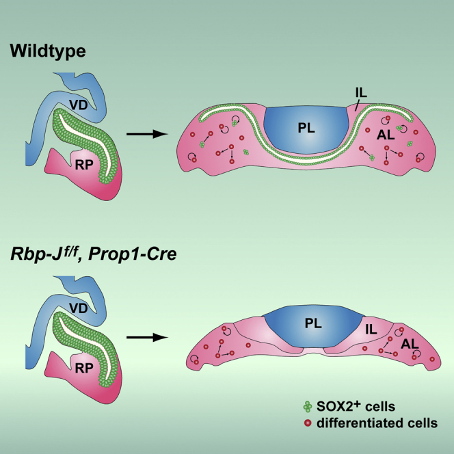
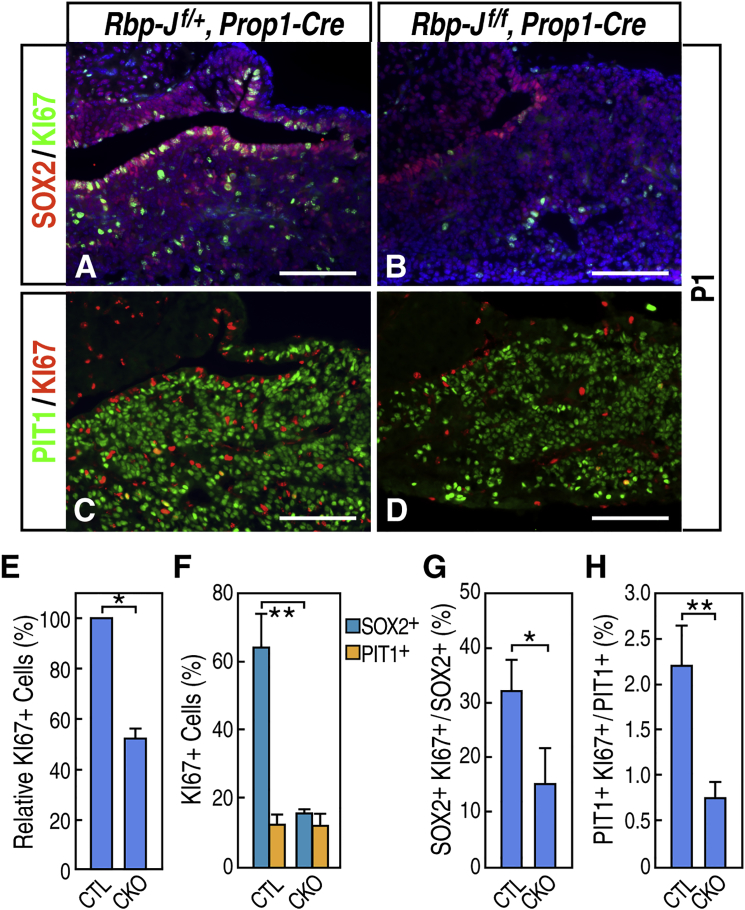
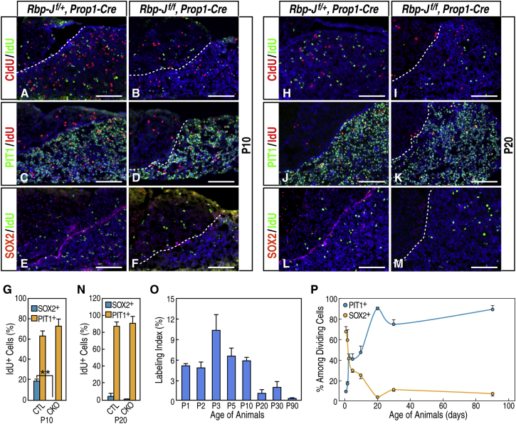
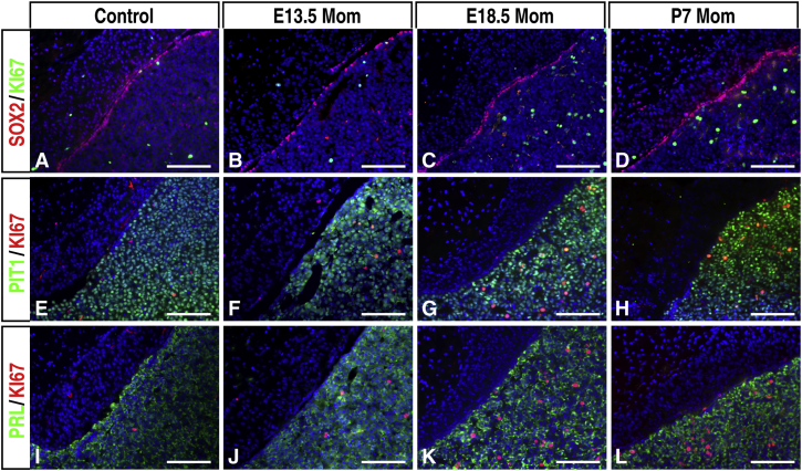
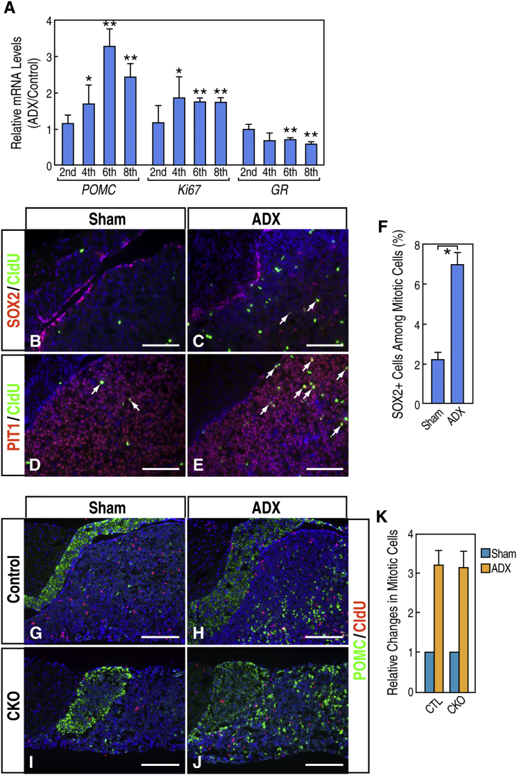
Although SOX2(+) stem cells are present in the postnatal pituitary gland, how they are regulated molecularly and whether they are required for pituitary functions remain unresolved questions. Using a conditional knockout animal model, here we demonstrate that ablation of the canonical Notch signaling in the embryonic pituitary gland leads to progressive depletion of the SOX2(+) stem cells and hypoplastic gland. Furthermore, we show that the SOX2(+) stem cells initially play a significant role in contributing to postnatal pituitary gland expansion by self-renewal and differentiating into distinct lineages in the immediate postnatal period. However, we found that within several weeks postpartum, the SOX2(+) stem cells switch to an essentially dormant state and are no longer required for homeostasis/tissue adaptation. Our results present a dynamic tissue homeostatic model in which stem cells provide an initial contribution to the growth of the neonatal pituitary gland, whereas the mature gland can be maintained in a stem cell-independent fashion.


Figures

None
Graphical abstract
Figure 1
Figure 1
Characterizations of Pituitary Stem Cells and Prop1-Cre Transgenic Mice (A) In situ hybridization of the Notch target gene Hes1 at E17.5 revealed that it is highly expressed in the periluminal region. (B–F) Immunofluorescence staining of Hes1 (B), SOX2/E-Cadherin (C), SOX2/LHX3 (D), SOX2/SOX9 (E), and SOX2/NES (F) in a 2-month-old pituitary gland. Arrows indicate representative double-positive cells. (G) The Prop1-Cre transgenic line is driven by the 2.2-kb promoter/enhancer region of the Prop1 gene. Blue boxes, exons 1 and 2; int1, intron 1. (H–J) LacZ reporter analysis of Prop1-Cre, Rosa-LacZ embryos in whole-mount (H) and middle sagittal frozen sections at E12.5 (I) and E14.5 (J). LacZ activity at E14.5 is detected in all cells in the anterior and intermediate lobes of the embryonic pituitary gland. It is noted that ectopic Cre activity is detected in the olfactory epithelium (H). (K) Immunofluorescence staining of LHX3 at E14.5. (L and M) Dual immunofluorescence staining of GFP/PIT1 and GFP/SOX2 in a Prop1-Cre, Z/EG postnatal day 22 pituitary gland. AL, anterior lobe; IL, intermediate lobe; PL, posterior lobe. Scale bars, 200 μm (A); 50 μm (B–F, L, and M), 500 μm (H), 100 μm (I–K).
Figure 2
Figure 2
Proliferating SOX2+ Progenitor Cells in the Embryonic Pituitary Gland Are Reduced in Rbp-Jf/f, Prop1-Cre Mutants (A–H) Double immunofluorescence labeling of SOX2 and KI67 of wild-type controls (A, C, E, and G) and Rbp-Jf/f, Prop1-Cre mutants (B, D, F, and H) at E14.5 (A–D) and E17.5 (E–H) showed reduced numbers of SOX2+ KI67+ cells in the Rbp-Jf/f, Prop1-Cre mutants, initially in the anterior lobe at E14.5 and later in the intermediate lobe at E17.5. Arrows indicate representative double-positive cells. (I–L) Double immunofluorescence labeling of POMC and KI67 at E17.5 revealed that periluminal KI67+ cells in the intermediate lobe (I and K) are absent in the Rbp-Jf/f, Prop1-Cre mutants (J and L). (M and N) Double immunofluorescence labeling of PIT1 and TSHβ at E17.5. The anterior lobe in the mutant (N) is smaller than in the wild-type control (M), with reduced numbers of PIT1+ and TSHβ+ cells. (O and P) Immunofluorescence labeling of GH in the wild-type control (O) and Rbp-Jf/f, Prop1-Cre mutants (P) at E17.5. The dashed lines indicate the lumen between the intermediate and anterior lobes. The dashed areas in (A), (B), (E), (F), (I), and (J) are also presented in (C), (D), (G), (H), (K), and (L), respectively. Scale bars, 100 μm (A, B, E, F, I, J, and M–P) and 50 μm (C, D, G, H, K, and L).
Figure 3
Figure 3
SOX2+ E-Cadherin+ Pituitary Stem Cells Are Depleted Gradually in Hypoplastic and Dysmorphic Pituitary Glands in Postnatal Rbp-Jf/f, Prop1-Cre Mutants (A–H) Double immunofluorescence labeling of SOX2/E-Cadherin (A, B, E, and F; magnification, 200×), SOX2/PROP1 (C and D; magnification, 400×), and SOX9/E-Cadherin (G and H; magnification, 200×) at P0 (A and B), P3 (C and D), and P10 (E–H) in wild-type controls (A, C, E, and G) and Rbp-Jf/f, Prop1-Cre mutants (B, D, F, and H). (I) qRT-PCR of a 3-month-old wild-type control and Rbp-Jf/f, Prop1-Cre mutant pituitary revealed reduced expression of Rbp-J, Hes1, Hey1, Prop1, Sox2, and E-Cadherin. Data are represented as mean ± SEM (n = 3 mice). p < 0.05, ∗∗p < 0.01. CKO/C, conditional knockout/control. (J–Q) Immunofluorescence labeling of GH/FSHβ (J and Κ), LHβ/TSHβ, and POMC/PRL (N–Q) in 1-month-old wild-type controls (J, L, N, and P) and Rbp-Jf/f, Prop1-Cre mutants (K, M, O, and Q) showed that all cell types are present in the mutant glands. Lower magnification (25×) of the gland (P and Q) revealed that, in the Rbp-Jf/f, Prop1-Cre mutants, the intermediate lobe is expanded laterally and discontinued in the ventral medial region, leading to a direct interaction of the anterior lobe with the posterior lobe (arrowhead). (R–T) Immunofluorescence labeling of SOX2 (R and S) and quantification (T) of pituitary spheres cultured from a 3-month-old wild-type control (CTL) and Rbp-Jf/f, Prop1-Cre mutant. Data are represented as mean ± SEM (n = 3 mice). p < 0.05, ∗∗p < 0.01. Scale bars, 100 μm (A, B, and E–H), 50 μm (C and D), 200 μm (J and O), 500 μm (P and Q), and 75 μm (R and S).
Figure 4
Figure 4
Both SOX2+ Stem Cells and PIT1+ Cells Exhibit Reduced Proliferation in the Anterior Lobe of Rbp-Jf/f, Prop1-Cre Mutants at P1 (A–D) Double immunofluorescence labeling of SOX2/KI67 (A and B) and PIT1/KI67 (C and D) in wild-type control (A and C) and Rbp-Jf/f, Prop1-Cre mutants (B and D) at P1. (E) Quantification of KI67+ proliferating cells in the anterior lobe in the control and mutant (CKO) at P1. Data are represented as mean ± SEM (n = 3 mice, p < 0.05). (F) Quantification of SOX2+ and PIT1+ cells among KI67+ proliferating cells in the control and mutant. Data are represented as mean ± SEM (n = 3 mice, ∗∗p < 0.01). (G) Quantification of SOX2+ KI67+ cells among SOX2+ cells. Data are represented as mean ± SEM (n = 3 mice, p < 0.05). (H) Quantification of PIT1+ KI67+ cells among PIT1+ cells. Data are represented as mean ± SEM (n = 3 mice, ∗∗p < 0.01). Scale bars, 100 μm.
Figure 5
Figure 5
Proliferation of SOX2+ Cells Is Diminished Gradually, and PIT1+ Cells Make up the Majority of Proliferating Cells in the Postnatal Pituitary Gland (A–F) Double immunofluorescence labeling of CldU/IdU (A and B), PIT1/IdU (C and D), and SOX2/IdU (E and F) in the wild-type control (A, C, and E) and Rbp-Jf/f, Prop1-Cre mutants (B, D, and F) at P10. (G) Quantification of PIT1+ or SOX2+ cells among dividing cells at P10. Data are represented as mean ± SEM (n = 3 mice, ∗∗p < 0.01). (H–M) Double immunofluorescence labeling of CldU/IdU (H and I), PIT1/IdU (J and K), and SOX2/IdU (L and M) in the wild-type control (H, J, and L) and Rbp-Jf/f, Prop1-Cre mutants (I, K, and M) at P20. (N) Quantification of PIT1+ or SOX2+ cells among dividing cells at P20. Data are represented as mean ± SEM (n = 3 mice). (O) Labeling index of the wild-type pituitary glands at different postnatal stages. Data are represented as mean ± SEM (n = 3 mice). (P) Respective quantification of PIT1+ and SOX2+ cells among IdU+ dividing cells in the anterior lobe of wild-type pituitaries at different postnatal stages. Data are represented as mean ± SEM (n = 3 mice). Scale bars, 100 μm.
Figure 6
Figure 6
Characterization of Proliferating Cells in 3-Month-Old Wild-Type Control Mice during Pregnancy and Lactation (A–L) Double immunofluorescence labeling of SOX2/KI67 (A–D), PIT1/KI67 (E–H), and PRL/KI67 (I–L) at different stages of pregnancy and lactation. Scale bars, 100 μm.
Figure 7
Figure 7
SOX2+ Cells Are Mobilized upon Target Organ Ablation but Contribute a Small Percentage of Overall Pituitary Gland Activation (A) qRT-PCR of Pomc, Ki67, and glucocorticoid receptor (GR) in the pituitary glands after bilateral ADX in wild-type control mice. Data are represented as mean ± SEM (n = 3 mice, p < 0.05, ∗∗p < 0.01). (B–E) Double immunofluorescence labeling of SOX2/CldU (B and C) and PIT1/CldU (D and E) in sham- (B and D) and ADX-operated (C and E) wild-type pituitary glands. The arrows indicate co-labeling. (F) Quantification of SOX2+ cells among mitotic cells in sham- and ADX-operated wild-type pituitary glands. Data are represented as mean ± SEM (n = 3 mice, p < 0.05). (G–J) Double immunofluorescence labeling of POMC/CldU in sham- (G) and ADX-operated (H) control animals and Rbp-Jf/f, Prop1-Cre mutants (CKO, I and J). (K) Quantification of relative changes in mitotic cells in response to ADX in wild-type control and Rbp-Jf/f, Prop1-Cre mutants. Data are represented as mean ± SEM (n = 3 mice). Scale bars, 100 μm (B–E) and 200 μm (G–J).

Similar articles

Cited by

References

    1. Andoniadou C.L., Matsushima D., Mousavy Gharavy S.N., Signore M., Mackintosh A.I., Schaeffer M., Gaston-Massuet C., Mollard P., Jacques T.S., Le Tissier P. Sox2(+) stem/progenitor cells in the adult mouse pituitary support organ homeostasis and have tumor-inducing potential. Cell Stem Cell. 2013;13:433–445. - PubMed
    1. Arnold K., Sarkar A., Yram M.A., Polo J.M., Bronson R., Sengupta S., Seandel M., Geijsen N., Hochedlinger K. Sox2(+) adult stem and progenitor cells are important for tissue regeneration and survival of mice. Cell Stem Cell. 2011;9:317–329. - PMC - PubMed
    1. Brennand K., Melton D. Slow and steady is the key to beta-cell replication. J. Cell. Mol. Med. 2009;13:472–487. - PMC - PubMed
    1. Carbajo-Pérez E., Watanabe Y.G. Cellular proliferation in the anterior pituitary of the rat during the postnatal period. Cell Tissue Res. 1990;261:333–338. - PubMed
    1. Castrique E., Fernandez-Fuente M., Le Tissier P., Herman A., Levy A. Use of a prolactin-Cre/ROSA-YFP transgenic mouse provides no evidence for lactotroph transdifferentiation after weaning, or increase in lactotroph/somatotroph proportion in lactation. J. Endocrinol. 2010;205:49–60. - PMC - PubMed

Publication types