. 2015 Dec 10:3:78.
doi: 10.1186/s40478-015-0258-3.

The domestic cat as a natural animal model of Alzheimer's disease

Affiliations

The domestic cat as a natural animal model of Alzheimer's disease

James K Chambers et al. Acta Neuropathol Commun. .

Abstract

Introduction: Alzheimer's disease (AD) is the most dominant neurodegenerative disorder that causes dementia, and no effective treatments are available. To study its pathogenesis and develop therapeutics, animal models representing its pathologies are needed. Although many animal species develop senile plaques (SP) composed of amyloid-β (Aβ) proteins that are identical to those found in humans, none of them exhibit neurofibrillary tangles (NFT) and subsequent neurodegeneration, which are integral parts of the pathology of AD.

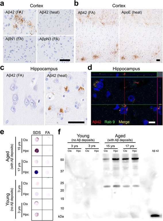
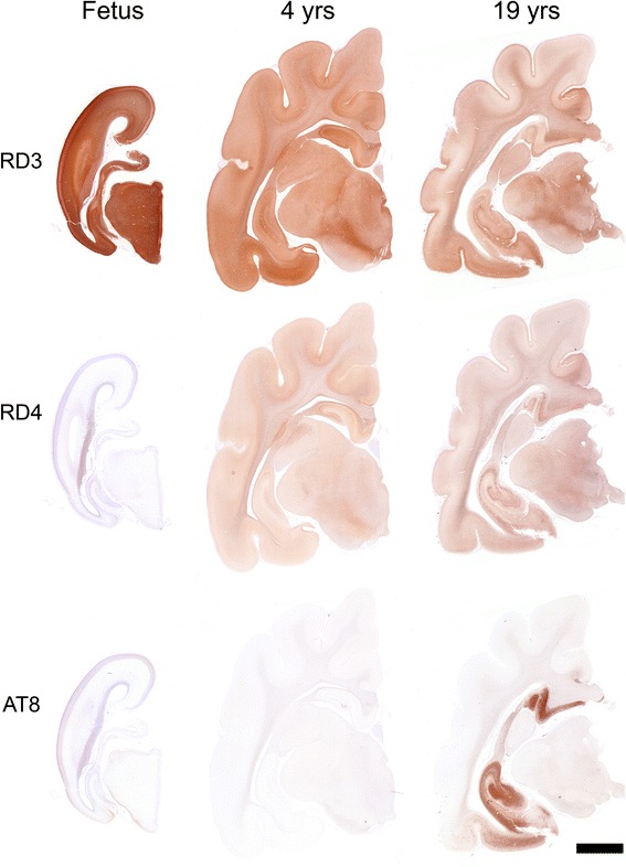
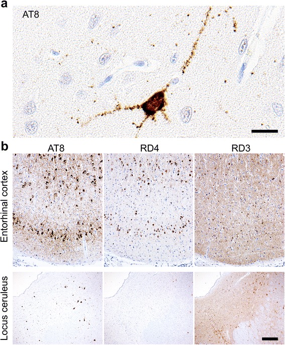
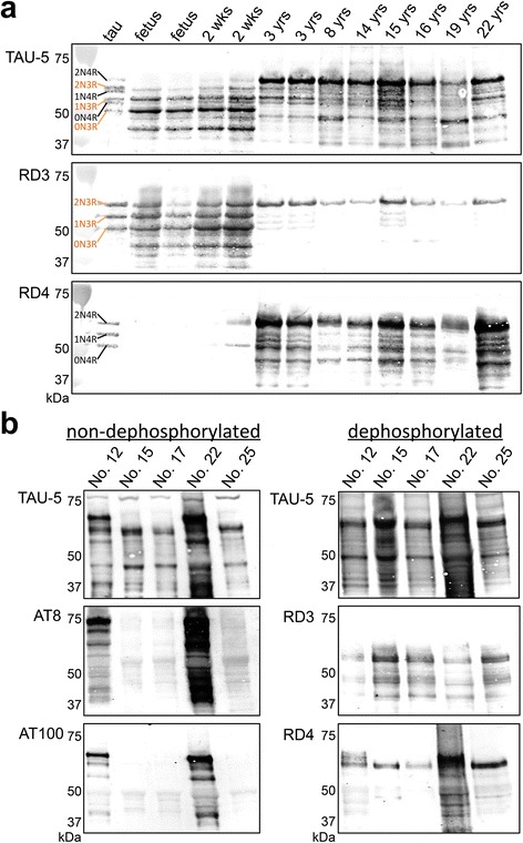
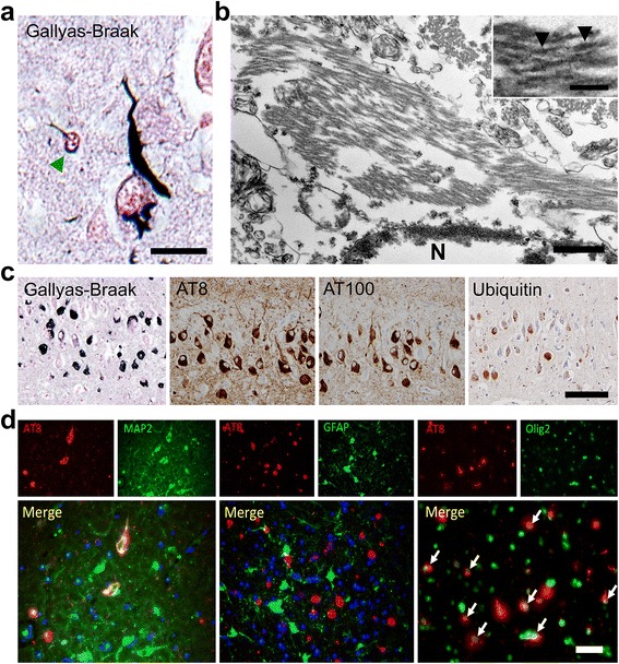
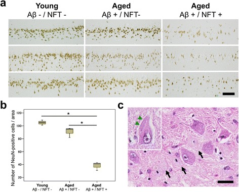
Results: The present study shows that Aβ accumulation, NFT formation, and significant neuronal loss all emerge naturally in the hippocampi of aged domestic cats. The NFT that form in the cat brain are identical to those seen in human AD in terms of their spatial distribution, the cells they affect, and the tau isoforms that comprise them. Interestingly, aged cats do not develop mature argyrophilic SP, but instead accumulate intraneuronal Aβ oligomers in their hippocampal pyramidal cells, which might be due to the amino acid sequence of felid Aβ.

Conclusions: These results suggest that Aβ oligomers are more important than SP for NFT formation and the subsequent neurodegeneration. The domestic cat is a unique animal species that naturally replicates various AD pathologies, especially Aβ oligomer accumulation, NFT formation, and neuronal loss.


Figures

Fig. 1
Fig. 1
Aβ deposits in cat brains. a Aβ42 aggregates are detected in the parenchyma of the cerebral cortex with anti-Aβ42 antibody (12 F4) on formic acid (FA)-pretreated sections but not on heat-pretreated sections. These aggregates are not detected with antibodies against the N-terminus of human Aβ (AβN1 and AβpN3). b Aβ42 aggregates in the cerebral cortex colocalized with ApoE. c Heat pretreatment revealed intracellular Aβ42 aggregates in the pyramidal cells of the hippocampus but not in the cortex. d Some of the intracellular Aβ42 (red) aggregates colocalized with Rab9 (green). Black bars = 50 μm, white bar = 10 μm. e Dot blot analysis of SDS fractions and FA fractions of cortex (Ctx) and hippocampus (Hpc) of young cats and aged cats. Aβ oligomers were detected with A11 antibody, predominantly in the SDS fraction from the hippocampus of aged cats. f Western blotting analysis of the SDS fraction of the Ctx and Hpc of young cats and aged cats. Two distinct bands were detected with anti-Aβ antibody 6E10 in the brains of aged cats: approximately 24 kDa and 54 kDa, indicating Aβ hexamers and dodecamers, respectively
Fig. 2
Fig. 2
Immunohistochemical analysis of tau isoforms and their phosphorylation status in cat brains. Immunohistochemistry for 3-repeat tau (RD3), 4-repeat tau (RD4), and hyperphosphorylated-tau (AT8) in the developing cat (Fetus, case No. 1 shown in Table 1), adult cat (4 years (yrs) old, case No. 7), and aged cat (19 yrs old, case No. 22) brains. Only the 3-repeat tau isoform is expressed in the fetal cat brain, whereas both 3-repeat and 4-repeat tau isoforms are expressed in the developed cat brains. Abundant AT8-positive hyperphosphorylated tau aggregates are observed in the hippocampus and entorhinal cortex of the aged cat brain. Bar = 5 mm
Fig. 3
Fig. 3
Hyperphosphorylated tau accumulation in the entorhinal cortex and locus ceruleus of cat brains. a Immunohistochemistry of the entorhinal cortex of a cat with mild hyperphosphorylated tau accumulation (15-year-old, case No. 13) for hyperphosphorylated-tau (AT8). The neuronal soma and dendrites are positively stained for hyperphosphorylated tau. Bar = 20 μm. b Immunohistochemistry of the entorhinal cortex and locus ceruleus of a cat with severe hyperphosphorylated tau accumulation (14-year-old, case No. 12) for AT8, 3-repeat tau (RD3), and 4-repeat tau (RD4). AT8-positive aggregates are also positively stained for 3-repeat tau and 4-repeat tau on consecutive sections. Bar = 300 μm
Fig. 4
Fig. 4
Western blotting analysis of tau isoforms and their phosphorylation status in cat brains. a Western blotting of TBS-soluble fractions obtained from the hippocampus of various ages and treated with alkaline phosphatase (AP). The left lane (tau) shows the six isoforms of human tau (recombinantly produced): three 3-repeat tau isoforms (2N3R, 1N3R, 0N3R) and three 4-repeat tau isoforms (2N4R, 1N4R, 0N4R). In the fetal brain, only the 3-repeat tau isoforms are expressed. In the adult cat brains, all six isoforms are detected using anti-tau antibody (TAU-5) and also 3-repeat tau (RD3), and 4-repeat tau (RD4) antibodies. b Western blotting of sarkosyl-insoluble guanidine HCl-soluble fractions obtained from the hippocampus of aged cat brains without AP treatment (left) and with AP treatment (right). In cat hippocampi that were immunohistochemically positive for hyperphosphorylated tau (cases No. 12 and 22), AT8- and AT100-positive tau proteins are detected. In these cats, the smear-like band pattern resolved into clear lower molecular weight bands consisting of both 3-repeat and 4-repeat tau isoforms after dephosphorylation treatment
Fig. 5
Fig. 5
NFTs in aged cats brains. a Gallyas-Braak staining-positive argyrophilic aggregates are observed mainly in the neuronal soma, neurites, and also in some oligodendroglial cells (green arrowhead) in the entorhinal cortex of aged cat brain. Bar = 20 μm. b Transmission electron microscopy of NFT in the hippocampus. Bundles of filaments are observed in the neuronal soma either in strait form or paired twisted form. For the paired twisted form, the lengths between the constrictions (arrowheads) were 80-100 nm. Bars = 500 nm and 100 nm (inset). N: nucleus. c Consecutive sections of hippocampus show AT8-, AT100-, and ubiquitin-immunopositivity for NFTs. Bar = 100 μm. d AT8-positive (red) hyperphosphorylated tau is observed in MAP2-positive (green) neurons (left) and Olig2-positive (green) oligodendrocytes (right, white arrows), but not in GFAP-positive (green) astrocytes (middle). Bar = 50 μm
Fig. 6
Fig. 6
Hippocampal neuronal loss in cats with NFTs. a Immunohistochemistry for NeuN in young cats (Aβ–/NFT–, n = 3, mean age 3.7 years old), aged cats with Aβ deposits but no NFTs in the hippocampus (Aβ+/NFT–, n = 3, mean age 18 years old), and aged cats with Aβ deposits and NFTs (Aβ+/NFT+, n = 3, mean age 17.6 years old). Bar = 100 μm. b The number of NeuN-positive pyramidal cells shown in (a). The number of hippocampal neurons is significantly decreased in aged cats with Aβ deposits and NFTs (Aβ+/NFT+) compared to young cats (Aβ–/NFT–) and aged cats with Aβ deposits but no NFTs in the hippocampus (Aβ+/NFT–). *P < 0.001. c Ghost tangles are observed on an HE-stained section (arrows). In some of the cells, inclusions (composed of hyperphosphorylated tau, confirmed on consecutive sections) were observed (inset, arrowheads). Bar = 25 μm

References

    1. Aho L, Pikkarainen M, Hiltunen M, Leinonen V, Alafuzoff I. Immunohistochemical visualization of amyloid-beta protein precursor and amyloid-beta in extra- and intracellular compartments in the human brain. J Alzheimers Dis. 2010;20:1015–28. - PubMed
    1. Arima K. Ultrastructural characteristics of tau filaments in tauopathies: immuno-electron microscopic demonstration of tau filaments in tauopathies. Neuropathology. 2006;26:475–83. doi: 10.1111/j.1440-1789.2006.00669.x. - DOI - PubMed
    1. Benilova I, Karran E, De Strooper B. The toxic Aβ oligomer and Alzheimer's disease: an emperor in need of clothes. Nat Neurosci. 2012;15:349–57. doi: 10.1038/nn.3028. - DOI - PubMed
    1. Bernstein SL, Dupuis NF, Lazo ND, Wyttenbach T, Condron MM, Bitan G, et al. Amyloid-β protein oligomerization and the importance of tetramers and dodecamers in the aetiology of Alzheimer's disease. Nat Chem. 2009;1:326–31. doi: 10.1038/nchem.247. - DOI - PMC - PubMed
    1. Brinkmalm G, Portelius E, Öhrfelt A, Mattsson N, Persson R, Gustavsson MK, et al. An online nano-LC-ESI-FTICR-MS method for comprehensive characterization of endogenous fragments from amyloid β and amyloid precursor protein in human and cat cerebrospinal fluid. J Mass Spectrom. 2012;47:591–603. doi: 10.1002/jms.2987. - DOI - PubMed

Publication types