. 2015 Dec 9;18(6):682-93.
doi: 10.1016/j.chom.2015.11.006.

A Single Bacterial Immune Evasion Strategy Dismantles Both MyD88 and TRIF Signaling Pathways Downstream of TLR4

Affiliations

A Single Bacterial Immune Evasion Strategy Dismantles Both MyD88 and TRIF Signaling Pathways Downstream of TLR4

Charles V Rosadini et al. Cell Host Microbe. .

Abstract

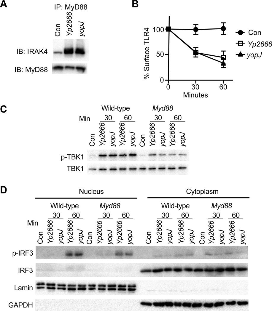
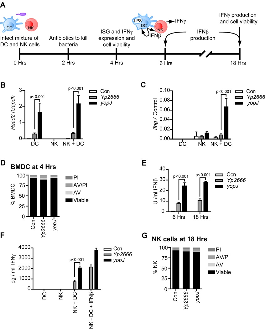
During bacterial infections, Toll-like receptor 4 (TLR4) signals through the MyD88- and TRIF-dependent pathways to promote pro-inflammatory and interferon (IFN) responses, respectively. Bacteria can inhibit the MyD88 pathway, but if the TRIF pathway is also targeted is unclear. We demonstrate that, in addition to MyD88, Yersinia pseudotuberculosis inhibits TRIF signaling through the type III secretion system effector YopJ. Suppression of TRIF signaling occurs during dendritic cell (DC) and macrophage infection and prevents expression of type I IFN and pro-inflammatory cytokines. YopJ-mediated inhibition of TRIF prevents DCs from inducing natural killer (NK) cell production of antibacterial IFNγ. During infection of DCs, YopJ potently inhibits MAPK pathways but does not prevent activation of IKK- or TBK1-dependent pathways. This singular YopJ activity efficiently inhibits TLR4 transcription-inducing activities, thus illustrating a simple means by which pathogens impede innate immunity.


Figures

Figure 1
Figure 1
Yptb inhibits TRIF-dependent gene expression via a YopJ-dependent mechanism. BMDCs (A, C, and D), iBMDMs (B) were infected with strains indicated at an MOI of 100. Gene expression relative to Gapdh was analyzed by qPCR at times indicated or 2 hrs post infection (C). Uninfected cells (Con) were included in all experiments. Analysis of cell viability was performed by flow cytometry using propidium iodide and annexin V stains at times indicated for BMDCs (E) or iBMDMs (F) and was performed in parallel with (A) or (B), respectively. For gene expression analysis, bars represent the average and error bars represent the standard deviation of triplicate readings from one representative experiment of three. Relative statistics were calculated using ANOVA with Bonferroni’s multiple comparison test. See also figure S1.
Figure 2
Figure 2
YopJ inhibits TRIF-dependent gene expression independent cytotoxicity. Cell viability (A, C, and F) and gene expression (B, D, and E) of WT (A and B) and Ripk3−/− BMDCs (C and D) Ripk3/Caspase 8−/− BMDCs, or (E and F) Ripk3−/− BMDMs. Cells were treated with either vehicle (DMSO) or pan-caspase inhibitor (zVAD-fmk) prior to infection at an MOI of 100. Gene expression relative to Gapdh was analyzed by qPCR at 4 hr (B and D) or 3 hr (E) after infection. Uninfected cells (Con) were used as a negative control. Analysis of cell viability was performed by flow cytometry using propidium iodide and annexin V staining. For gene expression analysis, bars represent the average and error bars represent the standard deviation of triplicate readings from one representative experiment of two (B and E) or three (D). Relative statistics were calculated using ANOVA with Bonferroni’s multiple comparison test. See also figure S2.
Figure 3
Figure 3
YopJ suppresses MyD88 and TRIF transcriptional responses downstream of TLR4. Gene expression induced during Yp2666 infection of WT vs TLR4-deficient (Tlr4) (A), MyD88-deficient (Myd88) (B) or Trif-deficient (Ticam1) BMDCs (C). BMDCs were infected with strains indicated at an MOI of 100. Gene expression relative to Gapdh was analyzed by qPCR at times indicated. Uninfected cells (Con) were used as a negative control. Bars represent the average and error bars represent the standard deviation of triplicate readings from one representative experiment of two (B) or three (A and C). Relevant statistics were calculated using ANOVA with Bonferroni’s multiple comparison test.
Figure 4
Figure 4
YopJ does not block TRIF-dependent IRF3 activation. iBMDMs were infected with strains indicated and monitored for Myddosome formation (A) or TLR4 endocytosis (B). Myddosome formation was assessed 1 hr after infection. TLR4 endocytosis was examined by flow cytometry. In (A) and (B) uninfected cells (Con) served as a negative control. WT or Myd88-deficient BMDCs (Myd88) were infected with strains indicated and monitored for TBK1 phosphorylation (C) and IRF3 phosphorylation and p-IRF3 nuclear translocation (D). Phosphorylation of TBK1 was assessed by immunoblot of cell lysates with a phospho-S172 specific antibody. Total TBK1 was monitored by immunoblot with an antibody specific for total TBK1. IRF3 activation was assessed after infection of BMDCs by isolating nuclear and cytoplasmic fractions and immunoblotting with antibodies specific for phospho-IRF3 or total IRF3. Immunoblotting for Lamin and GAPDH served as controls for nuclear and cytoplasmic fractions, respectively. Data are one representative of two independent experiments.
Figure 5
Figure 5
YopJ inhibits cytokine production in BMDCs by blocking MAPKs. (A) WT BMDCs or (B and C) WT and MyD88-deficient BMDCs (Myd88) were infected with strains indicated at MOI 100 and monitored for IĸBα degradation (A), p65 nuclear translocation (B), MKK phosphorylation (C), and MAPK phosphorylation (D) at times indicated. Nuclear and cytoplasmic fractions were isolated and immunoblotting for Lamin and GAPDH verified nuclear and cytoplasmic fractions, respectively (A). Whole cell lysates were collected and immunoblotting GAPDH or total MAPKs served as loading controls (A, C and D). BMDCs were pretreated with DMSO (vehicle control), CHX (50 ng/ml) or specific inhibitors for ERK, p38 or, JNK or a combination of inhibitors at the time of infection (A) or prior to infection with strains indicated (E and F). At 90 minutes post infection, gene expression relative to Gapdh was analyzed by qPCR (E and F). Uninfected cells served as a negative control in all experiments. Bars represent the average and error bars represent the standard deviation of triplicate readings from a one representative experiment. Data are representative of three independent experiments. Relevant statistics were calculated using ANOVA with Bonferroni’s multiple comparison test. See also figure S3 and Table S1.
Figure 6
Figure 6
Inhibition of TRIF-dependent responses by Yptb disrupts DC-mediated NK cell activation. Experimental layout: NK cells and DCs were mixed in 0.7:1 ratio and infected with strains indicated at an MOI of 100 (A). At 2 hr post infection, cells were treated with antibiotics to kill bacteria (A). Gene expression, cytokine production, and cell viability were monitored at times indicated (A). 4 hr post infection, gene expression relative to cell specific house-keeping genes (Control: Gapdh for DCs or Ncr1 for NK cells) was monitored by qPCR (B and C) and DC cell viability was analyzed by flow cytometry with propidum iodide and annexin V staining (D). IFNβ production was assessed by bioassay at times indicated (E). At 18 hrs post infection, IFNγ production was assessed by ELISA (F). At time 0, exogenous IFNβ was added at 200 U/ml where indicated. Viability of NK cells, infected in parallel with co-cultures in (F), was analyzed by flow cytometry using propidum iodide and annexin V staining (G). Uninfected cells (Con) were used in all experiments. For gene expression analysis, bars represent the average and error bars represent the standard deviation of triplicate readings from a one representative experiment of two. Statistics were calculated using ANOVA with Bonferroni’s multiple comparison test. See also figure S4.

References

    1. Balada-Llasat J-M, Mecsas J. Yersinia has a tropism for B and T cell zones of lymph nodes that is independent of the type III secretion system. 2006 - PMC - PubMed
    1. Baxt LA, Garza-Mayers AC, Goldberg MB. Bacterial subversion of host innate immune pathways. Science. 2013;340:697–701. - PubMed
    1. Bonham KS, Orzalli MH, Hayashi K, Wolf AI, Glanemann C, Weninger W, Iwasaki A, Knipe DM, Kagan JC. A promiscuous lipid-binding protein diversifies the subcellular sites of toll-like receptor signal transduction. Cell. 2014;156:705–716. - PMC - PubMed
    1. Brodsky IE, Medzhitov R. Reduced secretion of YopJ by Yersinia limits in vivo cell death but enhances bacterial virulence. PLoS pathogens. 2008;4:e1000067. - PMC - PubMed
    1. Brodsky IE, Palm NW, Sadanand S, Ryndak MB, Sutterwala FS, Flavell RA, Bliska JB, Medzhitov R. A Yersinia effector protein promotes virulence by preventing inflammasome recognition of the type III secretion system. Cell Host Microbe. 2010;7:376–387. - PMC - PubMed

MeSH terms

LinkOut - more resources