

Generation of contractile actomyosin bundles depends on mechanosensitive actin filament assembly and disassembly
- PMID: 26652273
- PMCID: PMC4714978
- DOI: 10.7554/eLife.06126
Generation of contractile actomyosin bundles depends on mechanosensitive actin filament assembly and disassembly
Abstract
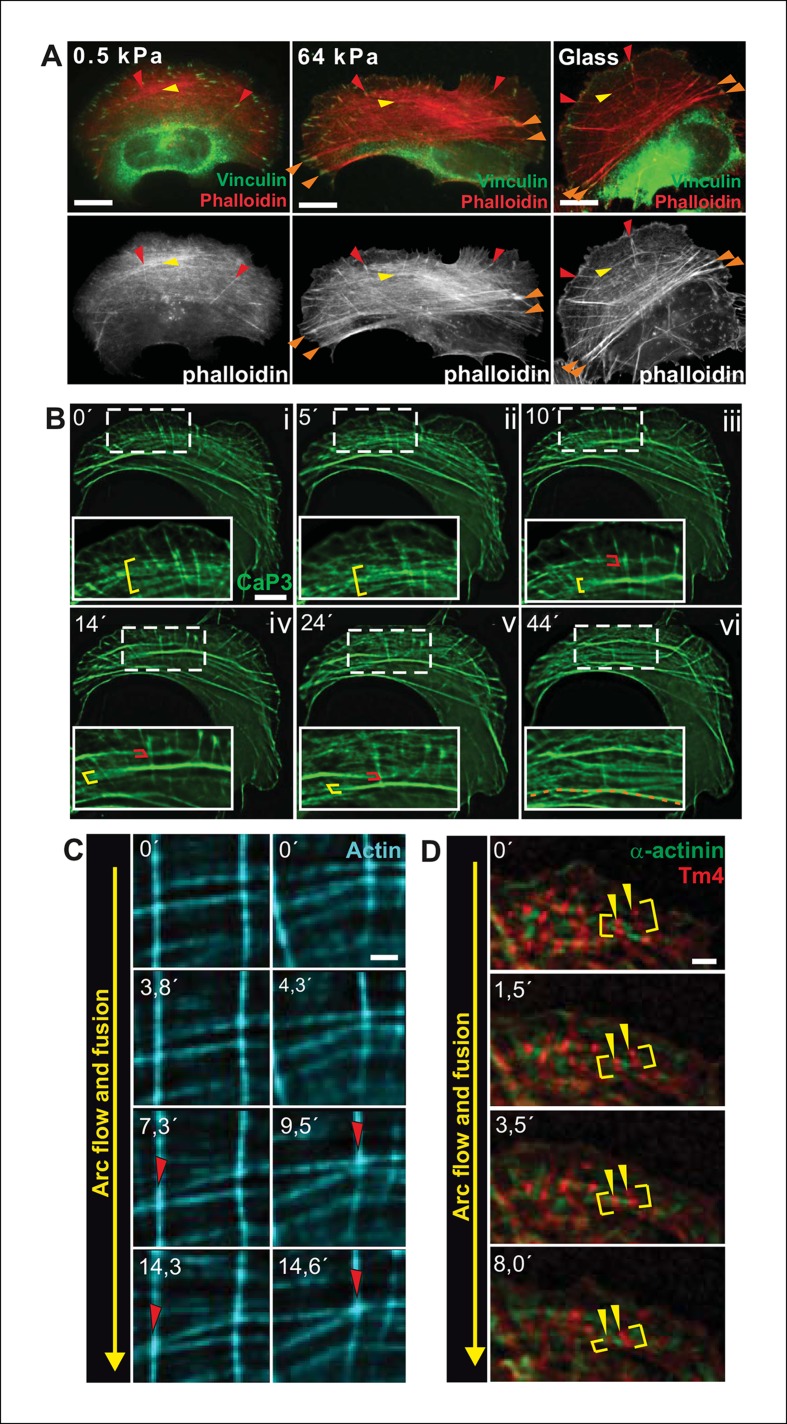
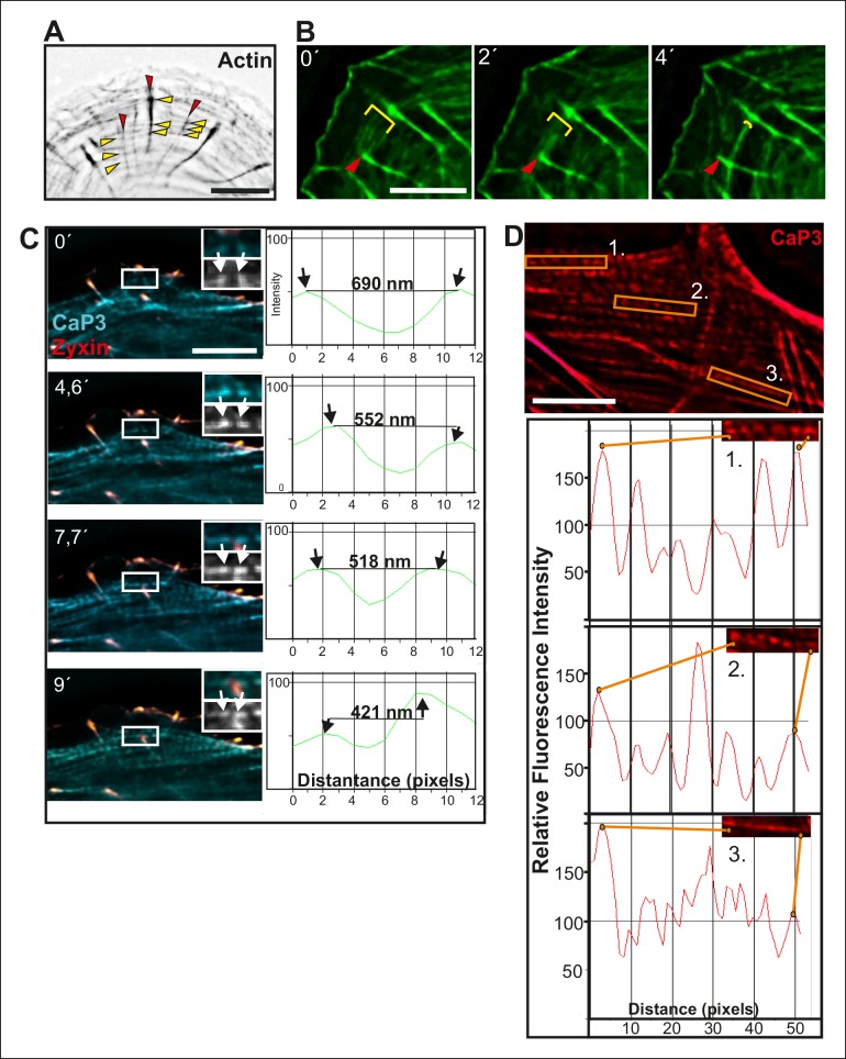
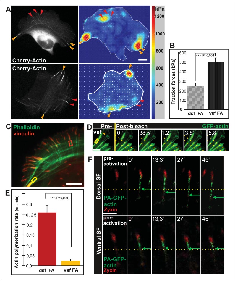
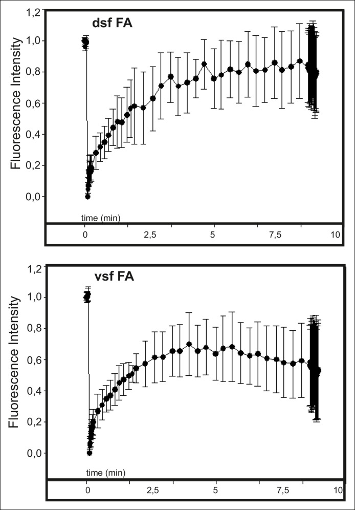
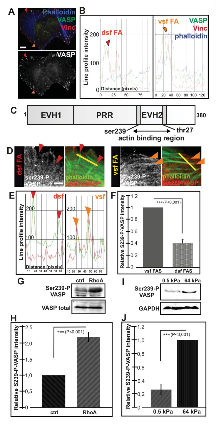
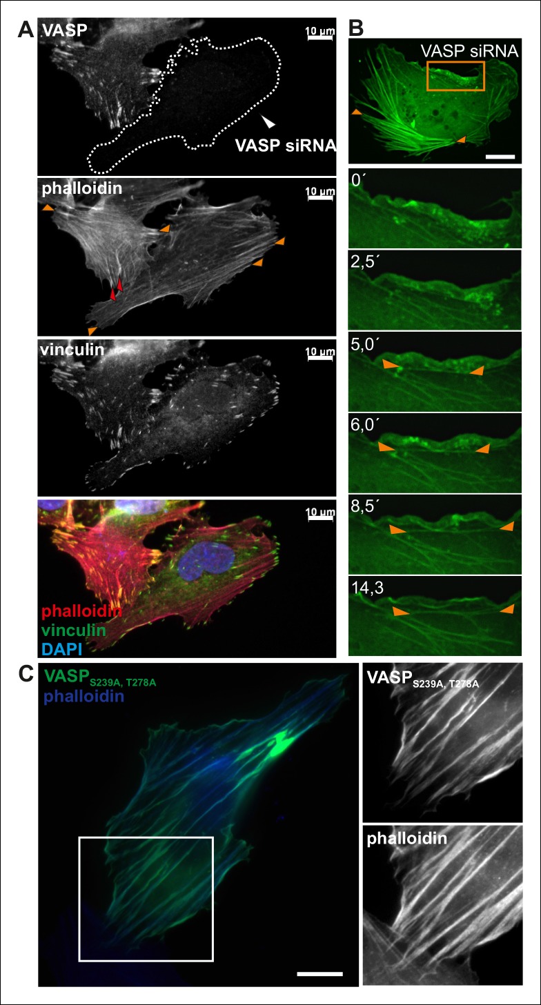
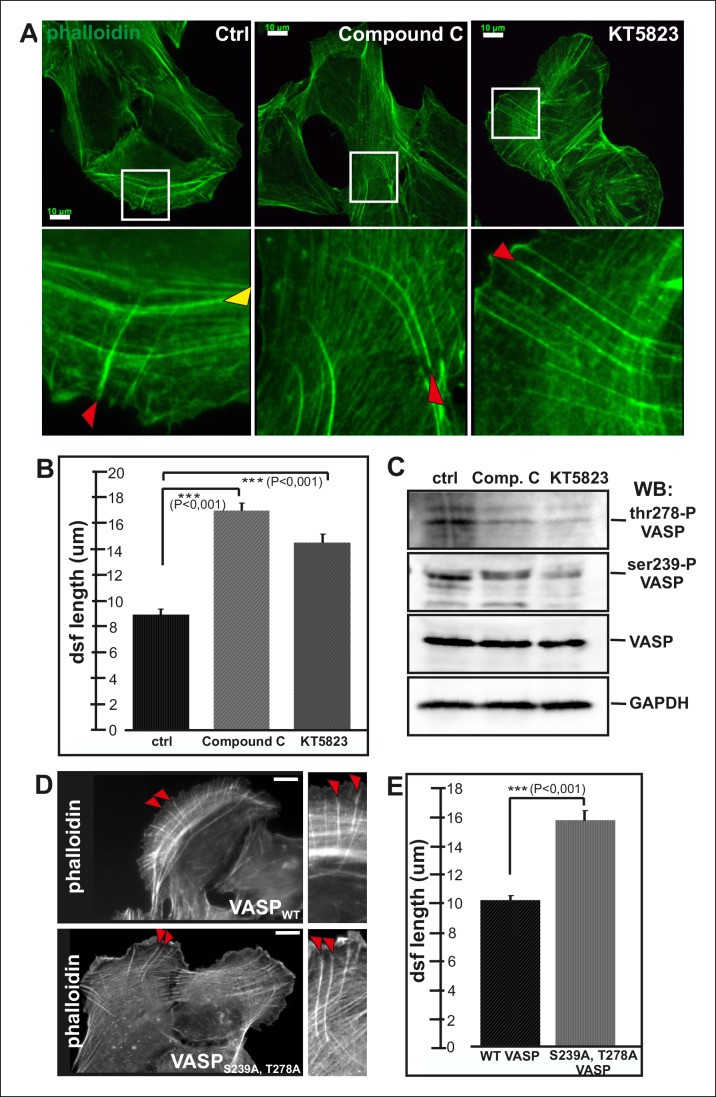
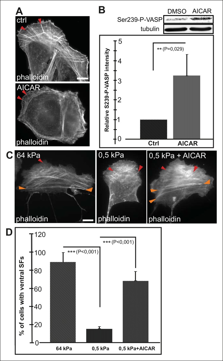
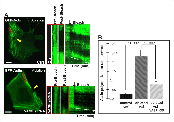
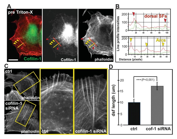
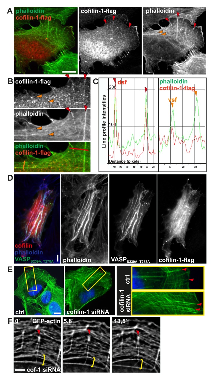
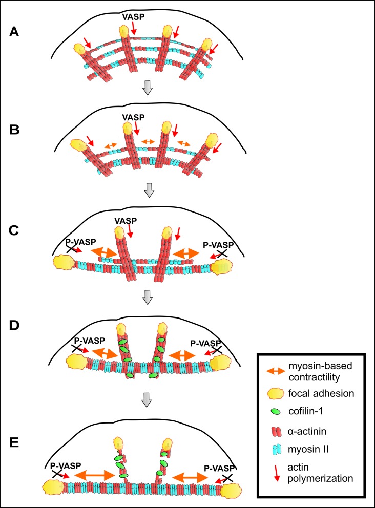
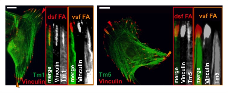
Adhesion and morphogenesis of many non-muscle cells are guided by contractile actomyosin bundles called ventral stress fibers. While it is well established that stress fibers are mechanosensitive structures, physical mechanisms by which they assemble, align, and mature have remained elusive. Here we show that arcs, which serve as precursors for ventral stress fibers, undergo lateral fusion during their centripetal flow to form thick actomyosin bundles that apply tension to focal adhesions at their ends. Importantly, this myosin II-derived force inhibits vectorial actin polymerization at focal adhesions through AMPK-mediated phosphorylation of VASP, and thereby halts stress fiber elongation and ensures their proper contractility. Stress fiber maturation additionally requires ADF/cofilin-mediated disassembly of non-contractile stress fibers, whereas contractile fibers are protected from severing. Taken together, these data reveal that myosin-derived tension precisely controls both actin filament assembly and disassembly to ensure generation and proper alignment of contractile stress fibers in migrating cells.
Keywords: actin; cell biology; human; mechanosensing; stress fibers.
Conflict of interest statement
The authors declare that no competing interests exist.
Figures

Similar articles
-
Generation of stress fibers through myosin-driven reorganization of the actin cortex.Elife. 2021 Jan 28;10:e60710. doi: 10.7554/eLife.60710. Elife. 2021. PMID: 33506761 Free PMC article.
-
CaMKK2 Regulates Mechanosensitive Assembly of Contractile Actin Stress Fibers.Cell Rep. 2018 Jul 3;24(1):11-19. doi: 10.1016/j.celrep.2018.06.011. Cell Rep. 2018. PMID: 29972773
-
A molecular pathway for myosin II recruitment to stress fibers.Curr Biol. 2011 Apr 12;21(7):539-50. doi: 10.1016/j.cub.2011.03.007. Epub 2011 Mar 31. Curr Biol. 2011. PMID: 21458264
-
The inner workings of stress fibers - from contractile machinery to focal adhesions and back.J Cell Sci. 2016 Apr 1;129(7):1293-304. doi: 10.1242/jcs.180927. J Cell Sci. 2016. PMID: 27037413 Review.
-
Assembly and mechanosensory function of focal adhesions: experiments and models.Eur J Cell Biol. 2006 Apr;85(3-4):165-73. doi: 10.1016/j.ejcb.2005.11.001. Epub 2005 Dec 19. Eur J Cell Biol. 2006. PMID: 16360240 Review.
Cited by
-
The GEF Trio controls endothelial cell size and arterial remodeling downstream of Vegf signaling in both zebrafish and cell models.Nat Commun. 2020 Oct 21;11(1):5319. doi: 10.1038/s41467-020-19008-0. Nat Commun. 2020. PMID: 33087700 Free PMC article.
-
MCM3AP-AS1: An Indispensable Cancer-Related LncRNA.Front Cell Dev Biol. 2021 Oct 7;9:752718. doi: 10.3389/fcell.2021.752718. eCollection 2021. Front Cell Dev Biol. 2021. PMID: 34692706 Free PMC article. Review.
-
Regulation of axon growth by myosin II-dependent mechanocatalysis of cofilin activity.J Cell Biol. 2019 Jul 1;218(7):2329-2349. doi: 10.1083/jcb.201810054. Epub 2019 May 23. J Cell Biol. 2019. PMID: 31123185 Free PMC article.
-
Extracellular Matrix Geometry and Initial Adhesive Position Determine Stress Fiber Network Organization during Cell Spreading.Cell Rep. 2019 May 7;27(6):1897-1909.e4. doi: 10.1016/j.celrep.2019.04.035. Cell Rep. 2019. PMID: 31067472 Free PMC article.
-
Ventral stress fibers induce plasma membrane deformation in human fibroblasts.Mol Biol Cell. 2021 Aug 19;32(18):1707-1723. doi: 10.1091/mbc.E21-03-0096. Epub 2021 Jun 30. Mol Biol Cell. 2021. PMID: 34191528 Free PMC article.
References
-
- Anderson TW, Vaughan AN, Cramer LP. Retrograde flow and myosin II activity within the leading cell edge deliver f-actin to the lamella to seed the formation of graded polarity actomyosin II filament bundles in migrating fibroblasts. Molecular Biology of the Cell. 2008;19:5006–5018. doi: 10.1091/mbc.E08-01-0034. - DOI - PMC - PubMed
-
- Blair DR, Funai K, Schweitzer GG, Cartee GD. A myosin II ATPase inhibitor reduces force production, glucose transport, and phosphorylation of AMPK and TBC1D1 in electrically stimulated rat skeletal muscle. AJP: Endocrinology and Metabolism. 2009;296:e06126. doi: 10.1152/ajpendo.91003.2008. - DOI - PMC - PubMed
Publication types
MeSH terms
Substances
LinkOut - more resources
Full Text Sources
Other Literature Sources
Molecular Biology Databases
Research Materials
