

Lactobacillus crispatus inhibits growth of Gardnerella vaginalis and Neisseria gonorrhoeae on a porcine vaginal mucosa model
- PMID: 26652855
- PMCID: PMC4675025
- DOI: 10.1186/s12866-015-0608-0
Lactobacillus crispatus inhibits growth of Gardnerella vaginalis and Neisseria gonorrhoeae on a porcine vaginal mucosa model
Abstract
Background: The vaginal microbiota can impact the susceptibility of women to bacterial vaginosis (BV) and sexually transmitted infections (STIs). BV is characterized by depletion of Lactobacillus spp., an overgrowth of anaerobes (often dominated by Gardnerella vaginalis) and a pH > 4.5. BV is associated with an increased risk of acquiring STIs such as chlamydia and gonorrhea. While these associations have been identified, the molecular mechanism(s) driving the risk of infections are unknown. An ex vivo porcine vaginal mucosal model (PVM) was developed to explore the mechanistic role of Lactobacillus spp. in affecting colonization by G. vaginalis and Neisseria gonorrhoeae.
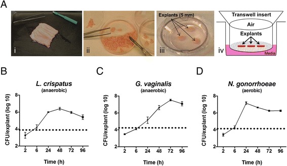
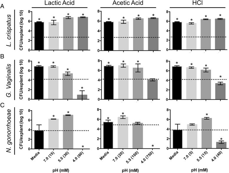
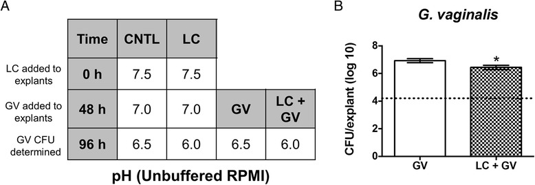
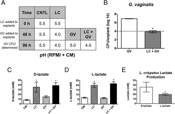
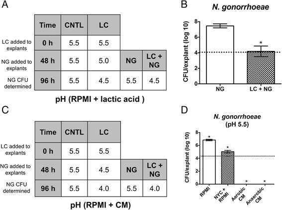
Results: The data presented here demonstrate that all organisms tested can colonize and grow on PVM to clinically relevant densities. Additionally, G. vaginalis and N. gonorrhoeae form biofilms on PVM. It was observed that lactic acid, acetic acid, and hydrochloric acid inhibit the growth of G. vaginalis on PVM in a pH-dependent manner. N. gonorrhoeae grows best in the presence of lactic acid at pH 5.5, but did not grow well at this pH in the presence of acetic acid. Finally, a clinical Lactobacillus crispatus isolate (24-9-7) produces lactic acid and inhibits growth of both G. vaginalis and N. gonorrhoeae on PVM.
Conclusions: These data reveal differences in the effects of pH, various acids and L. crispatus on the growth of G. vaginalis and N. gonorrhoeae on a live vaginal mucosal surface. The PVM is a useful model for studying the interactions of commensal vaginal microbes with pathogens and the mechanisms of biofilm formation on the vaginal mucosa.
Figures

Similar articles
-
Characteristics of Lactobacillus and Gardnerella vaginalis from women with or without bacterial vaginosis and their relationships in gnotobiotic mice.J Med Microbiol. 2012 Aug;61(Pt 8):1074-1081. doi: 10.1099/jmm.0.041962-0. Epub 2012 Apr 26. J Med Microbiol. 2012. PMID: 22539000
-
Glycogen availability and pH variation in a medium simulating vaginal fluid influence the growth of vaginal Lactobacillus species and Gardnerella vaginalis.BMC Microbiol. 2023 Jul 13;23(1):186. doi: 10.1186/s12866-023-02916-8. BMC Microbiol. 2023. PMID: 37442975 Free PMC article.
-
Vaginal Lactobacilli Reduce Neisseria gonorrhoeae Viability through Multiple Strategies: An in Vitro Study.Front Cell Infect Microbiol. 2017 Dec 6;7:502. doi: 10.3389/fcimb.2017.00502. eCollection 2017. Front Cell Infect Microbiol. 2017. PMID: 29270390 Free PMC article.
-
Cholesterol-Dependent Cytolysins Produced by Vaginal Bacteria: Certainties and Controversies.Front Cell Infect Microbiol. 2020 Jan 10;9:452. doi: 10.3389/fcimb.2019.00452. eCollection 2019. Front Cell Infect Microbiol. 2020. PMID: 31998661 Free PMC article. Review.
-
Bacterial vaginosis: what is physiological in vaginal bacteriology? An update and opinion.Acta Obstet Gynecol Scand. 2011 Dec;90(12):1302-6. doi: 10.1111/j.1600-0412.2011.01279.x. Epub 2011 Oct 17. Acta Obstet Gynecol Scand. 2011. PMID: 21916857 Review.
Cited by
-
Does Lactobacillus Exert a Protective Effect on the Development of Cervical and Endometrial Cancer in Women?Cancers (Basel). 2022 Oct 7;14(19):4909. doi: 10.3390/cancers14194909. Cancers (Basel). 2022. PMID: 36230832 Free PMC article. Review.
-
The Anti-Inflammatory and Curative Exponent of Probiotics: A Comprehensive and Authentic Ingredient for the Sustained Functioning of Major Human Organs.Nutrients. 2024 Feb 16;16(4):546. doi: 10.3390/nu16040546. Nutrients. 2024. PMID: 38398870 Free PMC article. Review.
-
Endocervical and vaginal microbiota in South African adolescents with asymptomatic Chlamydia trachomatis infection.Sci Rep. 2018 Jul 23;8(1):11109. doi: 10.1038/s41598-018-29320-x. Sci Rep. 2018. PMID: 30038262 Free PMC article.
-
Cervicovaginal Microbiome Factors in Clearance of Human Papillomavirus Infection.Front Oncol. 2021 Jul 28;11:722639. doi: 10.3389/fonc.2021.722639. eCollection 2021. Front Oncol. 2021. PMID: 34395294 Free PMC article. Review.
-
The Assessment of Knowledge About Cervical Cancer, HPV Vaccinations, and Screening Programs Among Women as an Element of Cervical Cancer Prevention in Poland.J Pers Med. 2024 Dec 4;14(12):1139. doi: 10.3390/jpm14121139. J Pers Med. 2024. PMID: 39728052 Free PMC article.
References
-
- Hillier SL, Krohn MA, Cassen E, Easterling TR, Rabe LK, Eschenbach DA. The role of bacterial vaginosis and vaginal bacteria in amniotic fluid infection in women in preterm labor with intact fetal membranes. Clin Infect Dis. 1995;20(Suppl 2):S276–8. doi: 10.1093/clinids/20.Supplement_2.S276. - DOI - PubMed
Publication types
MeSH terms
Substances
Grants and funding
LinkOut - more resources
Full Text Sources
Other Literature Sources
Molecular Biology Databases
Miscellaneous
