Review
. 2015 Dec 11:14:207.
doi: 10.1186/s12943-015-0474-2.

Novel drug targets for personalized precision medicine in relapsed/refractory diffuse large B-cell lymphoma: a comprehensive review

Affiliations
Review

Novel drug targets for personalized precision medicine in relapsed/refractory diffuse large B-cell lymphoma: a comprehensive review

Rosalba Camicia et al. Mol Cancer. .

Abstract

Diffuse large B-cell lymphoma (DLBCL) is a clinically heterogeneous lymphoid malignancy and the most common subtype of non-Hodgkin's lymphoma in adults, with one of the highest mortality rates in most developed areas of the world. More than half of DLBLC patients can be cured with standard R-CHOP regimens, however approximately 30 to 40 % of patients will develop relapsed/refractory disease that remains a major cause of morbidity and mortality due to the limited therapeutic options.Recent advances in gene expression profiling have led to the identification of at least three distinct molecular subtypes of DLBCL: a germinal center B cell-like subtype, an activated B cell-like subtype, and a primary mediastinal B-cell lymphoma subtype. Moreover, recent findings have not only increased our understanding of the molecular basis of chemotherapy resistance but have also helped identify molecular subsets of DLBCL and rational targets for drug interventions that may allow for subtype/subset-specific molecularly targeted precision medicine and personalized combinations to both prevent and treat relapsed/refractory DLBCL. Novel agents such as lenalidomide, ibrutinib, bortezomib, CC-122, epratuzumab or pidilizumab used as single-agent or in combination with (rituximab-based) chemotherapy have already demonstrated promising activity in patients with relapsed/refractory DLBCL. Several novel potential drug targets have been recently identified such as the BET bromodomain protein (BRD)-4, phosphoribosyl-pyrophosphate synthetase (PRPS)-2, macrodomain-containing mono-ADP-ribosyltransferase (ARTD)-9 (also known as PARP9), deltex-3-like E3 ubiquitin ligase (DTX3L) (also known as BBAP), NF-kappaB inducing kinase (NIK) and transforming growth factor beta receptor (TGFβR).This review highlights the new insights into the molecular basis of relapsed/refractory DLBCL and summarizes the most promising drug targets and experimental treatments for relapsed/refractory DLBCL, including the use of novel agents such as lenalidomide, ibrutinib, bortezomib, pidilizumab, epratuzumab, brentuximab-vedotin or CAR T cells, dual inhibitors, as well as mechanism-based combinatorial experimental therapies. We also provide a comprehensive and updated list of current drugs, drug targets and preclinical and clinical experimental studies in DLBCL. A special focus is given on STAT1, ARTD9, DTX3L and ARTD8 (also known as PARP14) as novel potential drug targets in distinct molecular subsets of DLBCL.


Figures

Fig. 1
Fig. 1
Origins of germinal center (GC)-derived and non-GC derived molecular subtypes of DLBCL. Germinal center (GC)-derived molecular subtypes of B-cell lymphoma originate from GC B cells that are blocked at different stages of development and have distinctive mechanisms of oncogenic activation and different clinical outcomes. T cell/histiocyte-rich large B-cell lymphoma. (T/HRLBCL) is thought to originate from a progenitor cell of germinal center origin. T/HRLBCLs are characterized by scattered large B cells immersed in a T-cell rich background with frequent presence of histiocytes. GC B cell (GCB)-like diffuse large B-cell lymphoma (DLBCL) originate from light zone B cells. Activated B cell-like (ABC) DLBCL shows characteristics of the late post–germinal center plasmablasts, a normally transient state that is committed to terminal plasmacytic differentiation. Primary mediastinal (thymic) large B-cell lymphomas (PMLBCL) shows characteristics of late post-germinal center thymic B cells and are thought to originate from thymic asteroid medulla B cells. Adapted from REF [3, 70, 86]. Information for this figure was gleaned from the following references: references: T/HRLBCL [, , , , –47, 111, 112, 116], GCB-DLBCL [, , , , , , , , –74, 86], ABC-DLBCL [2, 3, 13, 35, 39, 40, 42, 43, 65, 66, 70, 86, 87, 96] and PMLBCL [1, 3, 9, 35, 48, 49, 51, 52, 62, 63, 70, 86]. Abbreviations: FDC follicular dendritic cell, Ag antigen
Fig. 2
Fig. 2
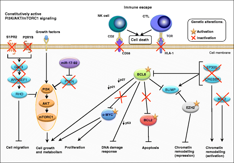
Oncogenic (deregulated) intracellular signaling pathways in germinal center B cell-like (GCB) diffuse large B-cell lymphoma (DLBCL). Germinal center B cell-like diffuse large B-cell lymphoma (GCB-DLBCL) are characterized by the inactivation or loss of the phosphatase and tensin homologue (PTEN) and S1PR2-Gα13-ARHGEF1 signaling pathways, which both negatively modulate GC B cell migration and phosphatidylinositol-3-kinase (PI3K) signaling [, –84, 324, 593]. In addition, activating mutations of the histone methyltransferase EZH2 (enhancer of zeste homologue 2) and master regulator of the GCB phenotype promote epigenetic changes that, at least partly, cooperate with the B-cell lymphoma 2 (BCL2) and BCL6 to mediate lymphomagenesis in GCB-DLBCL [77, 79]. A subset of GCB-DLBCL is characterized by translocations affecting the c-MYC and/or the BCL2 loci [80, 81, 87]. These translocations lead to constitutive activation of c-MYC and the anti-apoptotic BCL2 protein and to a malignant transformation by preventing terminal differentiation or blocking apoptosis. The different colors that are used in the figure indicate molecules that belong to a specific pathway and/or lead to a specific outcome. Adapted from REF: [3, 70, 86, 109]. Information for this figure was gleaned from the following references: [, , , –, , , , , –, , , , , , , –, , , , , –, –, , , , , –, –386, 593, 612]. Abbreviations: S1PR2 sphingosine-1-phosphate receptor-2, Gα13 (GNA13) heterotrimeric G protein alpha 13, ARHGEF1 Rho guanine nucleotide exchange factor (GEF) 1, PI3K phosphoinositide 3-kinase, mTORC1 mammalian target of rapamycin (mTOR) complex 1, BCL6 B cell lymphoma protein 6, BCL2 B cell lymphoma protein 2, EZH2 enhancer of zeste homologue 2
Fig. 3
Fig. 3
Oncogenic (deregulated) intracellular signaling pathways in activated B cell-like (ABC) diffuse large B-cell lymphoma (DLBCL). The oncogenic constitutive activation of canonical nuclear factor-kappa B (NF-κB) family members in activated B cell-like diffuse large B-cell lymphoma (ABC-DLBCL), together with a blockade in terminal B cell differentiation, which is in part mediated by BCL6, represent the hallmarks of ABC-DLBCL pathogenesis [, –18, 102]. The different colors that are used in the figure indicate molecules that belong to a specific pathway and/or lead to a specific outcome. Adapted from REF: [3, 70, 86, 109]. Information for this figure was gleaned from the following references: [, , , , –, , , , , , –, , , , , , –, , , , , , , , , , , , , –, –, , , , , –, –, –, , , , , –, –386, 572, 612, 620]. Abbreviations: BCR B cell receptor, BTK Bruton’s tyrosine kinase, SYK spleen tyrosine kinase, PI3K phosphoinositide 3-kinase, mTORC1 mammalian target of rapamycin (mTOR) complex 1, CD40L CD40 ligand, JAK Janus kinase, IRF4 interferon-regulatory factor 4, MALT1 mucosa-associated lymphoid tissue lymphoma translocation protein 1, BCL10 B cell lymphoma protein 10, TLR Toll-like receptor, MyD88 myeloid differentiation primary response 88, CARD11 caspase recruitment domain family, member 11, PKCβ protein kinase Cβ, STAT3 signal transducer and activator of transcription 3, BCL6 B cell lymphoma protein 6, BCL2 B cell lymphoma protein 2
Fig. 4
Fig. 4
Schematic comparison of the domain architecture of the macrodomain-containing ARTD family members ARTD7, ARTD8 and ARTD9 Domain architecture of the human diptheria toxin-like macro-domain containing mono-ADP-ribosyltransferases and ARTD family members ARTD7, ARTD8 and ARTD9. The following domains are indicated: The diptheria toxin-like ART(D) domain is the catalytic core required for basal (mono-)ADP-ribosyltransferae (ART) activity. The WWE domain is named after three conserved residues (W-W-E) and is predicted to mediate specific protein-protein interactions in ubiquitin- and ADP-ribose conjugation systems [555, 636, 637]. The macro or A1pp domains are structurally related to the catalytic domain of enzymes that process ADP-ribose-1-phosphate, a reaction product derived from ADP-ribose 1-2′cyclic phosphate generated by TpT [638]. The macrodomain can serve as ADP-ribose or O-acetyl-ADP-ribose binding module [638]. RRM is an RNA and poly-ADP-ribose-binding/recognition motif [639]
Fig. 5
Fig. 5
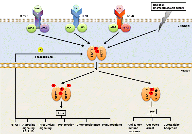
Proposed model for the postulated functions of STAT1 in HR-DLBCL-NOS. Chronic exposure to IFNs and chemotherapeutic agents such as alkylating agents, topoisomerase inhibitors, HDAC inhibitors, or proteasome inhibitors, can lead to constitutive oncogenic upregulation of STAT1 and interferon signaling genes (ISGs) during tumor development [–, , –643] and select tumor clones resistant to a cytotoxic microenvironment and concurrently to genotoxic therapy [518]. The overall population of tumor cells is sensitive to cytotoxic factors of microenvironment and committed to death because of the secretion of cytotoxic ligands by host cells (T lymphocytes, B lymphocytes, tumor-associated macrophages, dendritic cells etc.) [518]. The majority of tumor cells are relatively sensitive to radio- and/or chemotherapy. However, a small fraction of tumor cells (HR-DLBCL cells) constitutively express STAT1/ISGs and are resistant to the cytotoxic microenvironment and genotoxic therapy. Oncogenic constitutive expression of STAT1 can activate the prosurvival secretome, including the interleukines IL6 and IL10 [322, 468, 644] and in turn, can enhance selective advantages of surrounding tumor cells [518]. Moreover, STAT1 pathway can be also activated in the context of IL6 and IL10 [322, 645], thereby generating auto-stimulatory feedback loops (IL6-IL16R-STAT1 and IL10-IL10R-STAT1). Thus, IFN/JAK pathway and chemotherapeutic agents may act in concert with other HR-DLBCL specific intrinsic signaling pathways such as IL10/IL10R-JAK1-TYK2 and or IL6/IL6R-gp130-JAK1/2-TYK2 to induce chemotherapy resistance. In addition, oncogenic JAK/STAT1α-autocrine signaling in HR-DLBCL causes enhanced expression of STAT1α dependent proto-oncogenes (i.e., DTX3L ARTD9, IRF2, MCL1, BCL2, BCL6), thereby stimulating proliferation and survival pathways [470]. In addition, oncogenic STAT1α signaling helps the tumors to escape the attack by the immune system (immunoediting) [473, 497, 526]. Conversely, STAT1β repress the transcriptional activation of anti-proliferative and pro-apoptotic genes (including tumor the suppressors IRF1 and TP53) [470, 471] as well as genes involved in immune surveillance [473, 497, 526]. Adapted from REF: [470, 471, 518]. Abbreviations: HR-DLBCL host response DLBCL, ARTD ADP-ribosyltransferase Diphteria toxin-like, STAT signal transducer and activator of transcription, ISG interferon stimulated gene, IL interleukin, JAK Janus kinase, TYK2 tyrosine kinase 2, IRF interferon response factor, BCL6 B cell lymphoma protein 6, BCL2 B cell lymphoma protein 2
Fig. 6
Fig. 6
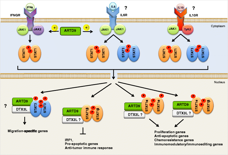
Proposed model for the postulated functions of ARTD9 and DTX3L in DLBCLs. ARTD9 and DTX3L act as novel oncogenic survival factors in relapsed or refractory DLBCL. Constitutively active IL6/IL6R-JAK1-STAT3, oncogenic IFN/IFNGR-JAK1/2-STAT1 and/or IL10/IL10R-JAK1-TYK2-autocrine signaling in HR-DLBCL causes overexpression of ARTD9 and DTX3L, which in turn, further stimulates the phosphorylation of STAT1 on Y701 and subsequently the enhanced expression of STAT1α dependent proto-oncogenes (i.e., ARTD8, ARTD9, DTX3L, IRF2, BCL6, BCL2). Both ARTD9 and DTX3L might also enhance the transcription of DNA damage repair/response genes upon treatment of tumors with genotoxic chemotherapeutic agents [548]. Conversely, ARTD9 and DTX3L repress together with STAT1β the transcriptional activation of tumor suppressor IRF1 and TP53 [470, 471]. Overexpression of ARTD9 inhibits intrinsic and extrinsic IFNγ-STAT1-IRF1/p53-mediated cell death pathways while simultaneously enhancing the STAT1-dependent IRF2-mediated proliferation and BCL6-BCL2 mediated survival pathways. As a consequence ARTD9 induces an oncogenic switch in STAT1 from a tumor suppressor to an oncogene and mediates proliferation, survival and chemo-resistance in HR-DLBCL. Pharmaceutical inhibition of ARTD9 in HR-DLBCL associated with constitutively active STAT1 signaling might therefore reactivate the anti-proliferative and pro-apoptotic IFNγ-STAT1-IRF1/p53 axes and reverses chemo-resistance in HR-DLBCL. In addition, endogenous overexpression of DTX3L and/or ARTD9 might also be associated with lymphocytes migration and may promote the dissemination of malignant B cells in high-risk DLBCL in vivo, potentially dependent on STAT3 or PI3K/AKT/mTORC1 signaling [471]. DTX3L and/or ARTD9 might form specific complexes with non-canonical STAT1:STAT3 heterodimers and activate a migration-specific signaling and gene expression program in HR-DLBCL. Finally, oncogenic. ARTD9/DTX3L-STAT1α signaling might upregulate immunomodulatory genes and may help the tumors to escape the attack by the immune system (immunoediting) [470, 471, 473]. Conversely, ARTD9 and DTX3L together with STAT1β might repress the transcriptional activation of genes involved in immune surveillance [470, 471, 473]. Adapted from REF: [470, 471]. Abbreviations: ARTD ADP-ribosyltransferase Diphteria toxin-like, DTX3L deltex (DTX)-3-like E3 ubiquitin ligase, STAT signal transducer and activator of transcription, ISG interferon stimulated gene, IL interleukin, JAK Janus kinase, TYK2 tyrosine kinase 2, IRF interferon-regulatory factor, BCL6 B cell lymphoma protein 6, BCL2 B cell lymphoma protein 2
Fig. 7
Fig. 7
Schematic comparison of the domain architecture of the DTX family. Domain architecture of the human Deltex-like E3 ubiquitin ligases and DTX family members DTX1, DTX2A, DTX2B, DTX3, DTX3L and DTX4. The domain organization is based on analogous functional motifs in murine DTX family members DTX1, DTX2, DTX2deltaE and DTX3 [589], human DTX1 [544, 646], and human DTX3L [542, 543]. DTX1, DTX2 and DTX4 have well-conserved domains I, II and III. DTX1, DTX2A, DTX2B and DTX4 contain two conserved tandem WWE modules in their domain I [646]. The N-terminal domains I of DTX1, DTX2A and DTX2B bind through their tandem WWE modules to the ankyrin repeat region of intracellular NOTCH1 [646]. The WWE domain is named after three conserved residues (W-W-E)and is predicted to mediate specific protein-protein interactions in ubiquitin- and ADP-ribose conjugation systems [555, 636, 637]. DTX3 lacks most of the site corresponding to the binding site for the ankyrin repeats of human NOTCH1. Of interest, the WWE domain of DTX1 serves also as a bona fide poly-ADP-ribose binding module [555]. Domains II of DTX1, DTX2A, DTX3 and DTX4 contain a proline-rich region (black boxes) that is not found in DTX3L. DTX2B is an alternatively spliced form of DTX2A, DTX2B lack a proline-rich motif (PRM), the fourth of nine predicted DTX2 exons. The most conserved region of all human DTX family members (DTX1, DTX2A, DTX2B, DTX3, DTX3L and DTX4) is their C-terminal part, domain III. The C-terminal domain III of DTX1, DTX2A, DTX2B and DTX4 contain a classic RING finger E3 ubiquitin ligase domain (RING-H2 finger motif; red box); while domain III of DTX3 and DTX3L contain a RING-C3HC4 finger motif found in many E3 ubiquitin ligases (indicated by an orange box). Human DTX3 has a short unique N-terminal domain (yellow box), and DTX3L contains a longer unique N-terminal region (grey box). Abbreviations: RING really interesting new gene, DTX deltex, DTC Deltex C-terminal domain
Fig. 8
Fig. 8
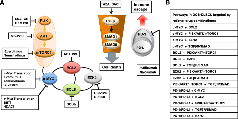
Mechanism-based combinatorial experimental regimens aimed at disrupting known oncogenic cooperation pathways in GCB-DLBCLs. Targets for rational mechanism-based combinatorial experimental regimens are shown for GCB-DLBCL (a and b). The black thick lines connecting the different drug targets pathways depict established biological interactions and/or dependencies. The single drugs listed in (a) have either proven clinical activity in patients with DLBCL or are currently in clinical trials for patients with B-cell lymphoma, including DLBCL. Pathways in GCB-DLBCL targeted by rational drug combinations using drugs tested as single agent in preclinical studies or clinical trials are shown in (b). Silvestrol inhibits RNA helicase and translation initiationfactor eIF4A thereby blocking translation (i.e., of oncogenic factors) [647]. Azacytidine (AZA) and decitabine/5-aza-2′-deoxycytidine (DAC/5-AZA) inhibit DNA methyltransferases (mainly DMNT1 and DMNT3) resulting in the re-expression of tumor-suppressor genes [419, 420]. Adapted from REF: [601, 602]. Information for this figure was gleaned from the following references: [, , , –, , , , , –, , , , , , , , , , , , –, –, , , , –, –, , , , –, , , , , , –650]. See also additional tables 2 and 5–9. Abbreviations: Gα13 (GNA13) heterotrimeric G protein alpha 13, ARHGEF1 Rho guanine nucleotide exchange factor (GEF) 1, PI3K phosphoinositide 3-kinase, mTORC1 mammalian target of rapamycin (mTOR) complex 1, BCL2 B-cell lymphoma protein 2, BCL6 B-cell lymphoma protein 6, EZH2 enhancer of zeste homologue 2, PD-1 programmed cell death, PD-L1 programmed cell death ligand, TGFβR transforming growth factor beta (TGFβ) receptor, HDACi inhibitors of histone deacetylases, BCL6i inhibitors of BCL6, AZA azacytidine, DAC decitabin
Fig. 9
Fig. 9
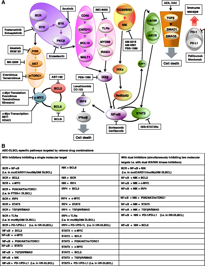
Mechanism-based combinatorial experimental regimens aimed at disrupting known oncogenic cooperation pathways in ABC-DLBCLs. Targets for rational mechanism-based combinatorial experimental regimens are shown for ABC-DLBCL (a and b). The black thick lines connecting the different drug targets pathways depict established biological interactions and/or dependencies. The single drugs listed in (a) have either proven clinical activity in patients with DLBCL or are currently in clinical trials for patients with B-cell lymphoma, including DLBCL. Pathways in ABC-DLBCL targeted by rational drug combinations using drugs tested as single agent in preclinical studies or clinical trials are shown in (b). Bortezomib and carfilzomib inhibit the 26S proteasome complex thereby blocking the degradation of negative regulators of cell cycle progression as well as of NF-κB inhibitory protein IκBα. Lenalidomide is an immunomodulatory agent directly binding to the E3 ubiquitin ligase substrate receptor CRBN and promoting the recruitment of its common substrates to the E3 ubiquitin ligase complex, thus leading to substrate ubiquitinylation and degradation [284] and subsequent repression of IRF4, a hallmark of ABC-DLBCL cells, thereby inhibiting the BCR-mediated canonical NF-κB-dependent pro-survival signaling pathways [90, 91]. Silvestrol inhibits RNA helicase and translation initiationfactor eIF4A thereby blocking translation (i.e., of oncogenic factors) [647]. Azacytidine (AZA) and decitabine/5-aza-2′-deoxycytidine (DAC/5-AZA) inhibit DNA methyltransferases (mainly DMNT1 and DMNT3) resulting in the re-expression of tumor-suppressor genes [419, 420]. Mechanism-based drug combinations targeting the biological interaction between MYC and BCL2 as well as MYC and the PI3K/AKT/mTORC1 pathway are aimed for both GCB- and ABC-DLBCL. Adapted from REF: [590, 591]. Information for this figure was gleaned from the following references: [, , , –, , , , , , , , , –, , , , , –, , , , , , , , , , , –, , , , , , , , , , , –, –, , , , , , –, , , , , –, –, , , , –, , –, , , , , –652]. See also Additional file 1: Table S2 and S5–S9. Abbreviations: BCR B cell receptor, BTK Bruton’s tyrosine kinase, CD40L CD40 ligand, JAK Janus kinase, CRBN cereblon, IRF4 interferon-regulatory factor 4, MALT1 mucosa-associated lymphoid tissue lymphoma translocation protein 1, BCL10 B cell lymphoma protein 10, TLR Toll-like receptor, MyD88 myeloid differentiation primary response 88, CARD11 caspase recruitment domain family, member 11, PKCβ protein kinase Cβ, IKK inhibitor kappa B (IκB) kinase, NF-κB nuclear factor-kappa B, NIK NF-κB inducing kinase, STAT signal transducer and activator of transcription, PI3K phosphoinositide 3-kinase, mTORC1 mammalian target of rapamycin (mTOR) complex 1, BCL2 B-cell lymphoma protein 2, BCL6 B-cell lymphoma protein 6, TGFβR transforming growth factor beta (TGFβ) receptor, HDACi inhibitors of histone deacetylases, BCL6i inhibitors of BCL6, PD-1 programmed cell death, PD-L1 programmed cell death ligand, AZA azacytidine, DAC decitabine

References

    1. Martelli M, Ferreri AJ, Agostinelli C, Di Rocco A, Pfreundschuh M, Pileri SA. Diffuse large B- cell lymphoma. Crit Rev Oncol Hematol. 2013;87(2):146–171. doi: 10.1016/j.critrevonc.2012.12.009. - DOI - PubMed
    1. Schneider C, Pasqualucci L, Dalla-Favera R. Molecular pathogenesis of diffuse large B-cell lymphoma. Semin Diagn Pathol. 2011;28(2):167–177. doi: 10.1053/j.semdp.2011.04.001. - DOI - PMC - PubMed
    1. Shaffer AL, 3rd, Young RM, Staudt LM. Pathogenesis of human B cell lymphomas. Annual review of immunology. 2012;30:565–610. doi: 10.1146/annurev-immunol-020711-075027. - DOI - PMC - PubMed
    1. De Paepe P, De Wolf-Peeters C. Diffuse large B-cell lymphoma: a heterogeneous group of non-Hodgkin lymphomas comprising several distinct clinicopathological entities. Leukemia. 2007;21(1):37–43. doi: 10.1038/sj.leu.2404449. - DOI - PubMed
    1. Fisher SG, Fisher RI. The epidemiology of non-Hodgkin's lymphoma. Oncogene. 2004;23(38):6524–6534. doi: 10.1038/sj.onc.1207843. - DOI - PubMed

Publication types

MeSH terms