Review
. 2015 Dec 7;25(23):R1143-50.
doi: 10.1016/j.cub.2015.11.001.

Shared and Distinct Mechanisms of Compartmentalized and Cytosolic Ciliogenesis

Affiliations
Review

Shared and Distinct Mechanisms of Compartmentalized and Cytosolic Ciliogenesis

Tomer Avidor-Reiss et al. Curr Biol. .

Abstract

Most motile and all non-motile (also known as primary) eukaryotic cilia possess microtubule-based axonemes that are assembled at the cell surface to form hair-like or more elaborate compartments endowed with motility and/or signaling functions. Such compartmentalized ciliogenesis depends on the core intraflagellar transport (IFT) machinery and the associated Bardet-Biedl syndrome complex (BBSome) for dynamic delivery of ciliary components. The transition zone (TZ), an ultrastructurally complex barrier or 'gate' at the base of cilia, also contributes to the formation of compartmentalized cilia. Yet, some ciliated protists do not have IFT components and, like some metazoan spermatozoa, use IFT-independent mechanisms to build axonemes exposed to the cytosol. Moreover, various ciliated protists lack TZ components, whereas Drosophila sperm surprisingly requires the activity of dynamically localized TZ proteins for cytosolic ciliogenesis. Here, we discuss the various ways eukaryotes use IFT and/or TZ proteins to generate the wide assortment of compartmentalized and cytosolic cilia observed in nature. Consideration of the different ciliogenesis pathways allows us to propose how three types of cytosol-exposed cilia (primary, secondary and tertiary), including cilia found in the human sperm proximal segment, are likely generated by evolutionary derivations of compartmentalized ciliogenesis.


Figures

Figure 1
Figure 1
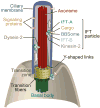
Evolutionarily conserved components of a compartmentalized basal body-cilium organelle. The centriole-derived basal body connects to the plasma membrane using transition fibers. These serve as docking sites for the intraflagellar transport (IFT) machinery, which is critical for cilium formation and functional maintenance. The IFT machinery consists of one or more Kinesin-2 anterograde molecular motors which mobilize two ‘core’ IFT subcomplexes (IFT-A and IFT-B) and a BBSome adaptor, and associated ciliary cargo. A Dynein-2 molecular motor, transported to the tip by the anterograde IFT machinery (not shown), brings the IFT components back to the base. A transition zone containing axoneme-to-ciliary membrane connectors (typically Y-shaped) serves as a selective membrane diffusion barrier. Together, the transition fibers, transition zone and IFT machinery help to compartmentalize the ciliary organelle and dynamically maintain its composition.
Figure 2
Figure 2
Types of cilia, ciliary components, and modes of cilium formation (compartmentalized and/or cytosolic) across eukaryotes. (A) Distribution of ciliary features throughout the main eukaryotic kingdoms. The major classes of cilia are motile and non-motile. Ciliary proteins used for cilium formation and functional maintenance are transition zone (TZ) proteins, as well as core intraflagellar transport (IFT) proteins and the associated BBS protein complex (BBSome). Compartmentalized ciliogenesis refers to the formation of an axoneme that protrudes from a basal body docked to the cell surface, and cytosolic ciliogenesis is the formation of an axoneme that also stems from a basal body but is at least in part exposed to the cytoplasm. (B) Ciliated organisms make variable use of TZ, core IFT, and BBSome machinery. Shown are all eight possible combinations of these machineries, and whether representative organisms possess such combinations or not.
Figure 3
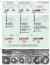
Figure 3. Compartmentalized and cytosolic ciliogenesis pathways, and model for cytosolic ciliogenesis evolution
(A) Non-ciliated cells (i) have centrioles (green) near the nucleus (n). Compartmentalized ciliogenesis (upper horizontal arrows) begins when a centrioles docks to the plasma membrane, or a vesicle that later fuses with the plasma membrane (not shown) and forms a transition zone (TZ; yellow) (ii). Intraflagellar transport (IFT) then mediates the formation of an elongated compartmentalized axoneme (red) ensheathed by a ciliary membrane (blue) (iii). Primary cilia are formed by this process, as are various motile cilia (e.g., in Chlamydomonas or vertebrate respiratory airway). Three types of cytosolic ciliogenesis pathways (vertical arrows) can each be recognized to begin at distinct points after, during, or before compartmentalized ciliogenesis. We refer to these three as forming primary, secondary, and tertiary cytosolic cilia, respectively. The three different types of cytosolic ciliogenesis pathways may represent gradual/distinct steps of evolution from an ancestral compartmentalized ciliogenesis pathway. First, as exemplified by the mammalian sperm tail and possibly Giardia (as well as Toxoplasma and Thalassiosira) flagella (step iii directly to iv), after completion of compartmentalized ciliogenesis, the centriole attaches to the nucleus, and the TZ migrates away from the centriole along the complete axoneme to expose it to the cytoplasm. Second, as exemplified by Drosophila sperm (step ii directly to v), after the centriole docks to the plasma membrane and forms a TZ, it then attaches to the nucleus. Then the TZ migrates away from the centriole, while a rudimentary axoneme forms. Here, the exposed axoneme completes ciliogenesis by recruiting proteins directly from the cytoplasm (axoneme maturation). Finally, as exemplified by Plasmodium flagella (step I directly to vi), the centriole forms the axoneme directly in the cytoplasm. In both the mammalian and Drosophila sperm, the centriole migrates toward the nucleus and attaches to it. The various cytosolic cilia undergo a final process of reattachment of the cytosolic axoneme to the plasma membrane. These processes are refered to as spermiation (iii), individualization (iv), or exflagellation (vi). (B) Example of a cytoplasmic cilium, from Drosophila. Transmission Electron Microscopy image showing the basal body, cytosol-exposed axoneme, and compartmentalized axoneme (i). Cross-sections reveal the microtubule architecture along the length: basal body with triplet microtubules (ii, iii), cytosol-exposed axoneme with doublet microtubules (iv, v), and compartmentalized (ciliary membrane-associated) axoneme with doublet microtubules (vi) or incomplete set of microtubules (vii). Image adapted from Gottardo et al.[50].

References

    1. Carvalho-Santos Z, Azimzadeh J, Pereira-Leal JB, Bettencourt-Dias M. Evolution: Tracing the origins of centrioles, cilia, and flagella. J Cell Biol. 2011;194(2):165–175. - PMC - PubMed
    1. Sung CH, Leroux MR. The roles of evolutionarily conserved functional modules in cilia-related trafficking. Nat Cell Biol. 2013;15(12):1387–1397. - PMC - PubMed
    1. Avidor-Reiss T, Maer AM, Koundakjian E, Polyanovsky A, Keil T, Subramaniam S, Zuker CS. Decoding cilia function: defining specialized genes required for compartmentalized cilia biogenesis. Cell. 2004;117:527–539. - PubMed
    1. Li JB, Gerdes JM, Haycraft CJ, Fan Y, Teslovich TM, May-Simera H, Li H, Blacque OE, Li L, Leitch CC, et al. Comparative genomics identifies a flagellar and basal body proteome that includes the BBS5 human disease gene. Cell. 2004;117:541–552. - PubMed
    1. Bloodgood RA. Sensory reception is an attribute of both primary cilia and motile cilia. J Cell Sci. 2010;123(Pt 4):505–509. - PubMed

Publication types

Substances

LinkOut - more resources