. 2015;14(23):3734-47.
doi: 10.1080/15384101.2015.1104441.

SIAH ubiquitin ligases regulate breast cancer cell migration and invasion independent of the oxygen status

Affiliations

SIAH ubiquitin ligases regulate breast cancer cell migration and invasion independent of the oxygen status

M Gordian Adam et al. Cell Cycle. 2015.

Abstract

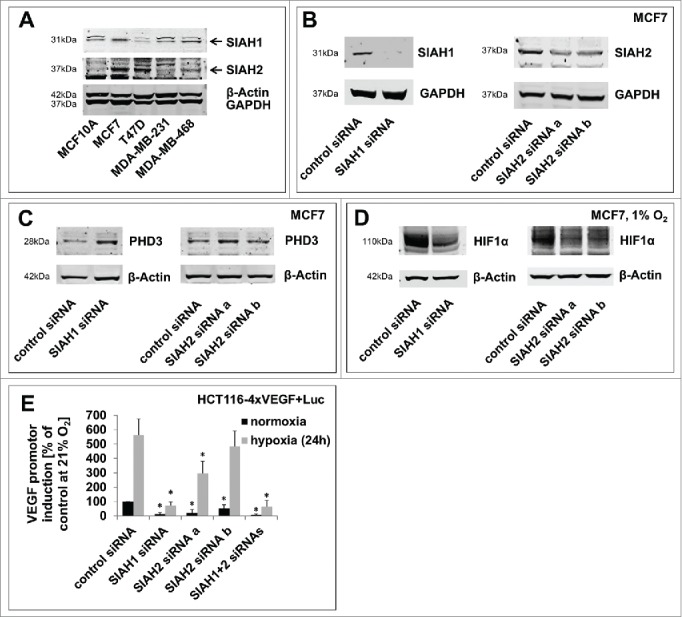
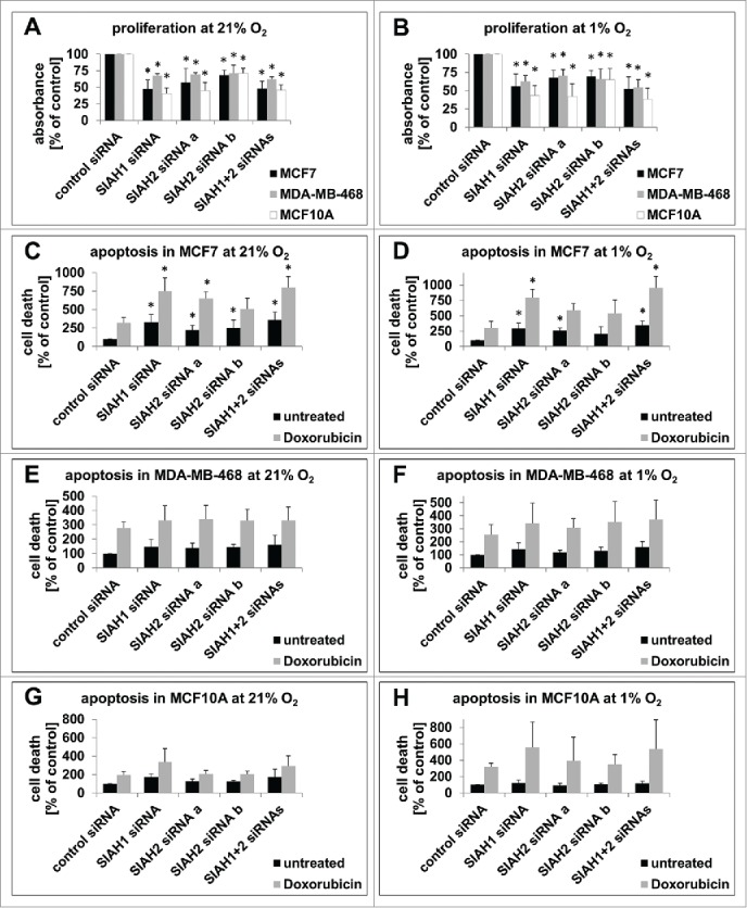
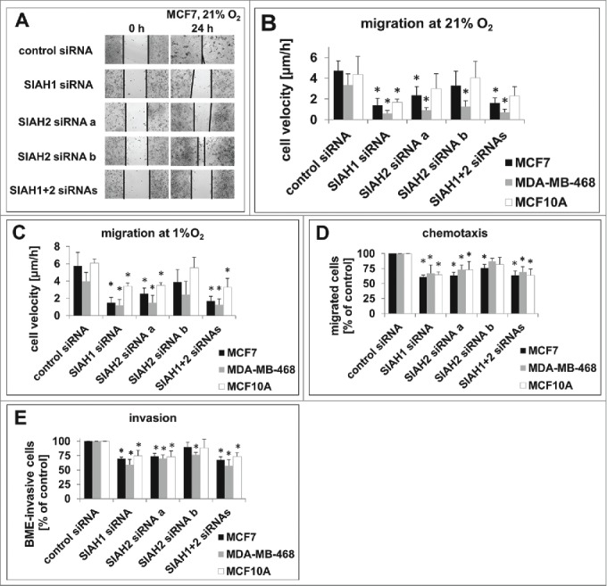
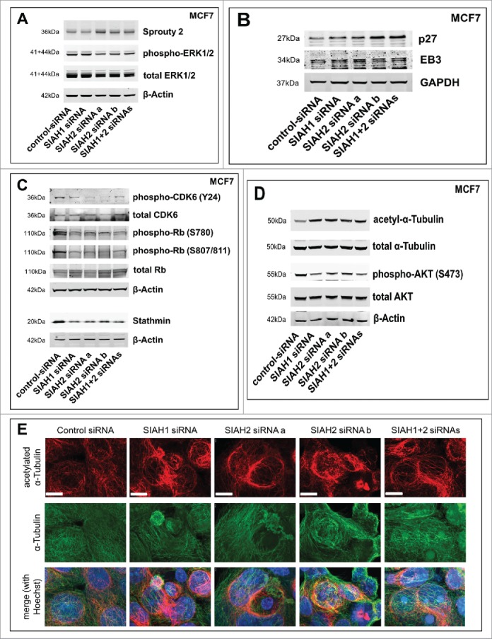
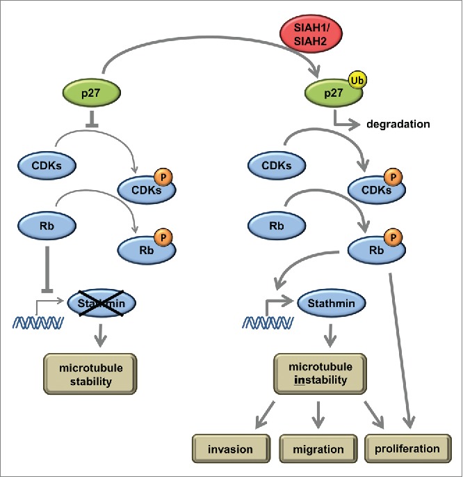
Seven-in-absentia homolog (SIAH) proteins are evolutionary conserved RING type E3 ubiquitin ligases responsible for the degradation of key molecules regulating DNA damage response, hypoxic adaptation, apoptosis, angiogenesis, and cell proliferation. Many studies suggest a tumorigenic role for SIAH2. In breast cancer patients SIAH2 expression levels correlate with cancer aggressiveness and overall patient survival. In addition, SIAH inhibition reduced metastasis in melanoma. The role of SIAH1 in breast cancer is still ambiguous; both tumorigenic and tumor suppressive functions have been reported. Other studies categorized SIAH ligases as either pro- or antimigratory, while the significance for metastasis is largely unknown. Here, we re-evaluated the effects of SIAH1 and SIAH2 depletion in breast cancer cell lines, focusing on migration and invasion. We successfully knocked down SIAH1 and SIAH2 in several breast cancer cell lines. In luminal type MCF7 cells, this led to stabilization of the SIAH substrate Prolyl Hydroxylase Domain protein 3 (PHD3) and reduced Hypoxia-Inducible Factor 1α (HIF1α) protein levels. Both the knockdown of SIAH1 or SIAH2 led to increased apoptosis and reduced proliferation, with comparable effects. These results point to a tumor promoting role for SIAH1 in breast cancer similar to SIAH2. In addition, depletion of SIAH1 or SIAH2 also led to decreased cell migration and invasion in breast cancer cells. SIAH knockdown also controlled microtubule dynamics by markedly decreasing the protein levels of stathmin, most likely via p27(Kip1). Collectively, these results suggest that both SIAH ligases promote a migratory cancer cell phenotype and could contribute to metastasis in breast cancer.

Keywords: SIAH ubiquitin ligases; SIAH1; SIAH2; breast cancer; invasion; metastasis; migration; p27Kip1; seven-in-absentia; stathmin.


Figures

Figure 1.
Figure 1.
SIAH1/2 silencing reduces hypoxic adaptation in breast cancer cells. (A) Comparison of SIAH1 and SIAH2 expression levels in different breast cancer cell lines. Four breast cancer cell lines and MCF10A as a non-cancer control cell line were lysed and immunoblotted for SIAH1 expression. The membrane was reprobed for SIAH2. GAPDH and β-Actin serve as a loading control. (B) Efficient SIAH knockdown in MCF7 cells. MCF7 breast cancer cells were transfected with siRNAs targeting SIAH1 or SIAH2. After 48 h the cells were lysed and Western Blot was performed with specific antibodies. Instead of reprobing the membrane was cut in advance to allow clear visualization of the results. (C) Lysates of MCF7 cells silenced for expression of SIAH1 or SIAH2 were examined for expression of PHD3. (D) MCF7 cells were transfected with SIAH1 and SIAH2 siRNA and cultivated at 1% O2 for 48 h. Lysates were probed for HIF1α accumulation. (E) SIAH1 and SIAH2 were silenced in HCT116–4xVEGF+Luc cells for 24 h. The cells were then incubated at 21% O2 or 1% O2 for another 24 h, lysed, and a luciferase assay was performed to assess HIF target gene expression. Relative light units from the luciferase assay were normalized to the cell number measured with CellTiter-Blue Cell Viability assay. Bar graphs show mean values, error bars indicate SD. Data were analyzed using 1-way ANOVA followed by Dunnett's post-hoc test. n = 3; *, p<0.05.
Figure 2.
Figure 2.
SIAH1/2 silencing inhibits proliferation and promotes apoptosis in breast cancer cells. (A, B) 48 h after transfection of MCF7, MDA-MB-468, and MCF10A cells with RNAi targeting SIAH1/2, cells were incubated in presence of BrdU for 2 h. BrdU incorporation into the DNA, signifying cell division, was quantified with an ELISA. Cells were either kept at 21% O2 (A) or 1% O2 (B) for the duration of the experiment. (C, D) 30 h post RNAi transfection, MCF7 cells were either left untreated or apoptosis was stimulated by adding the DNA-damage inducing chemotherapeutic drug Doxorubicin (1 µM) for 18 h. Then cells were lysed and apoptosis was measured with a TUNEL-based ELISA. The same experiments were performed with MDA-MB-468 (E, F) and MCF10A cells (G, H). Cells were either kept at 21% O2 (C, E, G) or 1% O2 (D, F, H) the whole time. Bar graphs show mean values, error bars indicate SD. Data were analyzed using 1-way ANOVA followed by Dunnett's post-hoc test. n = 3; *, p<0.05.
Figure 3.
Figure 3.
SIAH1/2 silencing inhibits migration and invasion in breast cancer cells. (A) Representative images of a wound healing assay in MCF7 cells silenced for SIAH1/2 expression. The gap was induced 48 h after RNAi transfection, cells were allowed to migrate for 24 h. Images were taken using a Zeiss Observer.Z1 microscope at 10-fold magnification. (B, C) Quantification of the cell migration speed in (A) employing 3 individual experiments with MCF7, MDA-MB-468, and MCF10A at 21% O2 (B) or 1% O2 (C). (D) Quantification of chemotaxis assays. MCF7, MDA-MB-468, and MCF10A cells were transfected with SIAH siRNAs and, after 48 h, seeded into the upper compartments of a modified Boyden chamber assay. The cells were allowed to migrate through a porous membrane toward the lower compartment filled with medium containing FCS for 24 h; the cells were then detached from the membrane undersurface, stained and quantified. (E) 48 h after RNAi transfection, the cells were seeded in a transwell coated with a rehydrated layer of basal membrane extract. Transmigrated cells were stained and quantified to assess the invasion capacity. Results are shown as means +SD. Data were analyzed using 1-way ANOVA followed by Dunnett's post-hoc test. n = 3; *, p<0.05.
Figure 4.
Figure 4.
SIAH1/2 silencing affects expression of p27 and stathmin in breast cancer cells. (A) Following siRNA transfection (48 h), MCF7 cells were lysed. Western blot was performed to assess expression levels of the SIAH substrate Sprouty2 and activation status of ERK1/2. (B) Western Blot showing expression levels of the SIAH substrates p27Kip1 and EB3 after silencing of SIAH1 or SIAH2. (C) Western blot demonstrating the effect of SIAH1/2 silencing on total CDK6 levels and CDK6 phosphorylation at tyrosine 24, total Rb levels and Rb phosphorylation at serines 780 and 807/811, as well as on stathmin protein levels. (D) Western blot to determine the stability of microtubules (via acetylated α-Tubulin) as well as the activity of AKT (phosphorylation at serine 473) downstream of stathmin. (E) Immunofluorescent staining to determine microtubule stability after knockdown of SIAH1 and SIAH2. Following RNAi transfection (48 h), MCF7 cells were fixed and stained for α-Tubulin (DyLight 488, green) and acetylated α-Tubulin (Cy3, red). Cell nuclei were stained with Hoechst dye (blue). Scale bars, 10 µm.
Figure 5.
Figure 5.
Summarizing model to explain the pathway how SIAH ubiquitin ligases influence migration, invasion, and proliferation according to our results. At low levels of active SIAH1 or SIAH2, p27 is present and inhibits phosphorylation of Rb via CDK inhibition. Non-phosphorylated Rb sequesters the E2F1 transcription factor and thereby inhibits stathmin expression. High levels of active SIAH1 and SIAH2 ligases induce ubiquitinylation of p27 and subsequent proteasomal degradation. Active CDKs phosphorylate Rb, inducing the release of E2F1 and stathmin expression. Stathmin depolymerizes microtubules, a prerequisite for increased migration, invasion, and proliferation.

References

    1. Hu G, Zhang S, Vidal M, Baer JL, Xu T, Fearon ER. Mammalian homologs of seven in absentia regulate DCC via the ubiquitin-proteasome pathway. Genes & Dev 1997; 11:2701-14; PMID:9334332; http://dx.doi.org/ 10.1101/gad.11.20.2701 - DOI - PMC - PubMed
    1. Roperch JP, Lethrone F, Prieur S, Piouffre L, Israeli D, Tuynder M, Nemani M, Pasturaud P, Gendron MC, Dausset J, et al.. SIAH-1 promotes apoptosis and tumor suppression through a network involving the regulation of protein folding, unfolding, and trafficking: identification of common effectors with p53 and p21(Waf1). Proc Natl Acad Sci U S A 1999; 96:8070-3; PMID:10393949; http://dx.doi.org/ 10.1073/pnas.96.14.8070 - DOI - PMC - PubMed
    1. Ahmed AU, Schmidt RL, Park CH, Reed NR, Hesse SE, Thomas CF, Molina JR, Deschamps C, Yang P, Aubry MC, et al.. Effect of disrupting seven-in-absentia homolog 2 function on lung cancer cell growth. J Natl Cancer Inst 2008; 100:1606-29; PMID:19001609; http://dx.doi.org/ 10.1093/jnci/djn365 - DOI - PMC - PubMed
    1. Behling KC, Tang A, Freydin B, Chervoneva I, Kadakia S, Schwartz GF, Rui H, Witkiewicz AK. Increased SIAH expression predicts ductal carcinoma in situ (DCIS) progression to invasive carcinoma. Breast Cancer Res Treat 2011; 129:717-24; PMID:21088888; http://dx.doi.org/ 10.1007/s10549-010-1254-8 - DOI - PMC - PubMed
    1. Qi J, Nakayama K, Cardiff RD, Borowsky AD, Kaul K, Williams R, Krajewski S, Mercola D, Carpenter PM, Bowtell D, et al.. Siah2-dependent concerted activity of HIF and FoxA2 regulates formation of neuroendocrine phenotype and neuroendocrine prostate tumors. Cancer Cell 2010; 18:23-38; PMID:20609350; http://dx.doi.org/ 10.1016/j.ccr.2010.05.024 - DOI - PMC - PubMed

MeSH terms