. 2015 Dec 12:15:289.
doi: 10.1186/s12870-015-0678-z.

The development of type VI glandular trichomes in the cultivated tomato Solanum lycopersicum and a related wild species S. habrochaites

Affiliations

The development of type VI glandular trichomes in the cultivated tomato Solanum lycopersicum and a related wild species S. habrochaites

Nick Bergau et al. BMC Plant Biol. .

Abstract

Background: Type VI glandular trichomes represent the most abundant trichome type on leaves and stems of tomato plants and significantly contribute to herbivore resistance, particularly in the wild species. Despite this, their development has been poorly studied so far. The goal of this study is to fill this gap. Using a variety of cell imaging techniques, a detailed record of the anatomy and developmental stages of type VI trichomes in the cultivated tomato (Solanum lycopersicum) and in a related wild species (S. habrochaites) is provided.

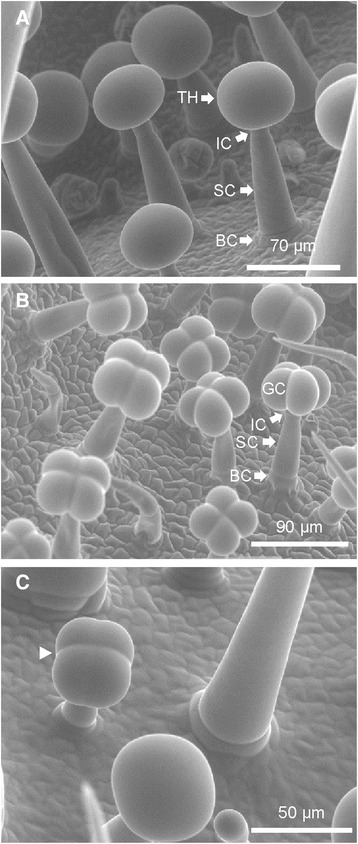
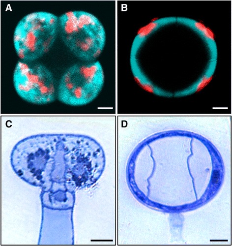
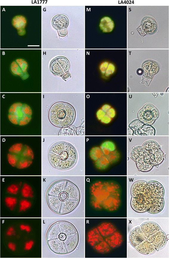
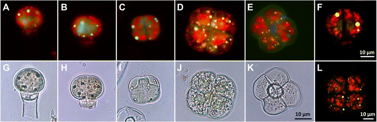
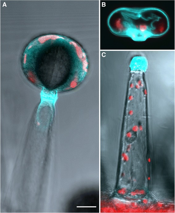
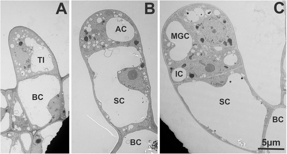
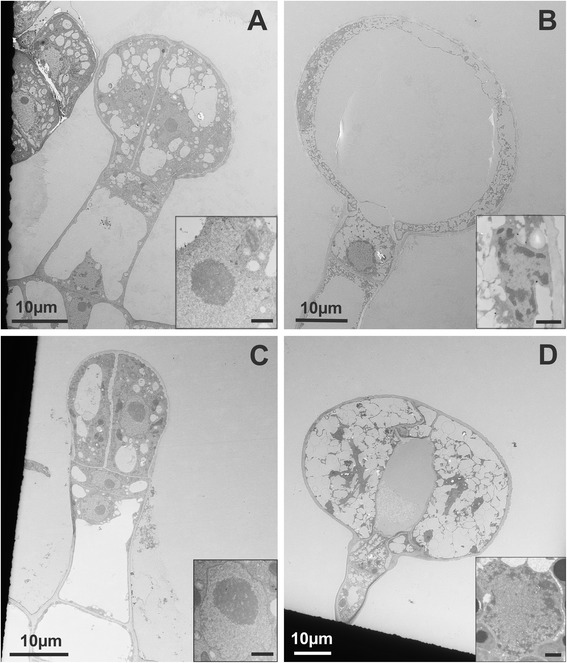
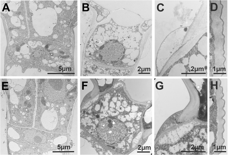
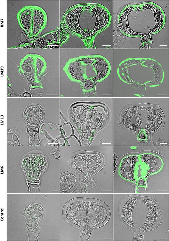
Results: In both species, the development of these structures follows a highly reproducible cell division pattern. The two species differ in the shape of the trichome head which is round in S. habrochaites and like a four-leaf clover in S. lycopersicum, correlating with the presence of a large intercellular cavity in S. habrochaites where the produced metabolites accumulate. In both species, the junction between the intermediate cell and the four glandular cells constitute a breaking point facilitating the decapitation of the trichome and thereby the quick release of the metabolites. A strongly auto-fluorescent compound transiently accumulates in the early stages of development suggesting a potential role in the differentiation process. Finally, immuno-labelling with antibodies recognizing specific cell wall components indicate a key role of pectin and arabinogalactan components in the differentiation of type VI trichomes.

Conclusions: Our observations explain the adaptive morphologies of type VI trichomes for metabolite storage and release and provide a framework for further studies of these important metabolic cellular factories. This is required to better exploit their potential, in particular for the breeding of pest resistance in tomato.


Figures

Fig. 1
Fig. 1
Scanning electron microscopy of type VI trichomes in S. habrochaites LA1777 (a and c) and S. lycopersicum LA 4024 (b). BC basal cell, SC stalk cell, IC intermediate cell, TH trichome head, GC glandular cell. The arrow head in (c) indicates the cell separation visible at an earlier development stage
Fig. 2
Fig. 2
Fluorescence and bright field microscopy of type VI trichome heads from S. habrochaites and S. lycopersicum. a-b Fluorescence microscopy images of trichome heads of S. lycopersicum LA 4024 (a) and S. habrochaites LA 1777 (b). Bright field microscopy images of sections of type VI trichomes attached to the leaves and stained with toluidine blue from S. lycopersicum LA 4024 (c) and S. habrochaites LA 1777 (d). The horizontal bars correspond to 10 μm in all panels
Fig. 3
Fig. 3
Fluorescence and bright field microscopy of detached type VI trichome heads from S. habrochaites LA 1777 and S. lycopersicum LA 4024. af Fluorescence microscopy (excitation 450–490 nm, emission 515 nm) of detached type VI trichomes from S. habrochaites. gl Corresponding bright field microscopy of the fluorescence images shown in panels a-f. m-r Fluorescence microscopy of detached type VI trichome heads of S. lycopersicum. sx Corresponding bright field microscopy of the fluorescence images shown in panels mr. The horizontal bar in A represents 20 μm and applies for all images of the figure
Fig. 4
Fig. 4
Fluorescence and bright field microscopy of detached type VI trichome heads from S. lycopersicum LA 1049 carrying a mutation in a chalcone isomerase gene. ae Fluorescence microscopy (excitation 450–490 nm, emission 515 nm) of detached type VI trichomes from S. lycopersicum LA1049. gk Corresponding bright field microscopy of the fluorescence images shown in panels ae. f and l laser scanning microscopy images of a 2-cell stage (f) and a 4-cell stage (l) trichome head of LA1049
Fig. 5
Fig. 5
Fluorescence microscopy of live S. habrochaites type VI trichomes. a Whole type VI trichome. b Detached type VI trichome head. c Stalk and intermediate cells of a decapitated type VI trichome. The horizontal white bar represents 20 μm
Fig. 6
Fig. 6
Electron microscopy of early stages of type VI glandular trichome development in S. habrochaites. a Trichome initial with a basal cell (BC) and the initial cell (TI). At this stage it is not possible to distinguish the various trichome types. b Trichome initial with one apical cell (AC) and a stalk cell (SC). c young trichome with an intermediate cell (IC) and two mothers of glandular cells (MGC)
Fig. 7
Fig. 7
Electron microscopy of type VI trichomes at different stages of development. a-b S. habrochaites LA 1777 trichomes. a a young 4-cell stage trichome. b mature trichome. c-d S. lycopersicum LA 4024 trichomes. c: a young 2-glandular cell trichome. d Mature trichome. Inserts in a-d: ultrastructure of nuclei of glandular cells in the corresponding stage of development. The scale bar in the inserts represents 1 μM
Fig. 8
Fig. 8
Electron microscopy of type VI trichomes of S. habrochaites and S. lycopersicum. ad S. habrochaites. eh S. lycopersicum. Intermediate cell at an early 4-cell stage (a) or 2-cell stage (e) and at the mature 4-cell stage (b, f). c and g Junction between the intermediate cell and the glandular cells highlighting the deposit of extra-cellular material and the breaking point. d and g External cell wall and cuticle of the glandular cells
Fig. 9
Fig. 9
Immuno-labelling of cell wall components of type VI trichomes from S. habrochaites LA1777. The antibodies used are indicated on the left (for details see text). Left panels: 1 to 2-cell stage trichomes. Middle panels: immature 4-cell stage trichomes. Right panels: mature 4-cell stage trichomes. The control is from labelling done with the secondary antibody alone. The white horizontal bar represents 10 μm

References

    1. Payne W. A glossary of plant hair terminology. Brittonia. 1978;30(2):239–255. doi: 10.2307/2806659. - DOI
    1. Tissier A. Glandular trichomes: what comes after expressed sequence tags? Plant J. 2012;70(1):51–68. doi: 10.1111/j.1365-313X.2012.04913.x. - DOI - PubMed
    1. Luckwill LC. The genus lycopersicon: a historical, biological, and taxonomic survey of the wild and cultivated tomatoes. Aberdeen: Aberdeen University Press; 1943.
    1. Uhrig JF, Hulskamp M. Trichome development in Arabidopsis. Methods Mol Biol. 2010;655:77–88. doi: 10.1007/978-1-60761-765-5_6. - DOI - PubMed
    1. Shepherd RW, Bass WT, Houtz RL, Wagner GJ. Phylloplanins of tobacco are defensive proteins deployed on aerial surfaces by short glandular trichomes. Plant Cell. 2005;17(6):1851–1861. doi: 10.1105/tpc.105.031559. - DOI - PMC - PubMed

Publication types