Observational Study
. 2015 Dec 10:10:157.
doi: 10.1186/s13023-015-0372-0.

Novel grading system for quantification of cystic macular lesions in Usher syndrome

Affiliations
Observational Study

Novel grading system for quantification of cystic macular lesions in Usher syndrome

Ieva Sliesoraityte et al. Orphanet J Rare Dis. .

Abstract

Background: To evaluate novel grading system used to quantify optical coherence tomography (OCT) scans for cystic macular lesions (CML) in Usher syndrome (USH) patients, focusing on CML associated alterations in MOY7A and USH2A mutations.

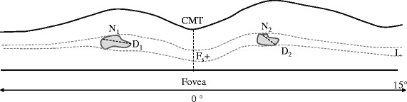
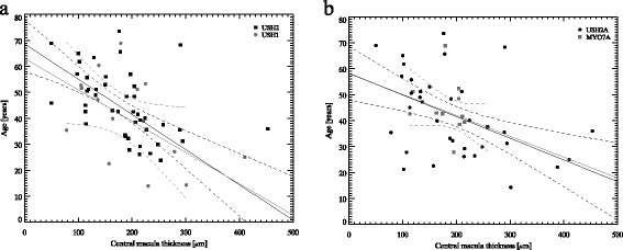
Methods: Two readers evaluated 76 patients' (mean age 42 ± 14 years) data prospectively uploaded on Eurush database. OCT was used to obtain high quality cross-sectional images through the fovea. The CML was graded as none, mild, moderate or severe, depending on the following features set: subretinal fluid without clearly detectable CML boundaries; central macular thickness; largest diameter of CML; calculated mean of all detectable CML; total number of detectable CML; retinal layers affected by CML. Intra-and inter-grader reproducibility was evaluated.

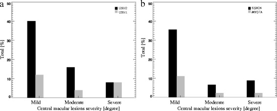
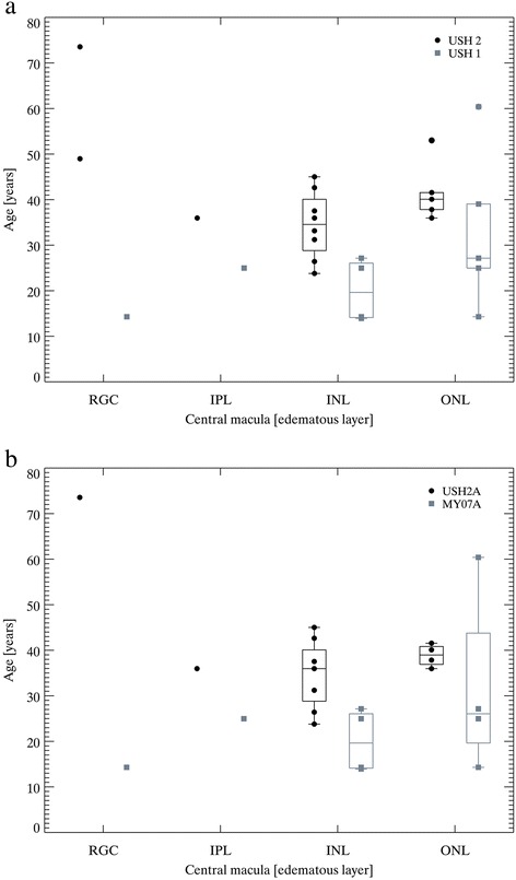
Results: CML were observed in 37 % of USH eyes, while 45 % were observed in MYO7A and 29 % in USH2A cases. Of those with CML: 52 % had mild, 22 % had moderate and 26 % had severe changes, respectively. CML were found in following retinal layers: 50 % inner nuclear layer, 44 % outer nuclear layer, 6 % retinal ganglion cell layer. For the inter-grader repeatability analysis, agreements rates for CML were 97 % and kappa statistics was 0.91 (95 % CI 0.83-0.99). For the intra-grader analysis, agreement rates for CML were 98 %, while kappa statistics was 0.96 (95 % CI 0.92-0.99).

Conclusions: The novel grading system is a reproducible tool for grading OCT images in USH complicated by CML, and potentially could be used for objective tracking of macular pathology in clinical therapy trials.


Figures

Fig. 1
Fig. 1
The schematic grading of cystic macular lesions in optical coherence tomography images. Retinal cystic lesions were quantified through the fovea line scan within to 15° eccentricity. Evaluated parameters were: total quantity of cystic macular lesions (N = N1 + N2 + […] + Nn); averaged largest diameter of all cystic lesions (D = (D1 + D2 + […] + Dn)/n); layer affected by cystic lesions (L); presence/absence of foveal retinal layer/-s swelling (Fs+/ Fs-), if boundaries of cystic lesions were not detectable (e.g. media opacities); central macular thickness (CMT)
Fig. 2
Fig. 2
The prevalence of cystic macular lesions severity in Usher syndrome cases. The prevalence of cystic macular lesions in USH 1 and USH 2 a, the prevalence of cystic macular lesions in USH2A and MYO7A b
Fig. 3
Fig. 3
Cystic macular lesions distribution by retinal layers distribution in Usher syndrome cases. The prevalence of CML by affected retinal layers in USH1 vs USH2 a, the prevalence of CML by affected retinal layers in USH2A vs MYO7A b
Fig. 4
Fig. 4
The linear regression analysis between age and central macula thickness in all Usher cohort. The statistically significant negative association was observed between CMT and patient age both in USH1 and USH2: r = −0.51 and r = −0.47 a; and as following in USH2A and MYO7A: r = −0.58 and r = −0.44 b

Similar articles

Cited by

References

    1. Sahel J, Bonnel S, Mrejen S, Paques M. Retinitis pigmentosa and other dystrophies. Dev Ophthalmol. 2010;47:160–167. doi: 10.1159/000320079. - DOI - PubMed
    1. Bonnet C, Grati M, Marlin S, Levilliers J, Hardelin JP, Parodi M, et al. Complete exon sequencing of all known Usher syndrome genes greatly improves molecular diagnosis. Orphanet J Rare Dis. 2011;6:21. doi: 10.1186/1750-1172-6-21. - DOI - PMC - PubMed
    1. Vaché C, Besnard T, Le Berre P, García-García G, Baux D, Larrieu L, et al. Usher syndrome type 2 caused by activation of an USH2A pseudoexon: implications for diagnosis and therapy. Hum Mutat. 2012;33(1):104–108. doi: 10.1002/humu.21634. - DOI - PubMed
    1. Aparisi MJ, Aller E, Fuster-García C, García-García G, Rodrigo R, Vázquez-Manrique RP, et al. Targeted next generation sequencing for molecular diagnosis of Usher syndrome. Orphanet J Rare Dis. 2014;9:168. doi: 10.1186/s13023-014-0168-7. - DOI - PMC - PubMed
    1. Blanco-Kelly F, Jaijo T, Aller E, Avila-Fernandez A, López-Molina MI, Giménez A, et al. Clinical Aspects of Usher Syndrome and the USH2A Gene in a Cohort of 433 Patients. JAMA Ophthalmol. 2015;133(2):157–164. doi: 10.1001/jamaophthalmol.2014.4498. - DOI - PubMed

Publication types