. 2016 Feb 27;428(5 Pt A):777-789.
doi: 10.1016/j.jmb.2015.11.026. Epub 2015 Dec 2.

Structural Analysis of Multi-Helical RNAs by NMR-SAXS/WAXS: Application to the U4/U6 di-snRNA

Affiliations

Structural Analysis of Multi-Helical RNAs by NMR-SAXS/WAXS: Application to the U4/U6 di-snRNA

Gabriel Cornilescu et al. J Mol Biol. .

Abstract

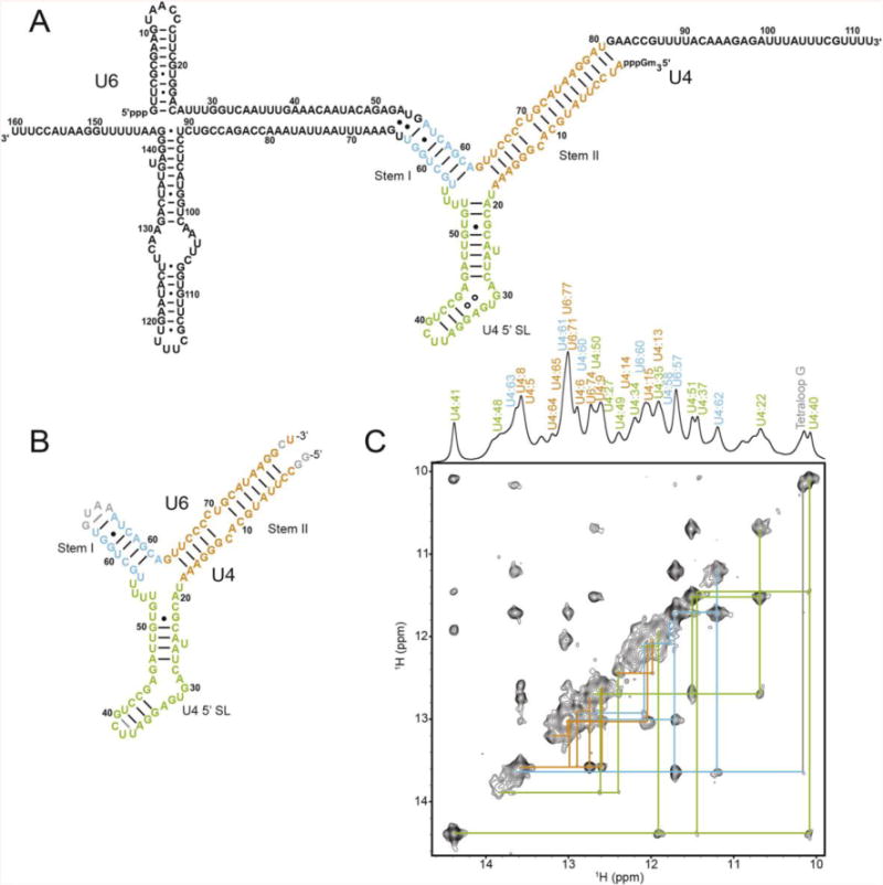
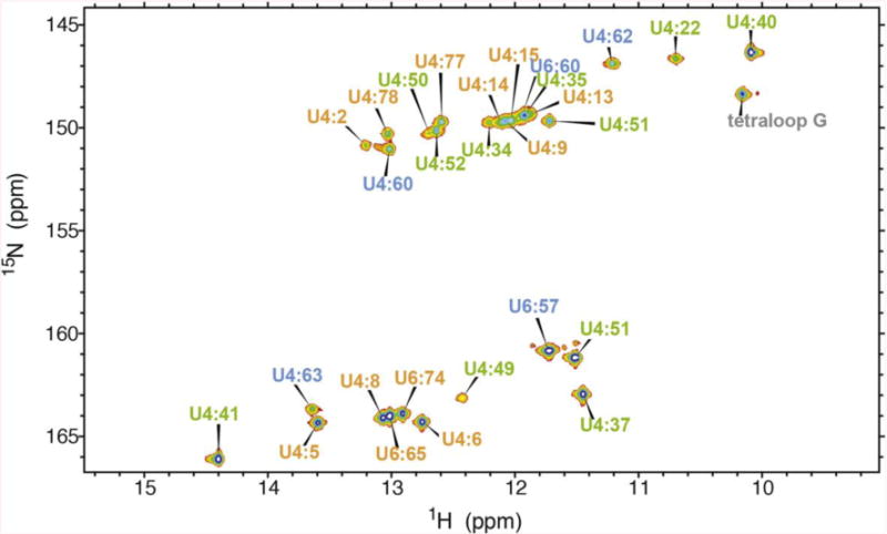
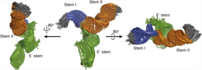
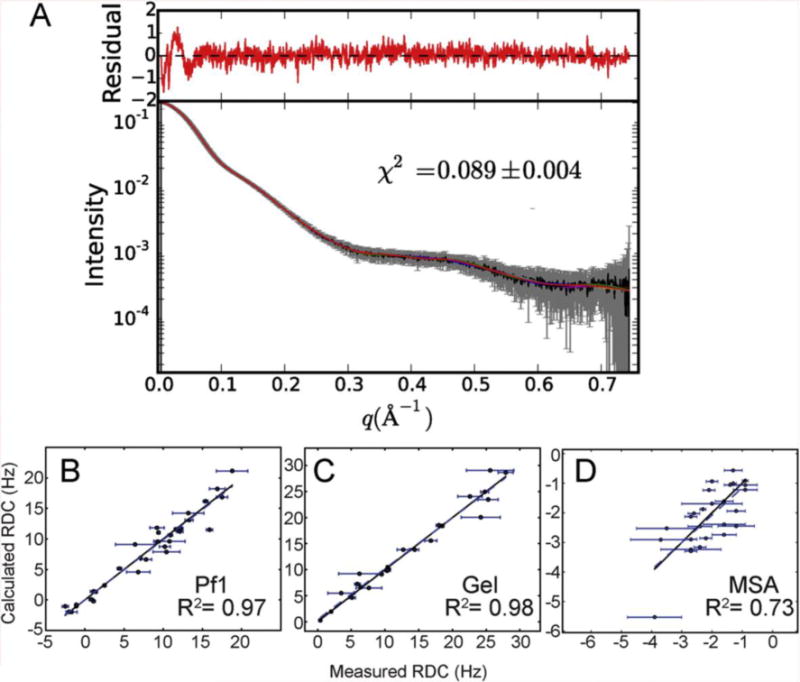
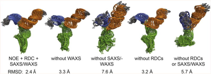
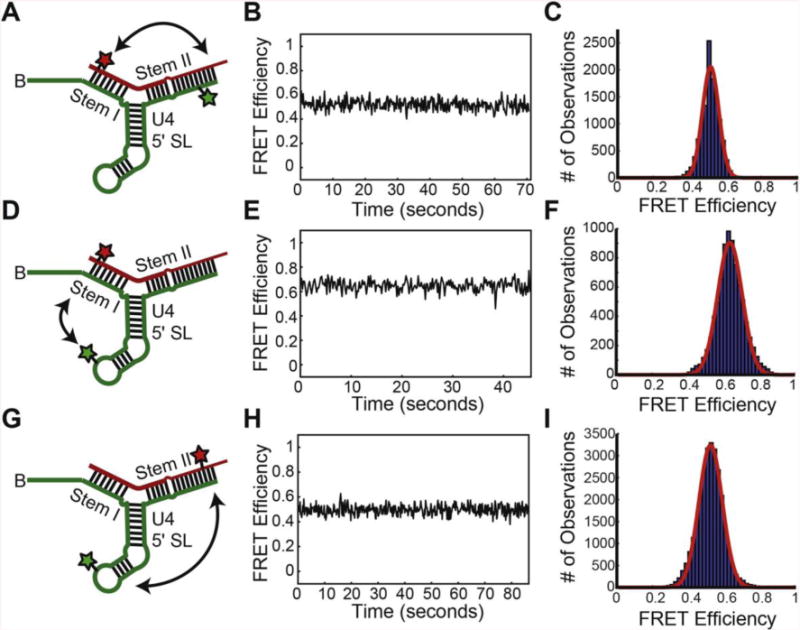
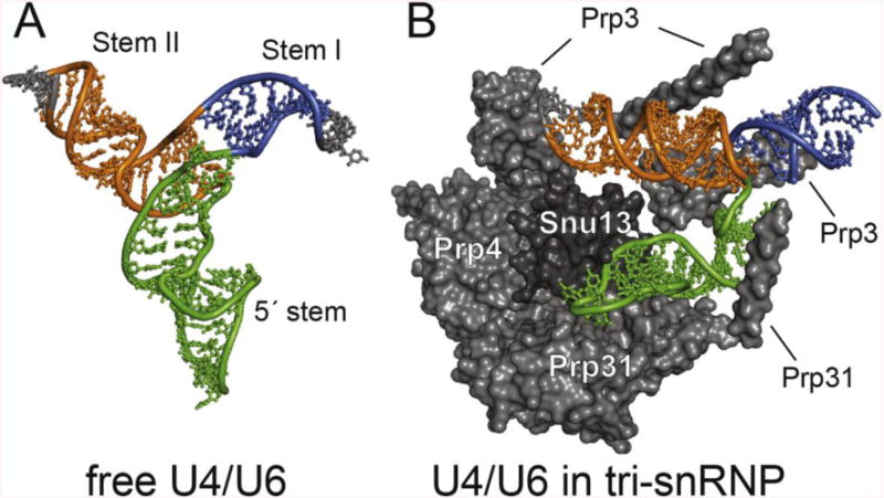
NMR and SAXS (small-angle X-ray scattering)/WAXS (wide-angle X-ray scattering) are highly complementary approaches for the analysis of RNA structure in solution. Here we describe an efficient NMR-SAXS/WAXS approach for structural investigation of multi-helical RNAs. We illustrate this approach by determining the overall fold of a 92-nt 3-helix junction from the U4/U6 di-snRNA. The U4/U6 di-snRNA is conserved in eukaryotes and is part of the U4/U6.U5 tri-snRNP, a large ribonucleoprotein complex that comprises a major subunit of the assembled spliceosome. Helical orientations can be determined by X-ray scattering data alone, but the addition of NMR RDC (residual dipolar coupling) restraints improves the structure models. RDCs were measured in two different external alignment media and also by magnetic susceptibility anisotropy. The resulting alignment tensors are collinear, which is a previously noted problem for nucleic acids. Including WAXS data in the calculations produces models with significantly better fits to the scattering data. In solution, the U4/U6 di-snRNA forms a 3-helix junction with a planar Y-shaped structure and has no detectable tertiary interactions. Single-molecule Förster resonance energy transfer data support the observed topology. A comparison with the recently determined cryo-electron microscopy structure of the U4/U6.U5 tri-snRNP illustrates how proteins scaffold the RNA and dramatically alter the geometry of the U4/U6 3-helix junction.

Keywords: NMR; RNA; SAXS; spliceosome; structure modeling.


Figures

Figure 1
Figure 1
Secondary structure of U4/U6. A. Proposed secondary structure diagram of the S. cerevisiae U4/U6 di-snRNA. B. NMR construct of U4/U6. Numbering corresponds to the yeast numbering in (A). Only the base pairs that could be experimentally determined by NMR are shown as lines for Watson-Crick pairs and dots for wobble pairs. C. 2D NOESY with NOE walk color coded to match (B).
Figure 2
Figure 2
2D 1H-15N HSQC-TROSY spectrum of the U4/U6 imino correlations. Assignments are indicated and color coded to match Figure 1.
Figure 3
Figure 3
NMR-SAXS/WAXS structure of U4/U6. The ensemble of the 10 lowest energy structures (out of 100 calculated) are shown.
Figure 4
Figure 4
Agreement between structure models and the experimental SAXS/WAXS and RDC data. A) Experimental SAXS/WAXS data (gray) were merged from 10 individual 0.5 second exposures and are plotted with error bars. Back calculated SAXS/WAXS data from the models are shown as colored lines. Residuals are plotted above, in red. B–D) Agreement between measured and predicted RDC for Pf1 phage (B), negatively charged stretched polyacrylamide gels (C) and magnetic susceptibility anisotropy (D).
Figure 5
Figure 5
Impact of SAXS, WAXS and NMR restraints on structure models. The lowest 5 energy models out of 48 calculated are shown. RMSDs for the ensembles are indicated.
Figure 6
Figure 6
smFRET data from U4/U6 di-RNAs containing fluorophores in either U4/U6 stem I, U4/U6 stem II, or the U4 5′ stemloop. (A) Diagram of a U4/U6 di-RNA for reporting on dynamics between U4/U6 stem I and U4/U6 stem II (double-headed arrow). The U4 RNA (green) contains a Cy3 FRET-donor fluorophore (green star) and a biotin tether for surface immobilization (“B”). The U6 RNA (red) contains a Cy5 FRET-acceptor fluorophore. (B) FRET efficiency (EFRET) trajectory for a single molecule the di-RNA shown in (A). (C) Histogram of EFRET calculated from N = 76 molecules of the di-RNA shown in (A). (D) Diagram of a U4/U6 di-RNA for reporting on dynamics between U4/U6 stem I and the U4 5′ stemloop (double-headed arrow). (E) EFRET trajectory for a single molecule the di-RNA shown in (D). (F) Histogram of EFRET calculated from N = 84 molecules of the di-RNA shown in (D). (G) Diagram of a U4/U6 di-RNA for reporting on dynamics between U4/U6 stem II and the U4 5′ stemloop (double-headed arrow). (H) EFRET trajectory for a single molecule the di-RNA shown in (G). (I) Histogram of EFRET calculated from N = 98 molecules of the di-RNA shown in (G). Red lines in panels C,F, and I represent the fitting results of each histogram to single exponential Gaussian functions.
Figure 7
Figure 7
Structural comparison of U4/U6 in the presence and absence of spliceosomal proteins. A) NMR/SAXS structure of free U4/U6. B) Cryo-EM structure of U4/U6 in the yeast tri-snRNP. Associated protein cofactors in tri-snRNP promote a conformation that is not favored in the free RNA, consistent with their essential role in driving RNA structural rearrangements during spliceosome assembly and disassembly.

References

    1. Cate JH, Gooding AR, Podell E, Zhou K, Golden BL, Kundrot CE, et al. Crystal structure of a group I ribozyme domain: principles of RNA packing. Science. 1996;273:1678–85. - PubMed
    1. Rose PW, Prlic A, Bi C, Bluhm WF, Christie CH, Dutta S, et al. The RCSB Protein Data Bank: views of structural biology for basic and applied research and education. Nucleic Acids Res. 2015;43:D345–56. - PMC - PubMed
    1. Mustoe AM, Brooks CL, 3rd, Al-Hashimi HM. Topological constraints are major determinants of tRNA tertiary structure and dynamics and provide basis for tertiary folding cooperativity. Nucleic Acids Res. 2014;42:11792–804. - PMC - PubMed
    1. Zuo X, Wang J, Yu P, Eyler D, Xu H, Starich MR, et al. Solution structure of the cap-independent translational enhancer and ribosome-binding element in the 3′ UTR of turnip crinkle virus. Proc Natl Acad Sci U S A. 2010;107:1385–90. - PMC - PubMed
    1. Keane SC, Heng X, Lu K, Kharytonchyk S, Ramakrishnan V, Carter G, et al. RNA structure. Structure of the HIV-1 RNA packaging signal. Science. 2015;348:917–21. - PMC - PubMed

Publication types

MeSH terms

Substances