. 2015 Dec 14:5:18215.
doi: 10.1038/srep18215.

Akbu-LAAO exhibits potent anti-tumor activity to HepG2 cells partially through produced H2O2 via TGF-β signal pathway

Affiliations

Akbu-LAAO exhibits potent anti-tumor activity to HepG2 cells partially through produced H2O2 via TGF-β signal pathway

Chunmei Guo et al. Sci Rep. .

Abstract

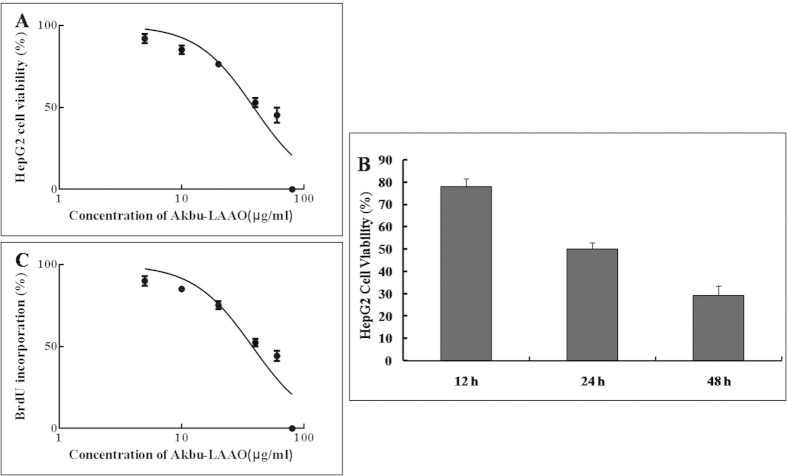
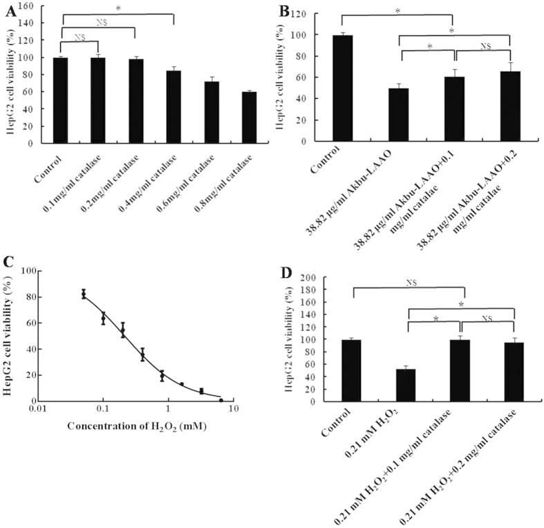
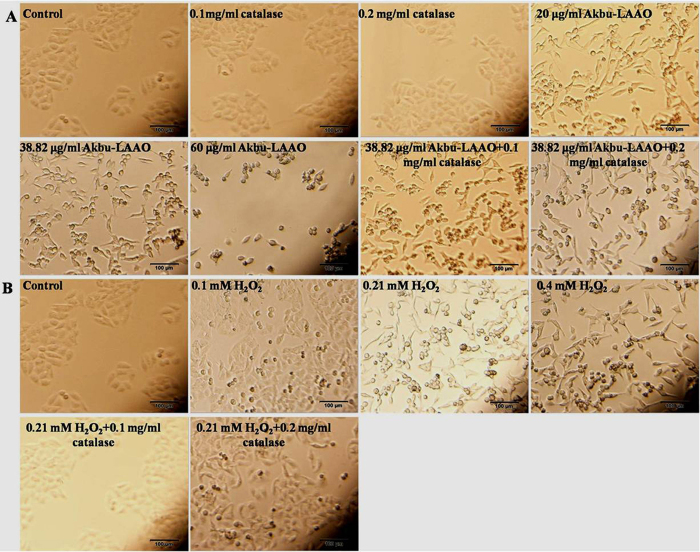
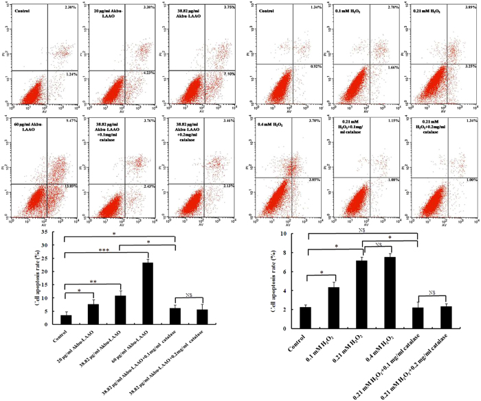
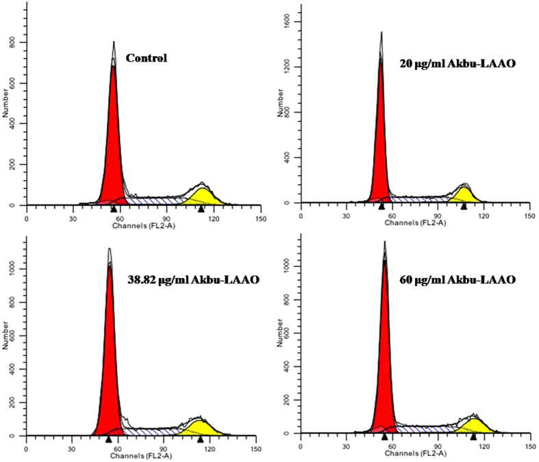
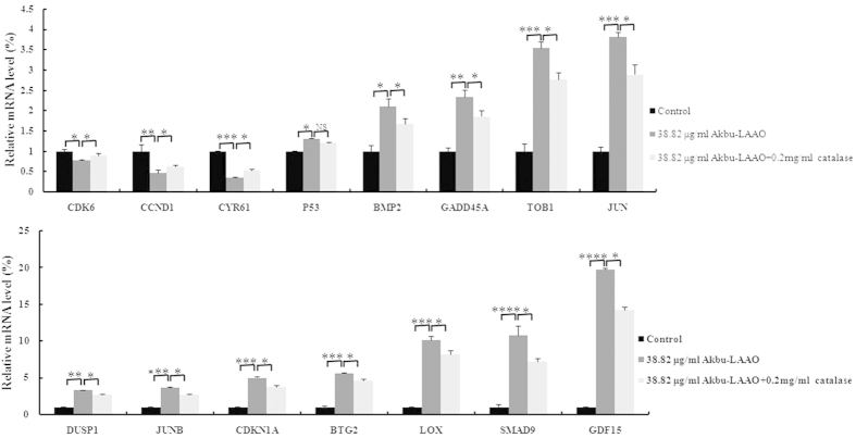
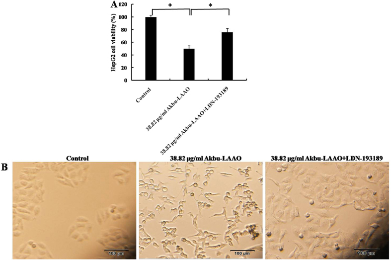
Previously, we characterized the biological properties of Akbu-LAAO, a novel L-amino acid oxidase from Agkistrodon blomhoffii ussurensis snake venom (SV). Current work investigated its in vitro anti-tumor activity and underlying mechanism on HepG2 cells. Akbu-LAAO inhibited HepG2 growth time and dose-dependently with an IC50 of ~38.82 μg/mL. It could induce the apoptosis of HepG2 cells. Akbu-LAAO exhibited cytotoxicity by inhibiting growth and inducing apoptosis of HepG2 as it showed no effect on its cell cycle. The inhibition of Akbu-LAAO to HepG2 growth partially relied on enzymatic-released H2O2 as catalase only partially antagonized this effect. cDNA microarray results indicated TGF-β signaling pathway was linked to the cytotoxicity of Akbu-LAAO on HepG2. TGF-β pathway related molecules CYR61, p53, GDF15, TOB1, BTG2, BMP2, BMP6, SMAD9, JUN, JUNB, LOX, CCND1, CDK6, GADD45A, CDKN1A were deregulated in HepG2 following Akbu-LAAO stimulation. The presence of catalase only slightly restored the mRNA changes induced by Akbu-LAAO for differentially expressed genes. Meanwhile, LDN-193189, a TGF-β pathway inhibitor reduced Akbu-LAAO cytotoxicity on HepG2. Collectively, we reported, for the first time, SV-LAAO showed anti-tumor cell activity via TGF-β pathway. It provides new insight of SV-LAAO exhibiting anti-tumor effect via a novel signaling pathway.


Figures

Figure 1
Figure 1. Akbu-LAAO inhibits the in vitro proliferation of HepG2.
(A) MTT assay indicated Akbu-LAAO treatment for 24 h dose-dependently inhibited HepG2 proliferation. (B) The administration of 38.82 μg/mL Akbu-LAAO time-dependently inhibited HepG2 growth. (C) BrdU assay showed Akbu-LAAO treatment for 24 h dose-dependently inhibited HepG2 proliferation.
Figure 2
Figure 2. Catalase scavenging influences on the cytotoxicities of Akbu-LAAO and exogenous H2O2.
(A) The effect of catalase on HepG2 proliferation. (B) The influence of catalase on Akbu-LAAO cytotoxicity to HepG2. (C) Exogenous H2O2 inhibited HepG2 proliferation. (D) The influence of catalase on exogenous H2O2 cytotoxicity to HepG2. All experiments were performed in triplicate, * denotes P < 0.05
Figure 3
Figure 3. The influences of Akbu-LAAO and exogenous H2O2 administrations on HepG2 morphology.
(A) HepG2 morphology observation following Akbu-LAAO administration in the presence and absence of catalase. (B) HepG2 morphology observation following exogenous H2O2 administration in the presence and absence of catalase. Cell images were taken using an inverted light microscope at the magnification of 100×.
Figure 4
Figure 4. Transmission electron microscopy ultrastructural characterization of HepG2 to Akbu-LAAO stimulation.
Images were obtained at a magnification of 12000×. formula image 
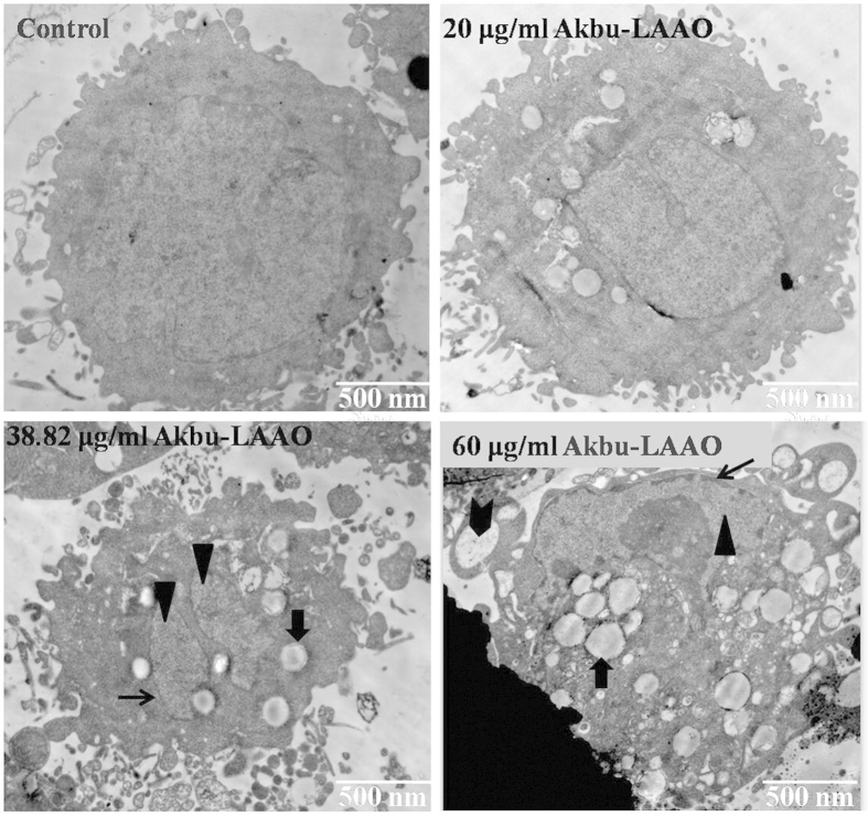
represents chromatin condensation, formula image represents cytoplasmic vacuolation, formula image represents nucleolus structure disorganization, formula image represents apoptotic bodies.
Figure 5
Figure 5. Hoechst 33258 staining assay of HepG2 apoptosis to the stimulations of Akbu-LAAO and exogenous H2O2.
HepG2 cells were incubated in the presence or absence of catalase for 1 h, treated with Akbu-LAAO or exogenous H2O2 for 24 h and stained Hoechst 33258. Nuclear condensation and/or fragmentation represent cell apoptosis. Images were taken at a magnification of 200×. Arrows marked the apoptotic cells for representing nuclear condensation/fragmentation.
Figure 6
Figure 6. Flow cytometry assay of HepG2 apoptosis induced by Akbu-LAAO and exogenous H2O2 administrations.
The propidium iodide (PI) and FITC-labeled AnnexinV antibody were used for sample labeling reagents. HepG2 cells were incubated in the presence or absence of catalase for 1 h and treated with Akbu-LAAO or exogenous H2O2 for 24 h. Cells from each group were incubated with Annexin V-FITC and PI, and immediately subjected to flow cytometry assay. Triplicate experiments were performed for each group. *, ** and *** denote P < 0.05, 0.01 and 0.001, respectively.
Figure 7
Figure 7. Akbu-LAAO treatment showed no effect on HepG2 cell cycle by flow cytometry assay.
Propidium iodide was used as the staining reagent. HepG2 cells were treated with 0, 20, 38.82 and 60 μg/mL of Akbu-LAAO for 24 h at 37 °C with 5% CO2. The cells stained with PI were subjected to flow cytometry foe measuring cell phase distributions.
Figure 8
Figure 8. qRT-PCR assay of TGF-β-pathway-related genes deregulated in HepG2 responding to Akbu-LAAO administration.
The comparisons of the level changes of targeted genes in HepG2 cells in absence of Akbu-LAAO, in presence of 38.82 μg/mL Akbu-LAAO, in presence of 38.82 μg/ml Akbu-LAAO plus 0.2 mg/ml catalase. ACTB was used as the internal reference. The fold changes were represented as mean ± SD from triplicate assays. *, **, *** and **** denote P < 0.05, <0.01, <0.001 and <0.0001, respectively.
Figure 9
Figure 9. The influence of TGF-β pathway inhibitor LDN-193189 on the cytotoxicity of Akbu-LAAO to HepG2 cells.
(A) LDN-193189 influence on the cytotoxicity of Akbu-LAAO to HepG2 by MTT assay. (B) LDN-193189 influence on the cytotoxicity of Akbu-LAAO to HepG2 cell morphology. HepG2 cells were pre- incubated with 10 μM LDN-193189 for 1 h and treated with 38.82 μg/mL Akbu-LAAO for 24 h. Experiments were performed in triplicate, * denotes P < 0.05
Figure 10
Figure 10. Potential action mechanism of Akbu-LAAO on HepG2 proliferation and apoptosis.
Red nodes represent upregulated genes, green nodes represent downregulated genes, formula image 
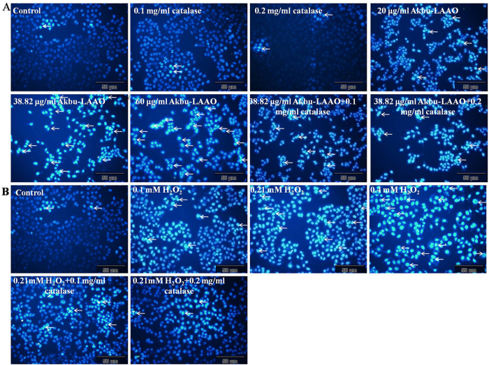
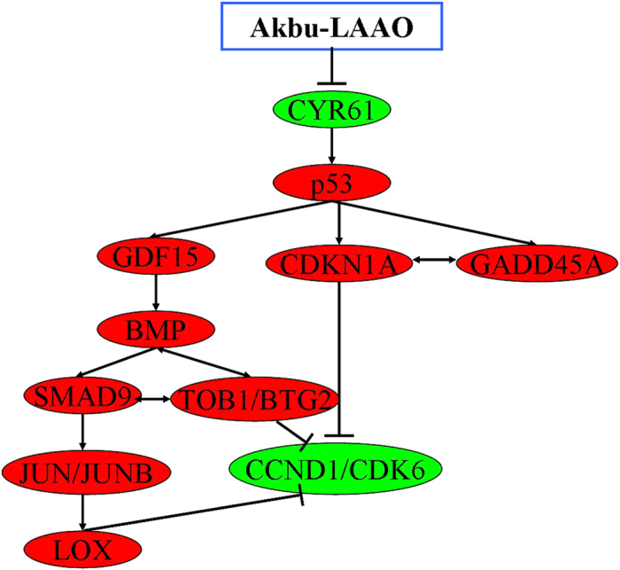
represents activation, ⊥ represents inhibition, ↔ represents interaction between genes.

References

    1. Li R. & Li A. Antibacterial efficacy of recombinant Siganus oramin L-amino acid oxidase expressed in Pichia pastoris. Fish Shellfish Immunol 41, 356–361 (2014). - PubMed
    1. Abdelkafi-Koubaa Z. et al. A thermoactive L-amino acid oxidase from Cerastes cerastes snake venom: purification, biochemical and molecular characterization. Toxicon 89, 32–44 (2014). - PubMed
    1. Yu Z. et al. Advances in detection methods of L-amino acid oxidase activity. Appl Biochem Biotechnol 174, 13–27 (2014). - PubMed
    1. Pollegioni L., Motta P. & Molla G. L-amino acid oxidase as biocatalyst: a dream too far? Appl Microbiol Biotechnol 97, 9323–9341 (2013). - PubMed
    1. Mitra J. & Bhattacharyya D. Irreversible inactivation of snake venom L-amino acid oxidase by covalent modification during catalysis of L-propargylglycine. FEBS Open Bio 3, 135–143 (2013). - PMC - PubMed

Publication types

MeSH terms