. 2015 Dec 11;11(12):e1005723.
doi: 10.1371/journal.pgen.1005723. eCollection 2015 Dec.

Tyro3 Modulates Mertk-Associated Retinal Degeneration

Affiliations

Tyro3 Modulates Mertk-Associated Retinal Degeneration

Douglas Vollrath et al. PLoS Genet. .

Abstract

Inherited photoreceptor degenerations (IPDs) are the most genetically heterogeneous of Mendelian diseases. Many IPDs exhibit substantial phenotypic variability, but the basis is usually unknown. Mutations in MERTK cause recessive IPD phenotypes associated with the RP38 locus. We have identified a murine genetic modifier of Mertk-associated photoreceptor degeneration, the C57BL/6 (B6) allele of which acts as a suppressor. Photoreceptors degenerate rapidly in Mertk-deficient animals homozygous for the 129P2/Ola (129) modifier allele, whereas animals heterozygous for B6 and 129 modifier alleles exhibit an unusual intermixing of degenerating and preserved retinal regions, with females more severely affected than males. Mertk-deficient mice homozygous for the B6 modifier allele display degeneration only in the far periphery, even at 8 months of age, and have improved retinal function compared to animals homozygous for the 129 allele. We genetically mapped the modifier to an approximately 2-megabase critical interval that includes Tyro3, a paralog of Mertk. Tyro3 expression in the outer retina varies with modifier genotype in a manner characteristic of a cis-acting expression quantitative trait locus (eQTL), with the B6 allele conferring an approximately three-fold higher expression level. Loss of Tyro3 function accelerates the pace of photoreceptor degeneration in Mertk knockout mice, and TYRO3 protein is more abundant in the retinal pigment epithelium (RPE) adjacent to preserved central retinal regions of Mertk knockout mice homozygous for the B6 modifier allele. Endogenous human TYRO3 protein co-localizes with nascent photoreceptor outer segment (POS) phagosomes in a primary RPE cell culture assay, and expression of murine Tyro3 in cultured cells stimulates phagocytic ingestion of POS. Our findings demonstrate that Tyro3 gene dosage modulates Mertk-associated retinal degeneration, provide strong evidence for a direct role for TYRO3 in RPE phagocytosis, and suggest that an eQTL can modify a recessive IPD.


Conflict of interest statement

The authors have declared that no competing interests exist.

Figures
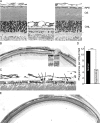
Fig 1
Fig 1. Genetic suppression of photoreceptor degeneration and enhanced RPE phagocytosis in C57BL/6 (B6) incipient congenic Mertk -/- mice.
(A) Retinal images taken at postnatal day (P) 60 of a wild-type B6 mouse (left), a mouse from the initial Mertk knockout line [17] (middle), and a Mertk -/- animal with a B6 modifier allele (right) demonstrate striking suppression of photoreceptor degeneration by the modifier. The retinal pigment epithelium (RPE), photoreceptor outer segment (OS) layer, and outer nuclear layer (ONL) are labeled. (B) A low magnification image illustrates intermixing of normal appearing (black bars) and degenerating retinal regions in a P60 Mertk -/- mouse heterozygous for a B6 modifier allele linked to Mertk. Insets show enlargements of regions of degenerating (1) and normal (2) areas. The ONL of the degenerating region is only 40–50% as thick as that in the normal region. The OS in the normal region are indistinguishable from those of wild-type mice (Fig 1A), whereas in the degenerating region the OS zone is vacuolated and contains disorganized membranous debris (d) characteristic of Mertk -/- mice [17]. (C) A higher magnification image from a different animal at P60 shows phagosomes (arrows) in the RPE of a normal-appearing region. (D) Phagosome counts made across full sections at the vertical meridian from five eyes of Mertk -/- mice (P104 –P108) heterozygous for the modifier demonstrate increased mean numbers in normal-appearing regions. *** P = 0.0004 by an unpaired two-tailed t-test. (E) A low magnification image of a retinal section from a Mertk -/- mouse homozygous for a B6 modifier allele at P255 demonstrates normal structure across a large extent of the retina, with photoreceptor degeneration in the far periphery (gray dashes) as evidenced by a thinner ONL and a thicker OS layer with vacuoles and debris.
Fig 2
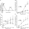
Fig 2. Preservation of photoreceptor function in Mertk -/- mice homozygous for a B6 modifier allele.
(A) Representative electroretinographic (ERG) waveforms at one light intensity for scotopic (dark-adapted) incipient congenic mice homozygous for either B6 or 129 modifier alleles. Comparison of mean amplitudes ± SD of (B) scotopic a-wave, (C) scotopic b-wave and (D) photopic b-wave at various light intensities for the two lines (n = 5 males at 10–11 weeks for each) demonstrates increased retinal responses in B6 homozygotes. **** P ≤ 0.0001, *** P ≤ 0.001, ** P ≤ 0.01 by a two-way ANOVA with Bonferroni’s correction for multiple comparisons.
Fig 3
Fig 3. Meiotic mapping identifies Tyro3 as a candidate modifier.
Haplotype map of chromosomes (rows) from 437 offspring of a backcross. Only chromosomes originating from a heterozygous parent are depicted. Black indicates a B6 allele, gray a 129 allele, and white an unscored marker. Retinal phenotyping (at right) localized the modifier between rs3669873 and rs3144592, a region of 2,099,167 bp (GRCm38 assembly). Marker rs27424653 (bold) is in an intron of Tyro3, which lies completely within and near the middle of the critical interval.
Fig 4
Fig 4. Tyro3 mRNA and protein levels vary as a function of modifier genotype.
(A) Quantitative RT-PCR of Tyro3 mRNA from the RPE of incipient B6 congenic Mertk -/- mice with different modifier genotypes. For each line, individual samples from two P36 males were assayed in triplicate. The normalized means ± SD are depicted. * P ≤ 0.05 by one-way ANOVA with Bonferroni’s correction for multiple comparisons. (B) An immunoblot shows variation in the level of TYRO3 protein among the same three incipient B6 congenic Mertk -/- lines plus a fourth non-congenic Mertk +/+ line homozygous for a 129 allele in the modifier region (leftmost lane). For each genotype, protein was isolated from eyecups pooled from two males, ages P40-45. (C) Quantification of the band intensities from (B) reveals a nearly linear relationship between genotypes and TYRO3 protein levels among the three lines, indicative of an expression quantitative trait locus (eQTL). TYRO3 and RLBP1 data were normalized to y-tubulin.
Fig 5
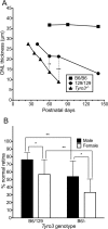
Fig 5. Genetic ablation of Tyro3 accelerates photoreceptor degeneration in Mertk -/- mice.
(A) Mean outer nuclear layer (ONL) thickness at various ages is shown for incipient B6 congenic Mertk -/- mice homozygous for either B6 or 129 modifier alleles, and also for Mertk -/- mice homozygous for a Tyro3 knockout allele and 129 alleles at all other candidate modifier genes (Tyro3 -/-). Multiple animals were measured at each age, including three per line at P60 and five per line at P75 for the statistical comparisons. ** P = 0.0046 and *** P = 0.00014, both by two-tailed t-tests. (B) Mertk -/-;Tyro3 B6/- mice have more severe degeneration than Mertk -/-;Tyro3 B6/129 animals, and females are more severely affected for both genotypes. Mean percentages ± SD of normal and degenerating retinal regions were obtained from both eyes of 54 offspring taken at P72-75 of a Mertk -/-;Tyro3 B6/B6 x Mertk -/-;Tyro3 129/- cross. * P ≤ 0.05 and ** P ≤ 0.01 by a two-way ANOVA with Bonferroni’s correction for multiple comparisons.
Fig 6
Fig 6. A central to peripheral gradient of TYRO3 expression correlates with regional variation in retinal preservation.
Immunostaining of frozen sections from P250 animals demonstrates higher expression of TYRO3 protein in the central RPE of a Mertk -/-;Tyro3 B6/B6 mouse compared to the periphery. The central retina of Mertk -/-;Tyro3 B6/B6 animals escapes degeneration, while the other areas pictured do not. As a control, the same sections were stained for the RPE-expressed protein, cytokeratin-18. Apical TYRO3 staining is evident in both regions of the B6 retina, whereas such staining is not apparent in 129. Scale bar = 20 μm.
Fig 7
Fig 7. TYRO3 localizes to nascent phagosomes and promotes photoreceptor outer segment (POS) phagocytosis.
(A) Confocal microscopy of primary human RPE cells challenged with bovine POS demonstrates co-localization of TYRO3 (green) with rhodopsin (red) after 1 hour. (B) Adenoviral vector-mediated overexpression of murine TYRO3 in NRK-49F cells stimulates POS phagocytosis, in particular POS ingestion (total minus bound). Adenoviral vectors encoding GFP and MERTK were used as negative and positive controls, respectively. Means ± SD are depicted. **** P ≤ 0.0001, ** P ≤ 0.01 by a two-way ANOVA with Bonferroni’s correction for multiple comparisons.

Similar articles

Cited by

References

    1. Kajiwara K, Berson EL, Dryja TP (1994) Digenic retinitis pigmentosa due to mutations at the unlinked peripherin/RDS and ROM1 loci. Science 264: 1604–1608. - PubMed
    1. Beales PL, Badano JL, Ross AJ, Ansley SJ, Hoskins BE, et al. (2003) Genetic interaction of BBS1 mutations with alleles at other BBS loci can result in non-Mendelian Bardet-Biedl syndrome. Am J Hum Genet 72: 1187–1199. - PMC - PubMed
    1. Ebermann I, Phillips JB, Liebau MC, Koenekoop RK, Schermer B, et al. (2010) PDZD7 is a modifier of retinal disease and a contributor to digenic Usher syndrome. J Clin Invest 120: 1812–1823. 10.1172/JCI39715 - DOI - PMC - PubMed
    1. Khanna H, Davis EE, Murga-Zamalloa CA, Estrada-Cuzcano A, Lopez I, et al. (2009) A common allele in RPGRIP1L is a modifier of retinal degeneration in ciliopathies. Nat Genet 41: 739–745. 10.1038/ng.366 - DOI - PMC - PubMed
    1. Louie CM, Caridi G, Lopes VS, Brancati F, Kispert A, et al. (2010) AHI1 is required for photoreceptor outer segment development and is a modifier for retinal degeneration in nephronophthisis. Nat Genet 42: 175–180. 10.1038/ng.519 - DOI - PMC - PubMed

Publication types