. 2015 Dec 14;10(12):e0144245.
doi: 10.1371/journal.pone.0144245. eCollection 2015.

Cisplatin Induces Overactivation of the Dormant Primordial Follicle through PTEN/AKT/FOXO3a Pathway which Leads to Loss of Ovarian Reserve in Mice

Affiliations

Cisplatin Induces Overactivation of the Dormant Primordial Follicle through PTEN/AKT/FOXO3a Pathway which Leads to Loss of Ovarian Reserve in Mice

Eun Mi Chang et al. PLoS One. .

Abstract

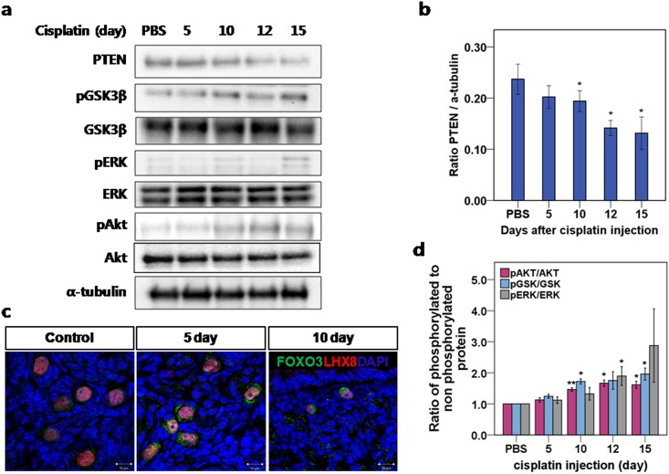
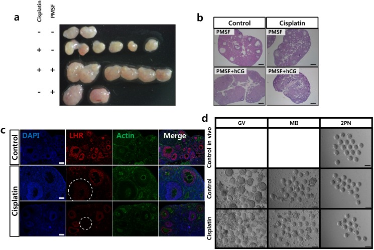
Cisplatin is a first-line chemotherapeutic agent for ovarian cancer that acts by promoting DNA cross links and adduct. However drug resistance and considerable side effects including reproductive toxicity remain a significant challenge. PTEN is well known as a tumor suppressor function which plays a fundamental role in the regulation of the cell cycle, apoptosis and development of cancer. At the same time PTEN has been revealed to be critically important for the maintenance of the primordial follicle pool. In this study, we investigated the role of PTEN/Akt/FOXO3 pathway in cisplatin-induced primordial follicle depletion. Cisplatin induced ovarian failure mouse model was used to evaluate how this pathway involves. In vitro maturation was used for oocyte rescue after cisplatin damage. We found that cisplatin treatment decreased PTEN levels, leading to a subsequent increase in the phosphorylation of key molecules in the pathway. The activation of the PTEN/Akt/FOXO3 pathway cascade increased cytoplasmic translocation of FOXO3a in cisplatin-treated follicles, which in turn increased the pool size of growing follicles, and rapidly depleted the number of dormant follicles. Once activated, the follicles were more prone to apoptosis, and their cumulus cells showed a loss of luteinizing hormone (LH) receptor expression, which leads to failure during final maturation and ovulation. In vitro maturation to rescue oocytes in a cisplatin-treated mouse model resulted in successful maturation and fertilization. This study is the first to show the involvement of the PTEN/Akt/FOXO3 pathway in premature ovarian failure after cisplatin treatment and the possibility of rescue through in vitro maturation.


Conflict of interest statement

Competing Interests: The authors have declared that no competing interests exist.

Figures

Fig 1
Fig 1. Body weight, mortality, ovarian weight, and gross morphology of the mice and ovaries after cisplatin daily injection.
(a) Bodyweight of the mice after 15 days of daily injections with various doses of cisplatin. (b) Mortality (bar) and bodyweight (continuous line) of the mice receiving 2.0mg/kg daily cisplatin. (c) Ovarian weight after 15 days of daily injections with various doses of cisplatin. (d) Gross morphology of mice and ovaries after 2.0mg/kg daily injections with cisplatin (at 0, 5, 10, and 15 days starting from the left in the picture). (a-c) mean values ± SEM; stars denote significant differences relative to control (*P<0.05, ** P<0.01).
Fig 2
Fig 2. Representative histological sections of mouse ovaries at different time points after daily injection of various doses of cisplatin or PBS control.
(a) Hematoxylin staining: Follicles at all stages were observed in control ovaries and in ovaries subjected to0.5mg/kg and 1.0mg/kg cisplatin. A significant decrease in the number of follicles beyond the antral stage was seen in the ovaries subjected to1.5mg/kg cisplatin after 15 days, and in those subjected to2.0mg/kg cisplatin after 12days(the inset at bottom right indicates day 12). An increase in the number of primary follicles was observed in the ovaries subjected to1.5mg/kg cisplatin after 15days, and in those subjected to2.0mg/kg cisplatin after 12 and 15 days. The primordial follicle depletion was definite after15 days of 2.0mg/kg cisplatin. (b) Differential counts of primordial, primary, secondary, and antral follicles and beyond. The total number of follicles at each stage and time point during 2.0mg/kg daily cisplatin treatment, revealed a significant increase in the total count of primary follicles after day 12. (c) The differential ratio of follicles at each stage and time point during cisplatin treatment showed a significant increase in the proportion of primary follicles after day 12. (b-c) mean values ± SEM; stars denote significant differences relative to control (*P <0.05, **P<0.01).
Fig 3
Fig 3. Cisplatin treatment triggered a decrease in PTEN levels and subsequent phosphorylation of key proteins in the PTEN/Akt/FOXO3 pathway.
(a) Western blot of key proteins involved in the PTEN/Akt/FOXO3 pathway. (b) Protein analysis comparing the concentrations of PTEN with α-tubulin used as loading control. (c) Immunofluorescence staining demonstrated extra-nucleation (cytoplasmic localization) of FOXO3a in activated Lhx8-stained primordial follicles. In the control ovary, small follicles stained with Lhx8 showed nuclear localization of FOXO3a. After 5 and 10 days of daily 2.0mg/kg cisplatin injections, FOXO3a showed overall nuclear exclusion. (d) Protein analysis comparing the concentrations of phosphorylated and total AKT, ERK, and GSK3β, respectively. The ratio of phosphorylated to nonphosphorylated protein was calculated for each protein. (b, d) mean values ± SEM; stars denote significant differences relative to control (*P <0.05, **P<0.01).
Fig 4
Fig 4. TUNEL staining of ovaries from cisplatin-injected mice.
(a) Ovary tissues from mice treated with 2mg/kg daily cisplatin. The sections were stained with TUNEL and counterstained with hematoxylin at each time point. The granulosa cells of secondary follicles after 15 days of cisplatin injections are positive for TUNEL while the oocyte is protected. (b) The number of TUNEL-positive follicles for each stage at different time points. Data represent means ± SEM. Stars denote significant differences relative to control (*P <0.05, **P<0.01).
Fig 5
Fig 5. Cleaved PARP expression in secondary follicles.
(a) Immunohistochemistry of ovarian tissue; cleaved PARP was observed in the granulosa cells of secondary follicles from cisplatin-treated mice. Cleaved PARP (red) counterstained with DAPI (blue) and actin (green). The arrow (yellow) indicates the cleaved PARP in secondary follicles. Scale bars = 100μm. (b) Ratio of cleaved PARP-positive follicles from PBS-injected control mice and cisplatin-treated mice. Data represent means ± SEM. Stars denote significant differences relative to control mice injected with PBS (*P <0.05)
Fig 6
Fig 6. PMSG injection induced follicular growth but failed to induce ovulation in cisplatin-treated ovaries.
Ovaries removed from ICR mice 48h after PMSG injection. (a) Comparison of the size of cisplatin-treated ovaries with or without PMSG. (b) Midline histological sections of ovaries removed after PMSG and hCG injections and stained with hematoxylin and eosin. The corpora lutea in the control ovary are shown after hCG injection. Scale bars = 100μm. (c) The expression of LH receptor was detected at 10 days after PBS or cisplatin injection. LH receptor (red) counterstained with DAPI(blue) and actin (green). In the control ovary, most of the follicles past the primary stage expressed LH receptor; however, half of the follicles from the cisplatin-treated mice did not express LH receptor(in circle). (d) In vivo and in vitro maturation of mouse oocytes after cisplatin treatment. Oocyte maturation (MII) and fertilization (two pronuclei, 2PN) of oocytes from ovaries after cisplatin treatment matured in vivo or in vitro. GV, germinal vesicle: MII. Scale bars = 100μm.

Similar articles

Cited by

References

    1. Meirow D. Reproduction post-chemotherapy in young cancer patients. Mol CellEndocrinol 2000;169:123–131. S0303-7207(00)00365-811155944 - PubMed
    1. Meirow D, Dor J, Kaufman B, Shrim A, Rabinovici J, Schiff E, et al. Cortical fibrosis and blood-vessels damage in human ovaries exposed to chemotherapy. Potential mechanisms of ovarian injury. Hum Reprod 2007;22:1626–1633. 10.1093/humrep/dem027 - DOI - PubMed
    1. Morgan S, Anderson RA, Gourley C, Wallace WH, Spears N. How do chemotherapeutic agents damage the ovary? Hum Reprod Update 2012;18:525–535. 10.1093/humupd/dms022 - DOI - PubMed
    1. Meirow D, Biederman H, Anderson RA, Wallace WH. Toxicity of chemotherapy and radiation on female reproduction.Clin Obstet Gynecol 2010;53:727–39. - PubMed
    1. Kalich-Philosoph L, Roness H, Carmely A, Fishel-Bartal M, Ligumsky H, Paglin S et al. Cyclophosphamide triggers follicle activation and "burnout"; AS101 prevents follicle loss and preserves fertility. Sci Transl Med 2013;5: 185ra162 10.1126/scitranslmed.3005402 - DOI - PubMed

Publication types

MeSH terms