. 2015 Dec 10;11(12):e1005727.
doi: 10.1371/journal.pgen.1005727. eCollection 2015 Dec.

Skp1 Independent Function of Cdc53/Cul1 in F-box Protein Homeostasis

Affiliations

Skp1 Independent Function of Cdc53/Cul1 in F-box Protein Homeostasis

Radhika Mathur et al. PLoS Genet. .

Abstract

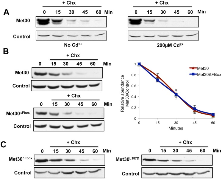
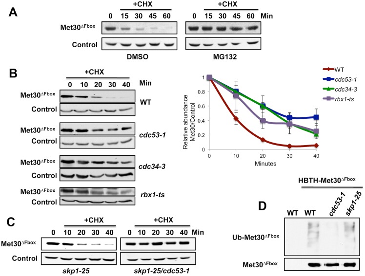
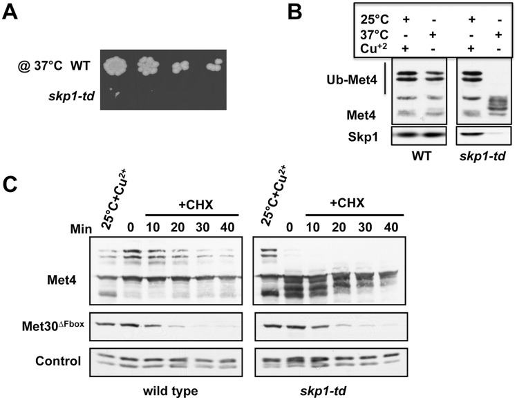
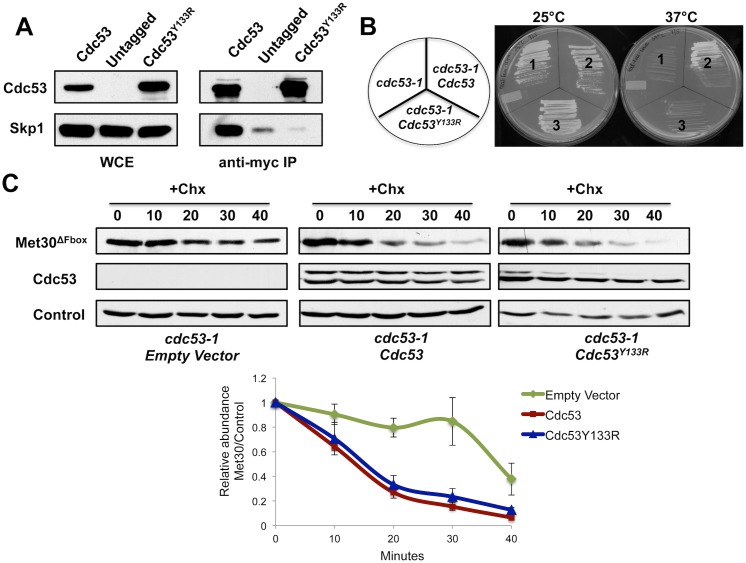
Abundance of substrate receptor subunits of Cullin-RING ubiquitin ligases (CRLs) is tightly controlled to maintain the full repertoire of CRLs. Unbalanced levels can lead to sequestration of CRL core components by a few overabundant substrate receptors. Numerous diseases, including cancer, have been associated with misregulation of substrate receptor components, particularly for the largest class of CRLs, the SCF ligases. One relevant mechanism that controls abundance of their substrate receptors, the F-box proteins, is autocatalytic ubiquitylation by intact SCF complex followed by proteasome-mediated degradation. Here we describe an additional pathway for regulation of F-box proteins on the example of yeast Met30. This ubiquitylation and degradation pathway acts on Met30 that is dissociated from Skp1. Unexpectedly, this pathway required the cullin component Cdc53/Cul1 but was independent of the other central SCF component Skp1. We demonstrated that this non-canonical degradation pathway is critical for chromosome stability and effective defense against heavy metal stress. More importantly, our results assign important biological functions to a sub-complex of cullin-RING ligases that comprises Cdc53/Rbx1/Cdc34, but is independent of Skp1.


Conflict of interest statement

The authors have declared that no competing interests exist.

Figures

Fig 1
Fig 1. Met30 dissociated from Skp1 is rapidly degradation.
(A) Cells expressing endogenous 12MycMet30 were grown at 30°C. Cadmium was added to a final concentration of 200μM and protein translation was inhibited by addition of 100μg/ml cycloheximide. Samples were collected at the time intervals indicated and Met30 stability was analyzed by immunoblotting with anti-myc antibodies. The proteasome subunit, Cim5 was detected as a loading control. (B) Experiment as in panel A, but experiment was performed with cells expressing either endogenous 12MycMet30 or 12mycMet30ΔFbox (residues 187–227 deleted). Quantification was performed a Fuji LAS-4000 imaging system followed by analyses with the Multi Gauge v3 software. Results are presented as mean ± standard error for three independent experiments (right panel) (C) Experiment as described for panel A, cells expressing endogenous 12MycMet30ΔFbox and 12MycMet30L187D were compared.
Fig 2
Fig 2. Degradation of ‘Skp1-free’ Met30 depends on the proteasome, Cdc53, Cdc34, and Rbx1, but is independent of Skp1.
(A) Cells expressing either endogenous 12MycMet30 or 12mycMet30ΔFbox were grown at 30°C. Proteasomes were inhibited with 50μM MG-132 for 45 min before cycloheximide was added to block translation. Cells carried a deletion of PDR5 to increase MG-132 permeability. (B) 12MycMet30ΔFbox stability was analyzed in wild type, cdc34-3, and cdc53-1 and rbx1-13myc temperature sensitive mutants by cycloheximide chase experiments and immunoblotting with anti-myc antibodies as described for Fig 1A. Cells were grown at 25°C, shifted to 37°C for 1.5 h to inactivate temperature sensitive mutants (C) Experiment as in panel B, but 12mycMet30ΔFbox stability was analyzed in skp1-25 single and skp1-25 cdc53-1 double mutants. (D) Cells expressing HBTHMet30ΔFbox under control of the GAL1 promoter were shifted to 37°C for 1.5 h to inactivate temperature sensitive alleles. HBTHMet30ΔFbox was purified on Ni2+-sepharose under denaturing conditions and analyzed by immunoblotting using antibodies directed against ubiquitin (upper panel) or the RGS6H epitope in the HBTH tag (lower panel). Cells expressing untagged Met30ΔFbox were processed as control.
Fig 3
Fig 3. Skp1 independent degradation of Met30.
(A) skp1-td mutants express an N-terminal temperature inducible degron (td) fused to Skp1 under the control of the CUP1 promoter. Wild type and skp1-td cells were serially diluted and spotted on YEPD plates without copper at 37°C for 2 days. (B) Wild type and skp1-td mutants were cultured at permissive (25°C + Cu2+) and non-permissive conditions (37°C for 1 h without Cu2+). Immunoblot analysis showed that Met4 ubiquitylation was blocked in skp1-td mutants indicative of inactivation of SCFMet30. (C) Wild type and skp1-td strains expressing endogenous 12mycMet30ΔFbox were cultured under permissive conditions and then shifted to non-permissive conditions for 1 h to deplete Skp1-td. Met30ΔFbox stability was analyzed using cycloheximide to block translation. Immunoblot analysis of Met4 confirmed inactivation of Skp1-td. Anti-PSTAIR detection of Cdc28 was used as loading control.
Fig 4
Fig 4. Cdc53 mutants unable to bind Skp1 can degrade Met30.
(A) Cells expressing GAL1 inducible 12MycCdc53 and 12MycCdc53Y133R or empty vector control were grown at 30°C in medium containing 2% galactose to induce Cdc53 expression. 12MycCdc53 was immunopurified and co-purified proteins were analyzed by immunoblotting. WCE: Whole cell extract (B) A cdc53-1 temperature sensitive strain containing Cdc53 expressing plasmids as indicated, were incubated on plates at 25°C or 37°C. (C) cdc53-1 temperature sensitive mutants expressing 12mycMet30ΔFbox under control of the native promoter and GAL1 inducible RGS6H-tagged CDC53 alleles as indicated or the empty vector control were grown at 25°C in medium containing 2% galactose to induce Cdc53 expression. Cultures were shifted to 37°C for 2 h to inactivate cdc53-1 and 12mycMet30ΔFbox stability was analyzed using cycloheximide to block translation. Quantification was performed as described for Fig 1. Results are presented as mean ± standard error for three independent experiments normalized to Cim5 (bottom panel).
Fig 5
Fig 5. Mutation of methionine 178 and isoleucine 179 in Met30 abolishes ‘Skp1-free’ Met30 degradation.
(A) Identification of a degron region for the ‘Skp1-free’ Met30 degradation pathway. Cells expressing either endogenous 12mycMet30ΔFbox or different Met30ΔFbox deletion mutants were grown at 30°C. Protein translation was inhibited by addition of cycloheximide and cells were collected at the time intervals indicated. Met30ΔFbox stability was analyzed by immunoblotting with anti-myc antibodies. (B & C) Experiment as in panel A, with cells expressing endogenous 12mycMet30ΔFbox, full-length 12mycMet30, or the respective degron point mutants. Results are presented as mean ± standard error for three independent experiments (right panels).
Fig 6
Fig 6. Cdc53/Rbx1 can bind Met30 in absence of Skp1.
(A) Yeast strains expressing endogenous Cdc53TAP and GAL1 inducible Skp1 were cultured in media containing 2% galactose to express Skp1 and then shifted to media containing 2% dextrose for 12 h to deplete Skp1. Skp1 was efficiently depleted from cells (left panel). Schematic of the tagged Met30 constructs used for binding experiments (right top panel). MBP, (MBP)-Met30(1–186) and (MBP)-Met30(1–186)M178E/I179E were expressed in E.coli and bound to amylose resin. The resin was then incubated with Skp1 depleted yeast lysates expressing Cdc53TAP. Beads were washed and bound proteins were eluted and analyzed by Western blotting (lower panel on right). Cdc53 levels were detected with a PAP antibody recognizing the TAP tag. (B) Amylose beads were bound with MBP, (MBP)-Met30(1–186) and (MBP)-Met30(1–186)M178E/I179E and were incubated with bacterial lysate expressing Cdc53267-851/Rbx1. Beads were washed, bound proteins eluted and analyzed by Western blotting. Cdc53 levels were detected with anti-Cdc53 antibodies. Quantification was performed as described for Fig 1. Results are presented for three independent experiments normalized to the Amido black signal for MBP tagged proteins.
Fig 7
Fig 7. Skp1-independent degradation of Ctf13 and Cdc4.
(A and B) skp1-td cells expressing 13mycCtf13 or 3mycCdc4 under control of the inducible GAL1 promoter were depleted of Skp1-td as described for Fig 3C. Dextrose was added to terminate transcription from the GAL1 promoter and degradation of Ctf13 and Cdc4 was analyzed by immunoblotting.
Fig 8
Fig 8. ‘Skp1-free’ Met30 degradation is important for cellular functions.
(A) Steady state protein levels for Met30 and Met30M178E/I179E strains were compared by immunoblotting. Anti-myc and anti-Met4 antibodies were used for detection of Met30 and Met4, respectively. Cim5 was used as loading control. (B) Met30 and Met30M178E/I179E strains were grown at 30°C to mid-log phase and serial dilutions of cells were spotted on YEPD plates with or without 50μM CdCl2. Plates were incubated at 30°C for 2 days. (C) Experiment performed as in panel B, except indicated strains were spotted on minimal medium (SC-LEU) plates with or without 25μM CdCl2. Different cadmium concentrations were used because cadmium sensitivity is dependent on media conditions [24] (D). Derepression of Met4 target genes in Met30M178E/I179E mutant. Met30 and Met30M178E/I179E strains were cultured in YEPD. Expression of the Met4 target genes, MET3 and GSH1 was analyzed by rt-qPCR (n = 3). Data are represented as mean ± SD. (E) Met30 and Met30M178E/I179E strains containing centromeric plasmid (YCpURA) were grown with and without selection in SC-URA and YPD media respectively. Equal number of cells were counted and plated on SC-URA and YPD plates for both conditions. Plates were incubated at 30°C for 2 days. Plasmid stability was determined by counting number of colonies on SC-URA and YPD plates. Data are represented as mean ± SD (n = 3).

Similar articles

Cited by

References

    1. Hershko A, Ciechanover A (1998) The ubiquitin system. Annu Rev Biochem 67: 425–479. - PubMed
    1. Deshaies RJ, Joazeiro CA (2009) RING domain E3 ubiquitin ligases. Annu Rev Biochem 78: 399–434. 10.1146/annurev.biochem.78.101807.093809 - DOI - PubMed
    1. Varshavsky A (2012) The ubiquitin system, an immense realm. Annu Rev Biochem 81: 167–176. 10.1146/annurev-biochem-051910-094049 - DOI - PubMed
    1. Finley D, Ulrich HD, Sommer T, Kaiser P (2012) The ubiquitin-proteasome system of Saccharomyces cerevisiae. Genetics 192: 319–360. 10.1534/genetics.112.140467 - DOI - PMC - PubMed
    1. Bosu DR, Kipreos ET (2008) Cullin-RING ubiquitin ligases: global regulation and activation cycles. Cell Div 3: 7 10.1186/1747-1028-3-7 - DOI - PMC - PubMed

Publication types

MeSH terms