. 2015 Dec 14:5:18133.
doi: 10.1038/srep18133.

A novel single-chain antibody redirects adenovirus to IL13Rα2-expressing brain tumors

Affiliations

A novel single-chain antibody redirects adenovirus to IL13Rα2-expressing brain tumors

Julius W Kim et al. Sci Rep. .

Abstract

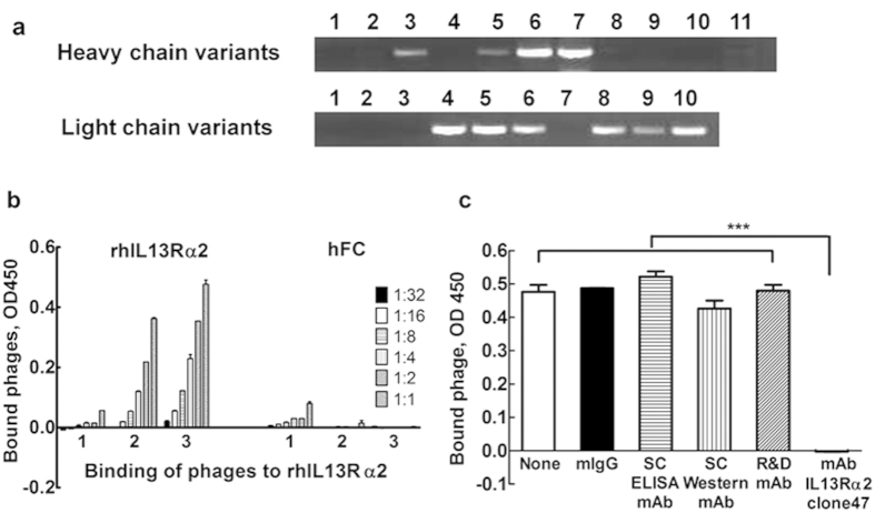
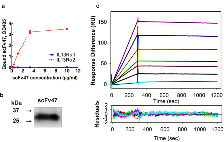
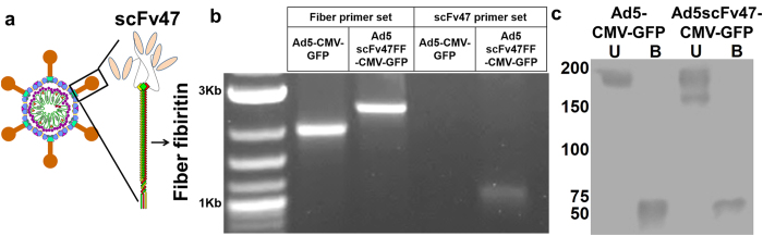
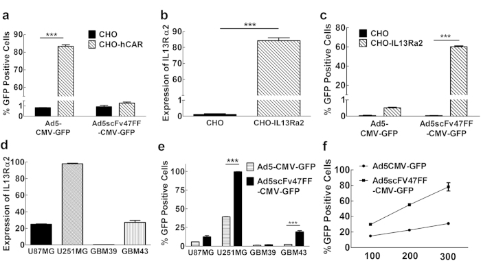
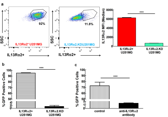
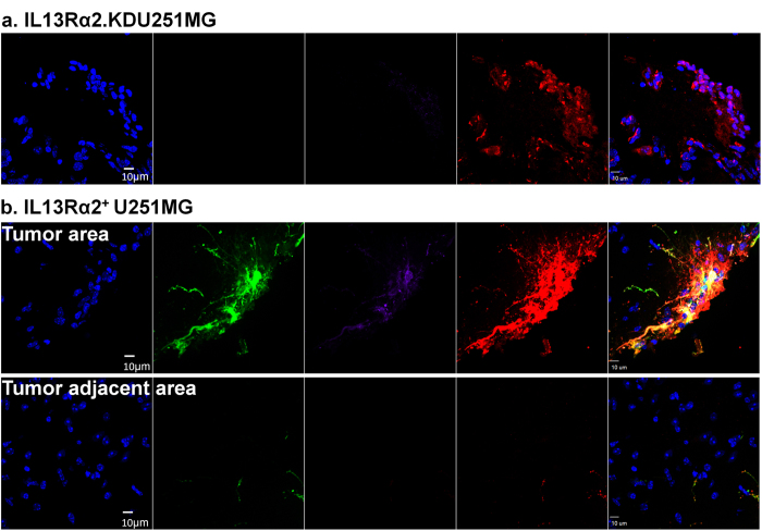
The generation of a targeting agent that strictly binds to IL13Rα2 will significantly expand the therapeutic potential for the treatment of IL13Rα2-expressing cancers. In order to fulfill this goal, we generated a single-chain antibody (scFv47) from our parental IL13Rα2 monoclonal antibody and tested its binding properties. Furthermore, to demonstrate the potential therapeutic applicability of scFv47, we engineered an adenovirus by incorporating scFv47 as the targeting moiety in the viral fiber and characterized its properties in vitro and in vivo. The scFv47 binds to human recombinant IL13Rα2, but not to IL13Rα1 with a high affinity of 0.9 · 10(-9) M, similar to that of the parental antibody. Moreover, the scFv47 successfully redirects adenovirus to IL13Rα2 expressing glioma cells both in vitro and in vivo. Our data validate scFv47 as a highly selective IL13Rα2 targeting agent and justify further development of scFv47-modified oncolytic adenovirus and other therapeutics for the treatment of IL13Rα2-expressing glioma and other malignancies.


Conflict of interest statement

IB and ML have patent applications in the field of IL13Rα2-targeted therapies.

Figures

Figure 1
Figure 1. Generation and characterization of scFv47.
(a) Screening of parental hybridoma IL13Rα2 cells mRNA for Vh and Vl using set Vh and Vl specific primers (Suppl. Table 1). (b) ELISA testing for phage binding enriched in three rounds of biopanning against rhIL13Rα2 protein and hFc as a negative control. (c) Competitive Assay. Plates coated with rhIL13Rα2 protein were treated with a panel of anti-IL13Rα2 mAbs that recognize non-overlapping epitopes with the parental mAb IL13Rα2 (clone 47). Binding of phages in the absence or presence of antibodies was analyzed via detection with anti-M13 phage antibody as described in the Material and Methods section. Each data point is an average of 3 independent replicates in all figures. Data presented as mean ± SEM. ***p < 0.001.
Figure 2
Figure 2. Binding characteristics of scFv47 to IL13Rα2.
(a) Binding of purified soluble scFv47 with rhIL13Rα2 and rhIL13Ra1 proteins was determined in plate ELISA. (b) Western blot analysis of soluble scFv47. The scFv47 protein runs under reducing conditions as a 30kDa protein in agreement with the predicted molecular weight. (c) The kinetics of interactions between the scFv47 and rhIL13Rα2 were visualized by SPR in a Biacore 3000. The scFv47 was injected at concentrations ranging from 1 to 50 nM (lower to upper curves) at a constant flow rate of 20 μL/min over immobilized rhIL13Rα2. The association phase was monitored for 30 sec, dissociation phage for 900 sec following by the change in SPR signal (colored curves), given in RU. Black curves represent the fit of the data to a one-site binding model. For derived kinetic parameters, see Table 1. Lower panels show residuals from the one-site binding model, indicating an excellent fit.
Figure 3
Figure 3. Design, Generation, and Confirmation of IL13Rα2 Tropic Virus Structure and Stability.
(a) Schematic diagram of anti-IL13Rα2 scFv-specific chimera fiber of Ad5FFscFv47-CMV-GFP. The fiber knob and shaft domains of Ad5 were replaced with a fiber fibritin trimerization domain, and anti-IL13Rα2 scFv47 was incorporated into the C-terminus of the chimeric fiber. (b) PCR confirmation of fiber modification. (c) Validation of the chimeric fiber structure. Western blot analysis detected the stable fiber trimerization when the chimeric fiber was unboiled (U: incubated at room temperature for 10 min), and detected denatured monomeric structures when the fiber was boiled (B: incubated at 95 °C for 10 min).
Figure 4
Figure 4. Confirmation of Tropism Modification of Ad5FFscFv47-CMV-GFP.
(a) CAR-independent infectivity of Ad5FFscFv47-CMV-GFP virus. CAR-negative CHO and CAR-positive CHO-hCAR cell lines were infected with Ad5CMV-GFP or Ad5FFscFv47-CMV-GFP virus. Cells were analyzed for GFP expression 72 hours post infection by flow cytometry. (b) The expression of IL13Rα2 on the surface of CHO-IL13Rα2 cell line detected using mAb IL13Rα2 (clone 47). (c) IL13Rα2-dependent infectivity of Ad5FFscFv47-CMV-GFP demonstrated by efficient transduction of CHO-IL13Rα2 cells and lack of transduction of IL13Rα2-negative CHO cells. (d) The IL13Rα2 expression on the surface of U87MG, U251MG, GBM39, and GBM43 glioma cell lines. Data presented as percent of positive cells. (e) The transduction efficiency of Ad5FFscFv47-CMV-GFP, but not Ad5CMV-GFP virus, strongly correlates with a level of IL13Rα2 expression in U87MG, U251MG, GBM39, and GBM43 glioma cells. Transduced glioma cells were analyzed by flow cytometry for GFP expression 72 hours post infection. (f) Steady increase in the infectivity of Ad5FFscFv47-CMV-GFP with an increase of MOI. U251MG cells were infected with Ad5FFscFv47-CMV-GFP or Ad5CMV-GFP at MOI: 100, 200, and 300 vp/cell. 72 hours post infection, a flow cytometric analysis for GFP expression in cells was performed. Each data point is an average of 3 independent replicates in all figures. Data presented as mean ± SEM. ***p < 0.001.
Figure 5
Figure 5. IL13Rα2-specific Infectivity of Ad5FFscFv47-CMV-GFP.
(a) Flow cytometry analysis of IL13Rα2 expression in U251MG cells following knockdown with control shRNA (IL13Rα2+ U251MG) or IL13Rα2-specific shRNA (IL13Rα2KDU251MG) presented as percent of positive cells (flow charts) and median fluorescent intensity (MFI). (b) IL13Rα2-dependent infectivity of Ad5FFscFv47 -CMV-GFP demonstrated by differential expression of GFP in in IL13Rα2+ U251MG and IL13Rα2.KDU251MG cell lines. (c) Competitive binding assay. U251MG cells were pre-treated with anti-IL13Rα2 mAb as described in the Material and Methods section. Control and treated cells were then infected with Ad5scFv47-CMV-GFP virus. Cells were analyzed for GFP transgene expression 72 hours later by flow cytometry. Each data point is an average of 3 independent replicates. Mean ± SEM is plotted. ***p < 0.001.
Figure 6
Figure 6. Infection of neurospheres by Ad5FFscFv47-CMV-GFP.
(a) Comparison of IL13Rα2 expression in U87MG cells growing as an adherent culture or as neurospheres. (b) Relative IL13Rα2 mRNA expression in U87MG glioma cells grown as adherent culture or as neurospheres was analyzed by RT-PCR. (c) Ad5FFscFv47-CMV-GFP infectivity of U87MG glioma cells grown as adherent culture versus neurospheres was determined by flow cytometry analysis for GFP-positive cells (d) Microscopic image of U87MG neurospheres (phase-contrast image-left panel) infected with Ad5FFscFv47-CMV-GFP. GFP expression (right panel) is shown in green fluorescence. Scale bar is 100 μm. Each data point is an average of 3 independent replicates. Mean ± SEM is plotted. ***p < 0.001, **p < 0.01.
Figure 7
Figure 7. IL13Rα2-specific infection in xenograft model of glioma.
Mouse brains were sectioned and stained for DAPI (Blue), GFP (viral infection), anti-GFP (Purple), and anti-human nestin (Red, tumor). (a) Immunohistochemistry analysis of IL13Rα2.KDU251MG cells implanted mice. There were no observable GFP-positive cells in tumor area. (b) Immunohistochemistry analysis of IL13Rα2+ U251MG cell implanted mice. While GFP positive cells were observed in tumor area, there was not observable virus infected cells in the tumor adjacent area, indicating the infectivity of Ad5FFscFv47-CMV-GFP is highly specific to the IL13Rα2 expression level. Scale bar is 10 μm.

Similar articles

Cited by

References

    1. Stupp R. et al. Radiotherapy plus concomitant and adjuvant temozolomide for glioblastoma. N Engl J Med 352, 987–996, 10.1056/NEJMoa043330 (2005). - DOI - PubMed
    1. Zheng S. et al. Fiber-knob modifications enhance adenoviral tropism and gene transfer in malignant glioma. The journal of gene medicine 9, 151–160, 10.1002/jgm.1008 (2007). - DOI - PubMed
    1. Van Houdt W. J. et al. The human survivin promoter: a novel transcriptional targeting strategy for treatment of glioma. J Neurosurg 104, 583–592, 10.3171/jns.2006.104.4.583 (2006). - DOI - PubMed
    1. Ulasov I. V. et al. Comparative evaluation of survivin, midkine and CXCR4 promoters for transcriptional targeting of glioma gene therapy. Cancer biology & therapy 6, 679–685 (2007). - PubMed
    1. Ulasov I. V. et al. Survivin-driven and fiber-modified oncolytic adenovirus exhibits potent antitumor activity in established intracranial glioma. Human gene therapy 18, 589–602, 10.1089/hum.2007.002 (2007). - DOI - PubMed

Publication types

MeSH terms

Substances