. 2015 Dec 9;90(4):1910-7.
doi: 10.1128/JVI.02685-15. Print 2016 Feb 15.

Crystal Structure of Feline Infectious Peritonitis Virus Main Protease in Complex with Synergetic Dual Inhibitors

Affiliations

Crystal Structure of Feline Infectious Peritonitis Virus Main Protease in Complex with Synergetic Dual Inhibitors

Fenghua Wang et al. J Virol. .

Abstract

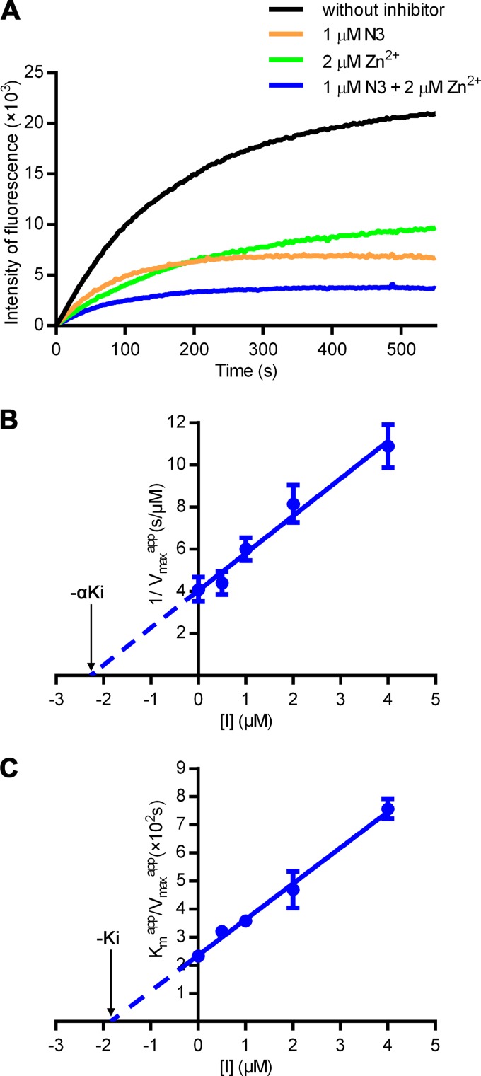
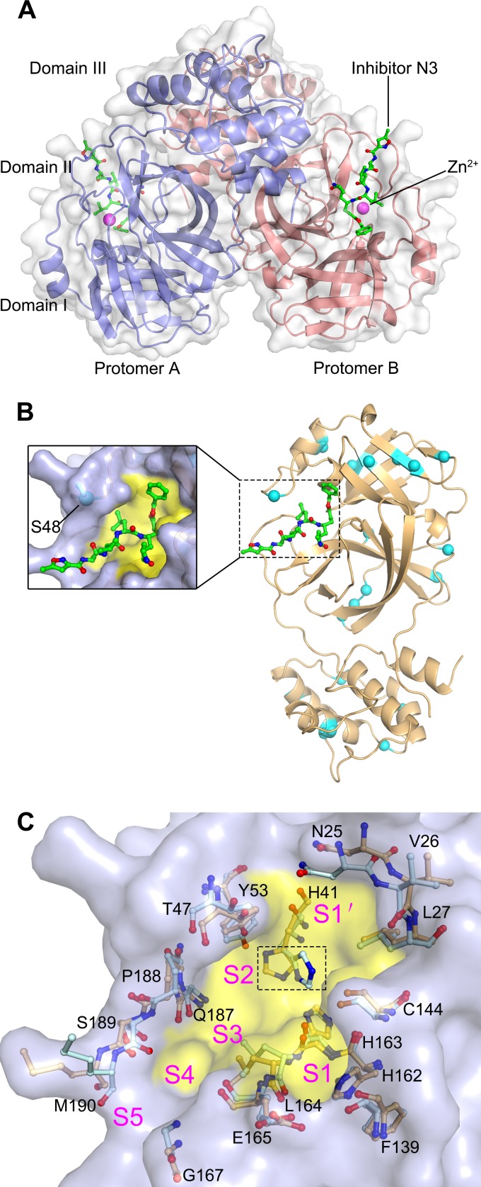
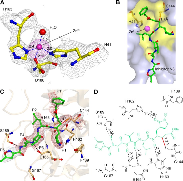
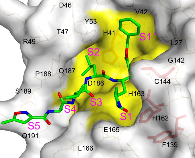
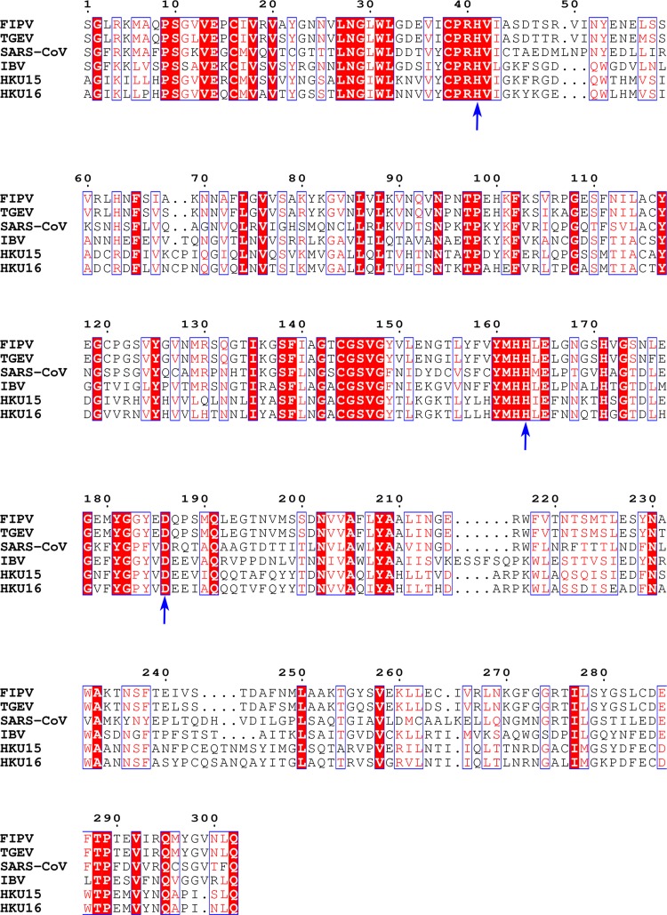
Coronaviruses (CoVs) can cause highly prevalent diseases in humans and animals. Feline infectious peritonitis virus (FIPV) belongs to the genus Alphacoronavirus, resulting in a lethal systemic granulomatous disease called feline infectious peritonitis (FIP), which is one of the most important fatal infectious diseases of cats worldwide. No specific vaccines or drugs have been approved to treat FIP. CoV main proteases (M(pro)s) play a pivotal role in viral transcription and replication, making them an ideal target for drug development. Here, we report the crystal structure of FIPV M(pro) in complex with dual inhibitors, a zinc ion and a Michael acceptor. The complex structure elaborates a unique mechanism of two distinct inhibitors synergizing to inactivate the protease, providing a structural basis to design novel antivirals and suggesting the potential to take advantage of zinc as an adjunct therapy against CoV-associated diseases.

Importance: Coronaviruses (CoVs) have the largest genome size among all RNA viruses. CoV infection causes various diseases in humans and animals, including severe acute respiratory syndrome (SARS) and Middle East respiratory syndrome (MERS). No approved specific drugs or vaccinations are available to treat their infections. Here, we report a novel dual inhibition mechanism targeting CoV main protease (M(pro)) from feline infectious peritonitis virus (FIPV), which leads to lethal systemic granulomatous disease in cats. M(pro), conserved across all CoV genomes, is essential for viral replication and transcription. We demonstrated that zinc ion and a Michael acceptor-based peptidomimetic inhibitor synergistically inactivate FIPV M(pro). We also solved the structure of FIPV M(pro) complexed with two inhibitors, delineating the structural view of a dual inhibition mechanism. Our study provides new insight into the pharmaceutical strategy against CoV M(pro) through using zinc as an adjuvant therapy to enhance the efficacy of an irreversible peptidomimetic inhibitor.


Figures

FIG 1
FIG 1
Synergetic inhibition of FIPV Mpro by Zn2+ and N3. (A) Inhibition of FIPV Mpro by different compounds. Fluorescence curve of FIPV Mpro free of inhibitors (black) or with 1 μM N3 (orange), 2 μM Zn2+ (green), or the dual inhibitors (blue), respectively. The fluorescence intensity is plotted against time to represent enzyme activity. (B and C) Secondary plots to determine the kinetic constants (αKi and Ki) of Zn2+ as a noncompetitive inhibitor. The values of αKi (B) and Ki (C) are calculated from the x intercept.
FIG 2
FIG 2
The structure of FIPV Mpro. (A) Overall structure of the FIPV Mpro-Zn2+-N3 complex in surface representation. The two protomers are colored slate and deep salmon, inhibitor N3 is shown as green sticks, and Zn2+ is shown as a magenta sphere. (B) Distribution of the nonconserved residues between FIPV and TGEV Mpros. The structure of FIPV Mpro is shown in cartoon representation (light orange). The nonconserved residues between FIPV Mpro and TGEV Mpro are shown as cyan spheres. The N3 molecule is shown as green sticks, and the substrate-binding pocket is colored in yellow. A zoomed view of the substrate-binding pocket is shown to the left. (C) Superimposition of the substrate-binding pocket of FIPV Mpro-Zn2+-N3 (light orange) with that of TGEV Mpro (pale cyan). The substrate-binding pocket of the FIPV Mpro complex is colored in yellow. The S1, S2, S4, S5, and S′ subsites are labeled. The key amino acids constituting the substrate-binding pockets of FIPV Mpro and TGEV Mpro are shown as sticks. The His41 residues specifically are marked by the dashed rectangular box.
FIG 3
FIG 3
Interactions between N3, Zn2+, and FIPV Mpro. (A) Coordination of the Zn2+ to the active center of FIPV Mpro. A simulated annealing mFo-DFc omit map for unbiased electron density shows the residues and Zn2+ in gray at 1 σ, and an anomalous difference Fourier map shows Zn2+ in magenta at 3 σ. The hydrogen bonds are displayed as dashed lines, and the distances are labeled in Å. (B) Surface representation of N3 and Zn2+ bound to the active site of FIPV Mpro. N3 is shown as sticks (green), and the substrate-binding pocket is colored in yellow. The catalytic dyad His41 and Cys144 are shown as light-orange sticks. (C) Interactions between the inhibitor N3 and FIPV Mpro. N3 and the key residues are shown as sticks. A sigma-sA-weighted 2mFo-DFc electron density map shows N3 in gray at 1 σ, and a simulated annealing 2mFo-DFc omit map shows N3 in red at 1 σ, respectively. The hydrogen bonds are shown by dashed lines, and the covalent bond by a solid line. The P1, P2, P4, P5, and P1′ sites are labeled. (D) Detailed view of the interactions between the inhibitor N3 and FIPV Mpro. The N3 inhibitor is shown in green. Hydrogen bonds are shown by dashed lines, and the covalent bond is a red solid line.
FIG 4
FIG 4
Structural insight into the mutagenesis analysis of the FIPV Mpro active center. Surface representation of FIPV Mpro complexed with inhibitor N3. The substrate-binding pocket is colored in yellow. The N3 molecule is shown as green sticks. Residues His41, Cys144, Tyr160, and His162 are shown as red sticks.
FIG 5
FIG 5
Sequence alignment of the Mpros from different CoVs. FIPV (GenBank accession no. AY994055) and TGEV (GenBank accession no. FJ755618) are from Alphacoronavirus, SARS-CoV (GenBank accession no. NC_004718) from Betacoronavirus, infectious bronchitis virus (IBV) (GenBank accession no. AY641576) from Gammacoronavirus, and porcine coronavirus HKU15 (GenBank accession no. KM012168) and white-eye coronavirus HKU16 (GenBank accession no. NC_016991) from Deltacoronavirus. The conserved residues that participate in coordinating the Zn2+ are labeled by arrows. Sequence alignment was performed with ClustalW and drawn using ESPript3. White letters with red backgrounds show identical residues, and red letters with white backgrounds show conservative variation. Blue arrows indicate residues involved in coordinating Zn2+ in FIPV Mpro and are well-conserved in Mpro sequences from multiple species.

References

    1. Lee N, Hui D, Wu A, Chan P, Cameron P, Joynt GM, Ahuja A, Yung MY, Leung CB, To KF, Lui SF, Szeto CC, Chung S, Sung JJ. 2003. A major outbreak of severe acute respiratory syndrome in Hong Kong. N Engl J Med 348:1986–1994. doi:10.1056/NEJMoa030685. - DOI - PubMed
    1. Zaki AM, van Boheemen S, Bestebroer TM, Osterhaus AD, Fouchier RA. 2012. Isolation of a novel coronavirus from a man with pneumonia in Saudi Arabia. N Engl J Med 367:1814–1820. doi:10.1056/NEJMoa1211721. - DOI - PubMed
    1. Kipar A, Meli ML. 2014. Feline infectious peritonitis still an enigma? Vet Pathol Online 51:505–526. doi:10.1177/0300985814522077. - DOI - PubMed
    1. Olsen CW. 1993. A review of feline infectious peritonitis virus: molecular biology, immunopathogenesis, clinical aspects, and vaccination. Vet Microbiol 36:1–37. doi:10.1016/0378-1135(93)90126-R. - DOI - PMC - PubMed
    1. Perlman S. 1998. Pathogenesis of coronavirus-induced infections. Review of pathological and immunological aspects. Adv Exp Med Biol 440:503–513. - PubMed

Publication types

Associated data

LinkOut - more resources