. 2015 Dec 9;90(4):1944-63.
doi: 10.1128/JVI.02698-15. Print 2016 Feb 15.

Mutations of Conserved Residues in the Major Homology Region Arrest Assembling HIV-1 Gag as a Membrane-Targeted Intermediate Containing Genomic RNA and Cellular Proteins

Affiliations

Mutations of Conserved Residues in the Major Homology Region Arrest Assembling HIV-1 Gag as a Membrane-Targeted Intermediate Containing Genomic RNA and Cellular Proteins

Motoko Tanaka et al. J Virol. .

Abstract

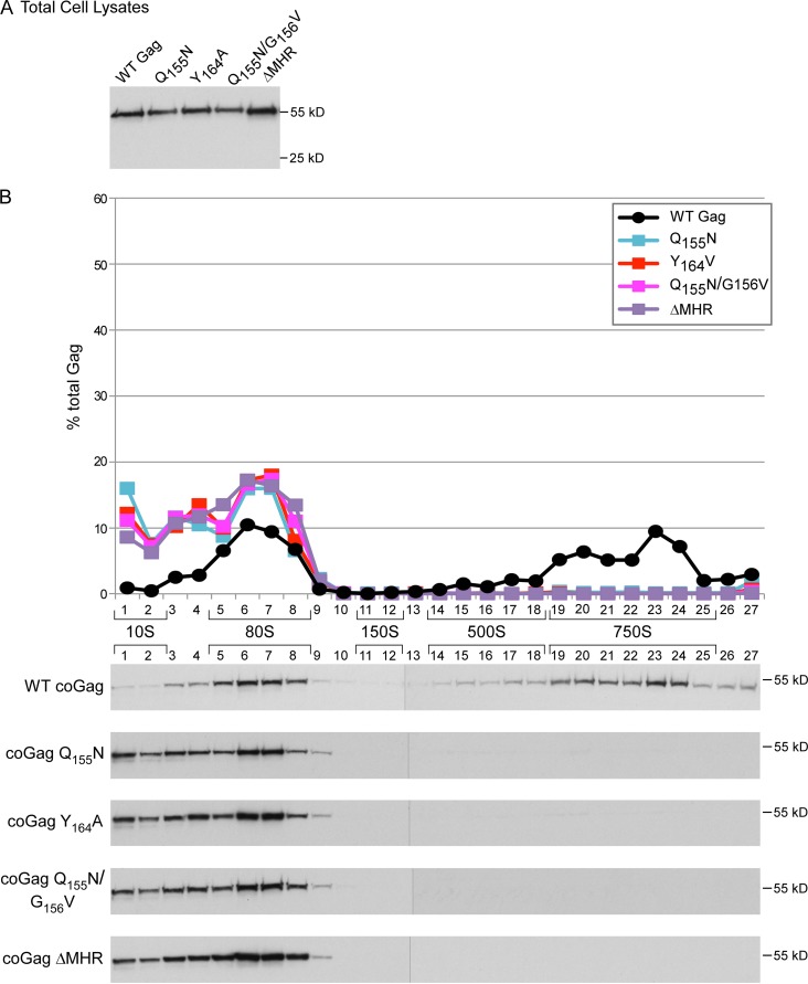
The major homology region (MHR) is a highly conserved motif that is found within the Gag protein of all orthoretroviruses and some retrotransposons. While it is widely accepted that the MHR is critical for assembly of HIV-1 and other retroviruses, how the MHR functions and why it is so highly conserved are not understood. Moreover, consensus is lacking on when HIV-1 MHR residues function during assembly. Here, we first addressed previous conflicting reports by confirming that MHR deletion, like conserved MHR residue substitution, leads to a dramatic reduction in particle production in human and nonhuman primate cells expressing HIV-1 proviruses. Next, we used biochemical analyses and immunoelectron microscopy to demonstrate that conserved residues in the MHR are required after assembling Gag has associated with genomic RNA, recruited critical host factors involved in assembly, and targeted to the plasma membrane. The exact point of inhibition at the plasma membrane differed depending on the specific mutation, with one MHR mutant arrested as a membrane-associated intermediate that is stable upon high-salt treatment and other MHR mutants arrested as labile, membrane-associated intermediates. Finally, we observed the same assembly-defective phenotypes when the MHR deletion or conserved MHR residue substitutions were engineered into Gag from a subtype B, lab-adapted provirus or Gag from a subtype C primary isolate that was codon optimized. Together, our data support a model in which MHR residues act just after membrane targeting, with some MHR residues promoting stability and another promoting multimerization of the membrane-targeted assembling Gag oligomer.

Importance: The retroviral Gag protein exhibits extensive amino acid sequence variation overall; however, one region of Gag, termed the major homology region, is conserved among all retroviruses and even some yeast retrotransposons, although the reason for this conservation remains poorly understood. Highly conserved residues in the major homology region are required for assembly of retroviruses; however, when these residues are required during assembly is not clear. Here, we used biochemical and electron microscopic analyses to demonstrate that these conserved residues function after assembling HIV-1 Gag has associated with genomic RNA, recruited critical host factors involved in assembly, and targeted to the plasma membrane but before Gag has completed the assembly process. By revealing precisely when conserved residues in the major homology region are required during assembly, these studies resolve existing controversies and set the stage for future experiments aimed at a more complete understanding of how the major homology region functions.


Figures

FIG 1
FIG 1
MHR alignment and location of known blocks in the HIV-1 assembly pathway. (A) Alignment of the amino acid sequences of the MHRs of retroviruses and the Ty3 yeast retrotransposon, with retrovirus or retrotransposon family and genus indicated. Amino acid numbering of the MHR within the HIV-1 capsid protein is shown above. The 19-residue consensus sequence for the MHR, based on the 13 sequences shown, is indicated with motif numbers above. Note that H represents hydrophobic residues. Invariant amino acids are shown in blue, highly conserved amino acids are in bold black, and the previously studied K158A mutation (7, 31) is indicated with an asterisk. SIV, simian immunodeficiency virus; EIAV, equine infectious anemia virus; VISNA, visna virus; RSV, Rous sarcoma virus; MPMV, Mason-Pfizer monkey virus; MMTV, mouse mammary tumor virus; HTLV-1, human T-cell lymphotropic virus type 1; BLV, bovine leukemia virus; MoMLV, Moloney murine leukemia virus; FLV, feline leukemia virus. (B) Diagram showing the hypothesis tested here, in the context of the HIV-1 assembly pathway. Newly synthesized Gag forms the ∼10S intermediate, which is recruited into the host precursor complex to form the ∼80S cytosolic assembly intermediate. The ∼80S intermediate likely traffics to the PM, where it forms the ∼150S and ∼500S assembly intermediates. The ∼80S, ∼150S, and ∼500S assembly intermediates are associated with the cellular facilitators of assembly ABCE1 and DDX6, while ∼10S Gag is not. Upon formation of the completely assembled ∼750S completed immature capsid, cellular proteins from the host precursor complex dissociate. Note that ∼10S Gag is also found at the PM and may be recruited into assembling ∼80S intermediates at the PM. Dashed lines indicate steps whose location or progression is hypothetical. For example, the pathway is ATP dependent, but exactly where ATP is required remains to be determined. Known assembly-defective Gag mutants are arrested at five different steps in the assembly pathway (classes 1 to 5, indicated by red bars showing points of blockade), as described in the text and reviewed in reference . Here, we hypothesized that MHR deletion and conserved residue mutations are class 4 mutations, like another MHR mutation tested previously (K158A).
FIG 2
FIG 2
HIV-1 proviruses expressing an MHR deletion or conserved residue mutations fail to form VLPs. (A) Diagram of HIV-1 proviral Gag proteins (LAI strain) analyzed in this study showing WT Gag domains (MA, CA, NC, and p6) and spacer peptides (sp1 and sp2), with CA in blue and the MHR in red. Amino acid sequences from the MHR region of WT Gag (residues 153 to 172 in CA) and MHR point mutants studied here are shown, with mutated residues in bold, deleted residues in parentheses, and an asterisk showing the location of the G2A mutation. (B) High-resolution structure of CA-CTD (PDB accession number 2KOD [48]) showing side chains of the five invariant or highly conserved MHR residues studied here shown in the colors indicated. (C and D) 293T cells (C) or COS-1 cells (D) were transfected with proviruses expressing WT Gag or Gag containing the indicated MHR deletion or mutations. VLPs and total cell lysates (TCL) were harvested and analyzed by WB assay for Gag, with migrations of full-length Gag (p55) and CA (p24) indicated. Data in each panel are from a single experiment that is representative of two independent experiments.
FIG 3
FIG 3
Assembly-defective MHR mutants associate with ABCE1 and DDX6. Human 293T cells were transfected with the indicated WT or mutant provirus. Lysates were immunoprecipitated with antiserum to ABCE1 (αA), antiserum to DDX6 (αD), or nonimmune IgG (N), followed by WB analysis for Gag. Equivalent inputs were also analyzed for Gag by WB analysis. Data are from a single experiment that is representative of three independent experiments.
FIG 4
FIG 4
Single point mutations of highly conserved MHR residues or MHR deletion arrest assembling Gag at the ∼80S intermediate. COS-1 cells were transfected with the indicated WT or mutant provirus. Equivalent aliquots of cell lysates were analyzed by WB assay for Gag (A) or subjected to velocity sedimentation, with gradient fractions analyzed by WB assay for Gag (B). The graph depicts relative amounts of Gag in each fraction of the blots below (as percentage of total Gag), with the approximate S value of each assembly intermediate indicated by brackets. Data are from a single experiment that is representative of three independent experiments.
FIG 5
FIG 5
Double or quadruple MHR point mutations arrest assembling Gag at the ∼80S intermediate. 293T cells were transfected with the indicated WT or mutant provirus. Equivalent aliquots of cell lysates were analyzed by WB assay for Gag (A) or subjected to velocity sedimentation, with gradient fractions analyzed by WB assay for Gag (B). The graph depicts relative amounts of Gag in each fraction of the blots below (as percentage of total Gag), with the approximate S value of each assembly intermediate indicated by brackets. Data are from a single experiment that is representative of two independent experiments.
FIG 6
FIG 6
MHR mutants target to membranes but fail to form the ∼500S membrane-bound intermediate. (A) 293T cells expressing the indicated WT and mutant proviruses were subjected to hypotonic lysis. Postnuclear supernatants were analyzed by WB assay for Gag (left) and subjected to membrane flotation followed by WB assay for Gag (right). Membrane (M) and nonmembrane (NM) fractions are indicated. The graph shows the relative amount of Gag in the membrane fraction, with the percentage of WT Gag in the membrane fraction (55.3%) set to 1. Data in the graph represent an average from two independent experiments ± SEM, with blots from a single representative experiment. (B) COS-1 cells expressing the indicated proviruses were harvested for membrane flotation followed by velocity sedimentation, as shown in the schematic. Inputs show WB analysis of Gag in equivalent aliquots of postnuclear supernatants (PNS) and membrane fractions (M). Membrane flotation and velocity sedimentation fractions were also analyzed by WB assay for Gag. Data are shown for COS-1 cells transfected with WT and ΔMHR proviruses, but similar results were also obtained for Q155N and Y164A (data not shown). Gag observed in fractions 24 to 27 likely represents denatured aggregates of Gag that are produced following repeated centrifugation of lysates, as described previously (31). Data are from a single experiment that is representative of two independent experiments.
FIG 7
FIG 7
Immunoelectron micrographs of MHR mutants arrested as assembly intermediates at the PM. COS-1 cells infected with proviruses expressing WT Gag or the indicated mutants were analyzed by double-label immuno-EM, using antibodies to Gag and ABCE1. Small gold particles indicate Gag labeling; large gold particles indicate ABCE1 labeling. Images were chosen from the quantitative immuno-EM experiments (Fig. 8 and Table 1) and represent the approximate distribution of targeted Gag, early assembly, and late assembly sites at the PM observed for each group. Dark arrows show Gag clusters at the PM that are colocalized with ABCE1; white arrowheads show Gag clusters at the PM that are not colocalized with ABCE1. Dashed lines are placed in some locations to help viewers identify the PM. Bars, 200 nm. As described previously (30), not all instances of colocalization are captured by immuno-EM since the 50-nm sections capture only a fraction of the ∼100-nm-diameter capsid. Targeted Gag, early assembly sites, and late assembly sites are defined in the text and in Table 1.
FIG 8
FIG 8
Quantitative immuno-EM confirms that MHR mutations arrest Gag after PM targeting. Immuno-EM images of infected COS-1 cells described in Fig. 7 were analyzed quantitatively (Table 1). For the indicated constructs, graphs show the average number of Gag clusters in 10 μm of PM (A) and the number of targeted Gag clusters, early assembly sites, and late assembly sites at the PM as a percentage of total Gag clusters at the PM (B). Targeted, early, and late sites are defined in the text and in Table 1. Error bars show standard errors of the means from two independent experiments. Brackets indicate two comparisons that were either not significant (NS) or significant (P < 0.002) using the unpaired Student t test.
FIG 9
FIG 9
Some MHR mutants are arrested as a labile ∼80S assembly intermediate. (A) Schematic showing that COS-1 cells expressing the indicated WT and mutant proviruses were analyzed by membrane flotation in the presence of standard intracellular (0.1 M NaCl) or high (0.25 to 0.375 M NaCl) salt concentrations; subsequently, membrane fractions (M fractions) were further analyzed by velocity sedimentation. The D197A and K158A mutants served as controls since they were previously found to be stable upon high-salt treatment (31). (B) Blots show membrane flotation fractions analyzed by Western blot (WB) assay for Gag, with membrane (M) and nonmembrane (NM) fractions indicated. PNS, postnuclear supernatant. (C) Shown are fractions analyzed by Western blotting for Gag from two experiments, with each experiment enclosed by vertical brackets. To the left are membrane flotation blots performed under standard or high-salt conditions, with M fractions and nonmembrane (NM) fractions indicated by horizontal brackets. To the right are blots from velocity sedimentation analyses of M fractions. Approximate S values are shown with horizontal brackets above blots. (D) Graph of the amount of Gag (as percentage of total Gag) present in small complexes (≤60S, i.e., fractions 1 to 4) in velocity sedimentation analyses of membrane fractions isolated under standard-salt or high-salt conditions. Western blots of inputs for the graphed constructs are shown in panel B; Western blots for Q155N and Q155N/E159D are not shown. Note that the repeated ultracentrifugations required for this experiment resulted in precipitation of some Gag, most likely due to aggregation, observed most prominently in fractions 20 to 28 under standard-salt conditions, as noted previously (31). Graphed data are from a single experiment that is representative of 2 to 4 independent experiments.
FIG 10
FIG 10
MHR deletion and point mutations of highly conserved MHR residues do not inhibit association of assembling Gag with HIV-1 genomic RNA. Total cell lysates from 293T cells expressing the indicated proviruses were subjected to immunoprecipitation (IP) with HIV immunoglobulin (αG) or a nonimmune control antibody (N). The graph shows the number of HIV-1 gRNA copies in immunoprecipitation eluates analyzed by RT-qPCR and in aliquots representing 10% of immunoprecipitation input (in). Also shown is a Western blot (WB) of immunoprecipitation eluates from total cell lysates, using HIV immunoglobulin, to confirm that αG immunoprecipitates Gag efficiently. Nonimmune antibody (N) and 5% of immunoprecipitation input are also shown in WB assays. The dashed line shows the limit of detection based on analysis of beads-alone control. Data in the graph are from a single experiment that is representative of three independent experiments. Error bars show standard deviations between duplicate samples.
FIG 11
FIG 11
MHR mutations introduced into a codon-optimized, type C Gag from a primary isolate arrest assembly at the ∼80S intermediate. COS-1 cells were transfected with the indicated WT or mutant constructs encoding codon-optimized Gag from a subtype C primary isolate (36). Equivalent aliquots of cell lysates were analyzed by WB assay for Gag (A) or subjected to velocity sedimentation, with gradient fractions analyzed by WB assay for Gag (B). The graph depicts relative amounts of Gag in each fraction of the blots below (as percentage of total Gag), with the approximate S value of each assembly intermediate indicated by brackets. Data are from a single experiment that is representative of two independent experiments.
FIG 12
FIG 12
Model showing where specific MHR mutations arrest the HIV-1 assembly pathway. (A) Based on data presented here and elsewhere (10), MHR mutations inhibit assembly at one of two points following membrane targeting of an assembling Gag oligomer. Mutation of the highly conserved residues Y164 and Q155 or MHR deletion arrests Gag as a membrane-targeted ∼80S oligomer that is disrupted by high-salt treatment (labile ∼80S), while mutation of the less conserved K158 residue arrests Gag at the next step in the pathway as a stable membrane-targeted ∼80S oligomer. (B) To show the location of the MHR residues analyzed here relative to residues important for intrahexameric CA-CTD interfaces in the three-dimensional structure, we generated a ribbon diagram using a PDB file provided by J. Briggs, showing selected residues along with their side chains on three CA-CTD domains. Residues critical for the CA-CTD interhexa meric (dimer) interface are in pink (Q176, WMT184/185/186, and TL188/189), residues critical for intrahexameric CA-CTD contacts are in blue (G156, P157, K158, T218, and Q221), and MHR residues studied here are in green (Q155, Y164, R167, and E159). Note that G156 and K158 were also studied here but are shown in blue rather than green because they are known to form intrahexameric CA-CTD contacts (10). The ribbon diagram shows the predicted orientations of residues (but not relative distances) and reveals that MHR residues studied here are either in the intrahexameric CA-CTD interface or very close to it. (C) The structure on the left is the same high-resolution CA-CTD structure shown in Fig. 2B (PDB accession number 2KOD [48]) but this time rotated to display an end-on view of the MHR. Side chains for the five invariant or highly conserved MHR residues studied here are shown in turquoise and include both uncharged or polar residues (Y164, Q155, and G156) and charged residues (E159 and R167). Side chains for other MHR residues not studied here are also shown, including uncharged and polar residues in red (V165, F168, Y169, T171, and L172) and charged residues in blue (R162, D163, D166, and K170). The location of each residue in the amino acid sequence of the HIV-1 MHR is shown below using the same color coding. The structure on the right shows the same view and color coding but with side chains indicated by space-filling dotted clouds. CA helix 8 is shown in pink, CA helix 9 in green, and CA helix 11 in wheat.

References

    1. Lingappa JR, Reed JC, Tanaka M, Chutiraka K, Robinson BA. 2014. How HIV-1 Gag assembles in cells: putting together pieces of the puzzle. Virus Res 193:89–107. doi:10.1016/j.virusres.2014.07.001. - DOI - PMC - PubMed
    1. Sundquist WI, Krausslich HG. 2012. HIV-1 assembly, budding, and maturation. Cold Spring Harb Perspect Med 2(7):a006924. doi:10.1101/cshperspect.a006924. - DOI - PMC - PubMed
    1. Kuzembayeva M, Dilley K, Sardo L, Hu WS. 2014. Life of psi: how full-length HIV-1 RNAs become packaged genomes in the viral particles. Virology 454–455:362–370. doi:10.1016/j.virol.2014.01.019. - DOI - PMC - PubMed
    1. Kingston RL, Vogt VM. 2005. Domain swapping and retroviral assembly. Mol Cell 17:166–167. doi:10.1016/j.molcel.2005.01.002. - DOI - PubMed
    1. Benit L, De Parseval N, Casella JF, Callebaut I, Cordonnier A, Heidmann T. 1997. Cloning of a new murine endogenous retrovirus, MuERV-L, with strong similarity to the human HERV-L element and with a gag coding sequence closely related to the Fv1 restriction gene. J Virol 71:5652–5657. - PMC - PubMed

Publication types

Substances

Associated data