. 2015 Dec 14:5:18136.
doi: 10.1038/srep18136.

Long-term xeno-free culture of human pluripotent stem cells on hydrogels with optimal elasticity

Affiliations

Long-term xeno-free culture of human pluripotent stem cells on hydrogels with optimal elasticity

Akon Higuchi et al. Sci Rep. .

Abstract

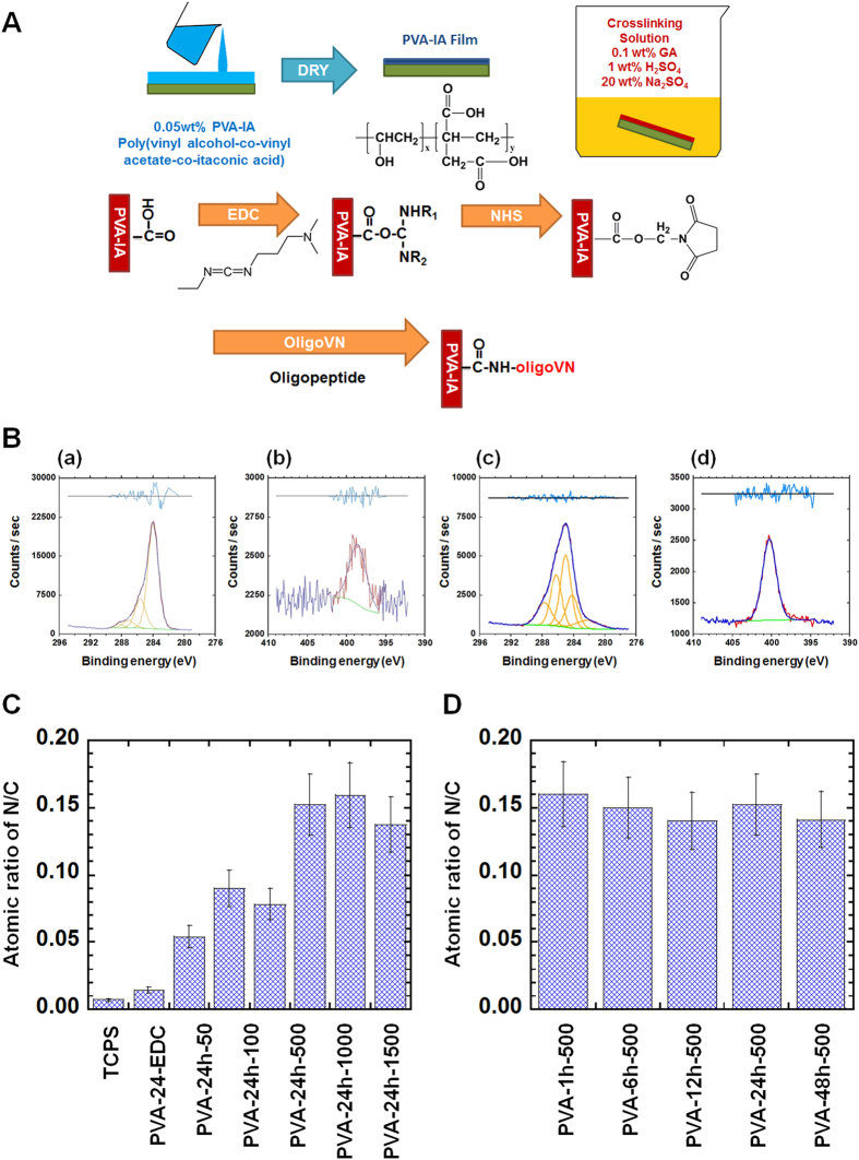
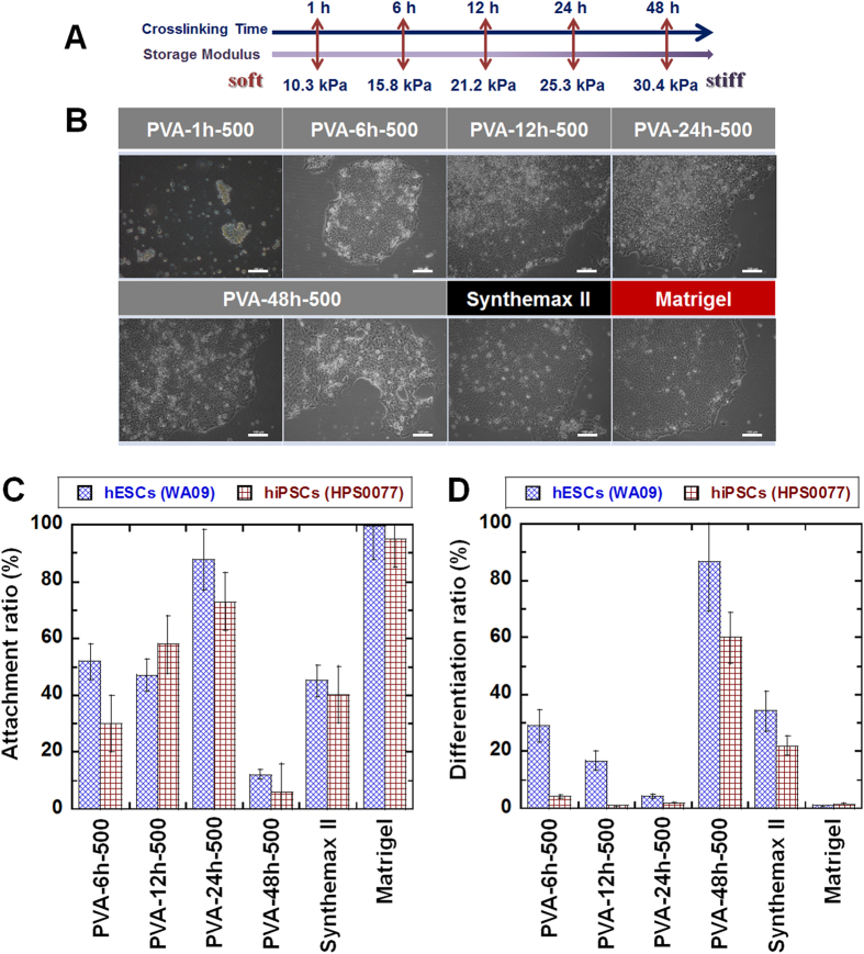
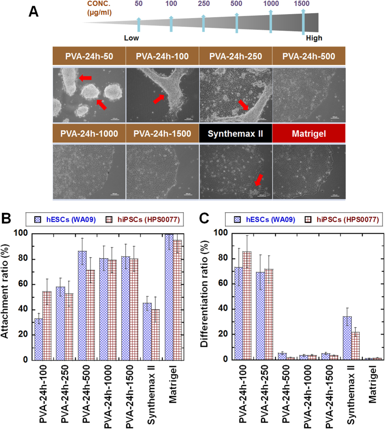
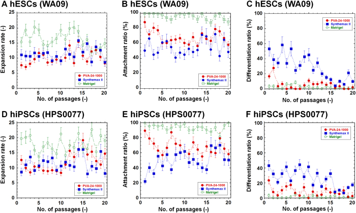
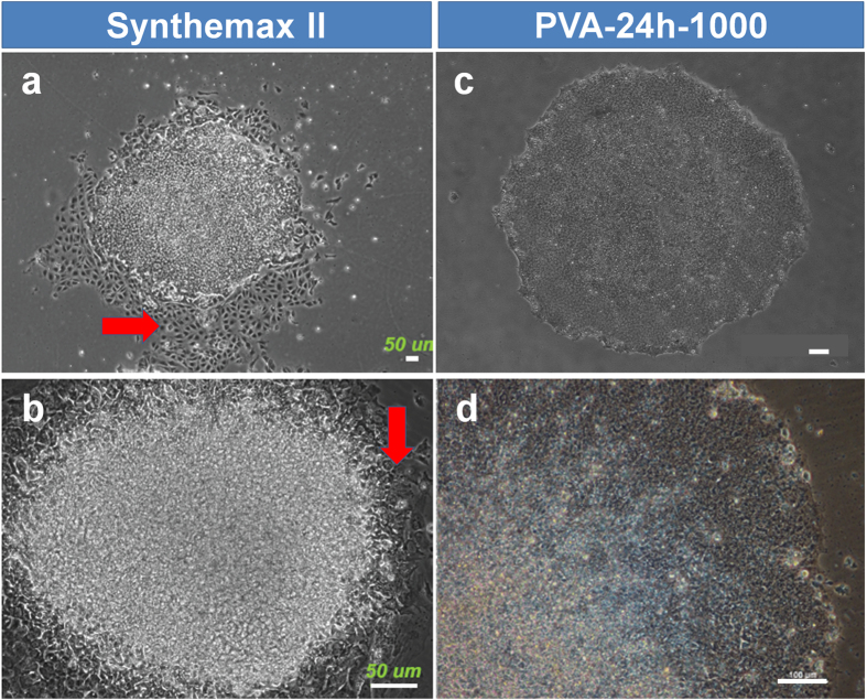
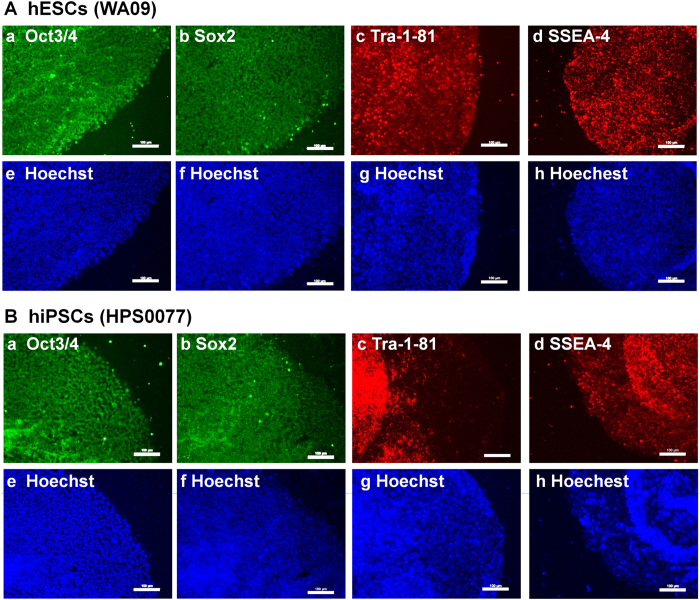
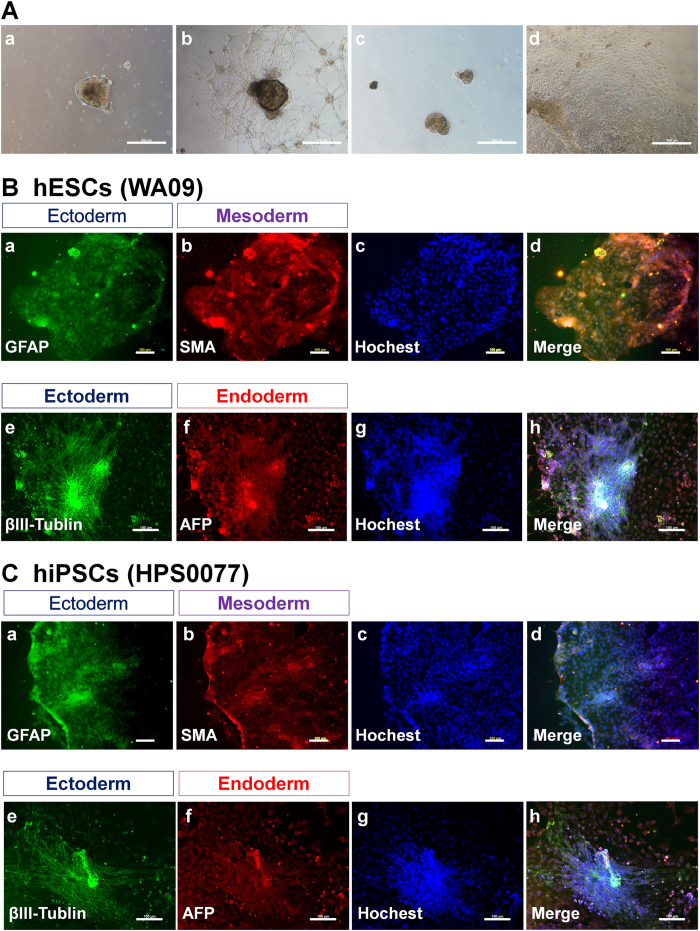
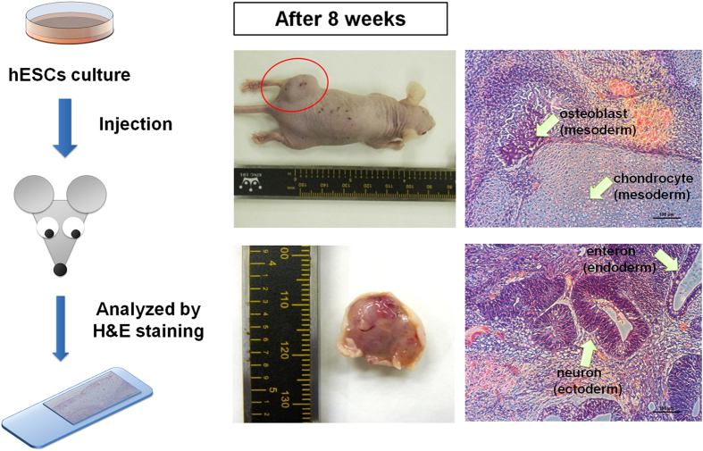
The tentative clinical application of human pluripotent stem cells (hPSCs), such as human embryonic stem cells and human induced pluripotent stem cells, is restricted by the possibility of xenogenic contamination resulting from the use of mouse embryonic fibroblasts (MEFs) as a feeder layer. Therefore, we investigated hPSC cultures on biomaterials with different elasticities that were grafted with different nanosegments. We prepared dishes coated with polyvinylalcohol-co-itaconic acid hydrogels grafted with an oligopeptide derived from vitronectin (KGGPQVTRGDVFTMP) with elasticities ranging from 10.3 to 30.4 kPa storage moduli by controlling the crosslinking time. The hPSCs cultured on the stiffest substrates (30.4 kPa) tended to differentiate after five days of culture, whereas the hPSCs cultured on the optimal elastic substrates (25 kPa) maintained their pluripotency for over 20 passages under xeno-free conditions. These results indicate that cell culture matrices with optimal elasticity can maintain the pluripotency of hPSCs in culture.


Figures

Figure 1
Figure 1. Preparation and characterization of PVA-IA hydrogels grafted with oligoVN.
(A) Reaction scheme for PVA-IA hydrogels grafted with oligoVN. (B) High-resolution XPS spectra of the C1s (a and c) and N1s (b and d) peaks obtained on the surface of unmodified PVA-24h (a and b) and PVA-24h-1000 (c and d) dishes. (C) The atomic ratios of nitrogen to carbon (N/C) in TCPS, PVA-24h-EDC, and PVA-24h hydrogels grafted with different concentration of oligoVN (PVA-24h-50, PVA-24h-100, PVA-24h-500, PVA-24h-1000, and PVA-24h-1500). (D) The atomic ratios of nitrogen to carbon (N/C) in PVA-24h hydrogels grafted with different reaction time of oligoVN (PVA-1h-500, PVA-6h-500, PVA-12h-500, PVA-24h-500, and PVA-48h-1500).
Figure 2
Figure 2. hPSC culture on PVA-oligoVN hydrogels with optimal elasticity.
(A) Elasticity (storage modulus) is regulated by the crosslinking time on PVA-oligoVN hydrogels. (B) Morphology of hESCs (WA09) cultured on PVA-oligoVN hydrogels with several elasticities (PVA-1h-500, PVA-6h-500, PVA-12h-500, PVA-24h-500, and PVA-48h-500), Synthemax II dishes, and Matrigel at passage 1. The bar indicates 100 μm. (C) Attachment ratio of PSCs (blue bar, hESCs [WA09] and red bar, hiPSCs [HPS0077]) on PVA-oligoVN hydrogels with several elasticities, Synthemax II dishes, and Matrigel at passage 3. (D) Differentiation ratio of hPSCs (blue bar, hESCs [WA09] and red bar, hiPSCs [HPS0077]) on PVA-oligoVN hydrogels with several elasticities, Synthemax II dishes, and Matrigel at passage 3.
Figure 3
Figure 3. hPSC culture on PVA-oligoVN hydrogels with different surface densities of oligoVN.
(A) Morphology of hESCs (WA09) cultured on PVA-oligoVN hydrogels with several surface densities of oligoVN (PVA-24h-50, PVA-24h-100, PVA-24h-250, PVA-24h-500, PVA-24h-1000, and PVA-24h-1500), Synthemax II dishes, and Matrigel at passage 1. The red arrows indicate detached cells. The bar indicates 100 μm. (B) Attachment ratio of hPSCs (blue bar, hESCs [WA09] and red bar, hiPSCs [HPS0077]) on PVA-oligoVN hydrogels with several surface densities of oligoVN, Synthemax II dishes, and Matrigel at passage 3. (D) Differentiation ratio of hPSCs (blue bar, hESCs [WA09] and red bar, hiPSCs [HPS0077]) on PVA-oligoVN hydrogels with several surface densities of oligoVN, Synthemax II dishes, and Matrigel at passage 3.
Figure 4
Figure 4. Long-term culture of hPSCs on PVA-oligoVN hydrogels with an optimal elasticity under xeno-free culture conditions.
(A) Expansion rate of hESCs (WA09) on PVA-24h-1000 dishes (closed red circle), Synthemax II dishes (closed blue square), and Matrigel (open green circle) for 20 passages. (B) Attachment ratio of hESCs (WA09) on PVA-24h-1000 dishes (closed red circle), Synthemax II dishes (closed blue square), and Matrigel (open green circle) for 20 passages. (C) Differentiation ratio of hESCs (WA09) on PVA-24h-1000 dishes (closed red circle), Synthemax II dishes (closed blue square), and Matrigel (open green circle) for 20 passages. (D) Expansion rate of hiPSCs (HPS0077) on PVA-24h-1000 dishes (closed red circle), Synthemax II dishes (closed blue square), and Matrigel (open green circle) for 20 passages. (E) Attachment ratio of hiPSCs (HPS0077) on PVA-24h-1000 dishes (closed red circle), Synthemax II dishes (closed blue square), and Matrigel (open green circle) for 20 passages. (F) Differentiation ratio of hiPSCs (HPS0077) on PVA-24h-1000 dishes (closed red circle), Synthemax II dishes (closed blue square), and Matrigel (open green circle) for 20 passages.
Figure 5
Figure 5. Comparison of hESC cultures on Synthemax II and PVA-oligoVN hydrogels.
The morphology of hESCs (WA09) cultured on Synthemax II (a,b) and PVA-24h-1000 (c,d) dishes at passage 1 when hPSCs were shifted from culture on MEFs into culture on Synthemax II or PVA-24h-1000 dishes. Red arrows indicate differentiated hESCs. The bar indicates 50 μm (a,b) and 100 μm (c,d).
Figure 6
Figure 6. Characterization of pluripotency of hPSCs (hESCs and hiPSCs) cultured on PVA-oligoVN hydrogels based on expression of pluripotent proteins.
(A) Pluripotent protein expression on hESCs (WA09) analyzed by immunostaining after culture on PVA-24h-1000 dishes under xeno-free conditions for 20 passages. (a) Oct3/4, (b) Sox2, (c) Tra-1-81, (d) SSEA-4 and (e–h) Hoechest staining of hESCs used in (a–d). The bar indicates 100 μm. (B) Pluripotent protein expression on hiPSCs (HPS0077) analyzed by immunostaining after culture on PVA-24h-1000 dishes under xeno-free conditions for 20 passages. (a) Oct3/4, (b) Sox2, (c) Tra-1-81, (d) SSEA-4 and (e–h) Hoechest staining of hESCs used in (a–d). The bar indicates 100 μm.
Figure 7
Figure 7. Characterization of the differentiation ability of hPSCs (hESCs and hiPSCs) in vitro after culture on PVA-oligoVN hydrogels for 20 passages.
(A) Morphology of EBs differentiated from hESCs (WA09, a,b) and hiPSCs (HPS0077, c,d) after culture on PVA-24h-1000 dishes under xeno-free conditions for 20 passages. (B) Immunostaining of an ectoderm protein (a, GFAP; e, βIII-tubulin), mesoderm protein (b, SMA), and endoderm (f, AFP) protein on hESCs (WA09) after culture on PVA-24h-1000 dishes under xeno-free conditions for 20 passages. (c) Hoechest staining of hESCs used in (a,b). (d) Merged picture of (a–c). (g) Hoechest staining of hESCs used in (e,f). (h) Merged picture of (e–g). The bar indicates 100 μm. (C) Immunostaining of an ectoderm protein (a, GFAP; e, βIII-tubulin), mesoderm protein (b, SMA), and endoderm (f, AFP) protein on hiPSCs (HPS0077) after culture on PVA-24h-1000 dishes under xeno-free conditions for 20 passages. (c) Hoechest staining of hESCs used in (a,b). (d) Merged picture of (a–c). (g) Hoechest staining of hESCs used in (e,f). (h) Merged picture of (e–g). The bar indicates 100 μm.
Figure 8
Figure 8. Characterization of the differentiation ability of hPSCs (hESCs and hiPSCs) in vivo after culture on PVA-oligoVN hydrogels for 20 passages.
Pluripotency of teratoma-forming hESCs (WA09) after culture on PVA-24h-1000 dishes under xeno-free conditions for 20 passages. Osteoblasts and chondrocytes (mesoderm), neurons (ectoderm), and enterons (endoderm) can be detected. The bar indicates 100 μm.

References

    1. Thomson J. A. et al. Embryonic stem cell lines derived from human blastocysts. Science 282, 1145–1147 (1998). - PubMed
    1. Takahashi K. et al. Induction of pluripotent stem cells from adult human fibroblasts by defined factors. Cell 131, 861–872 (2007). - PubMed
    1. Yu J. et al. Induced pluripotent stem cell lines derived from human somatic cells. Science 318, 1917–1920 (2007). - PubMed
    1. Pagliuca F. W. et al. Generation of functional human pancreatic beta cells in vitro. Cell 159, 428–439 (2014). - PMC - PubMed
    1. Rezania A. et al. Reversal of diabetes with insulin-producing cells derived in vitro from human pluripotent stem cells. Nat. Biotechnol. 32, 1121–1133 (2014). - PubMed

Publication types

MeSH terms