. 2016 Feb;48(2):176-82.
doi: 10.1038/ng.3470. Epub 2015 Dec 14.

Identification of focally amplified lineage-specific super-enhancers in human epithelial cancers

Affiliations

Identification of focally amplified lineage-specific super-enhancers in human epithelial cancers

Xiaoyang Zhang et al. Nat Genet. 2016 Feb.

Abstract

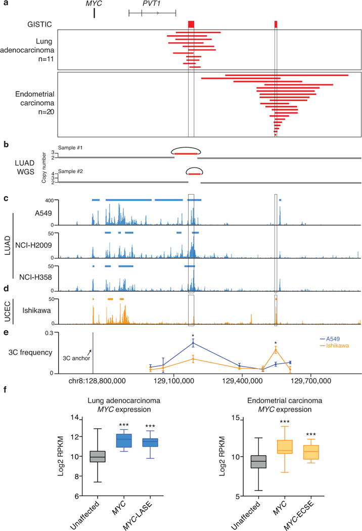
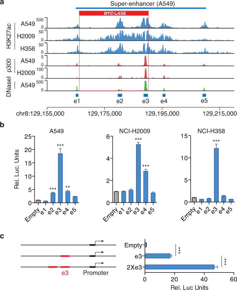
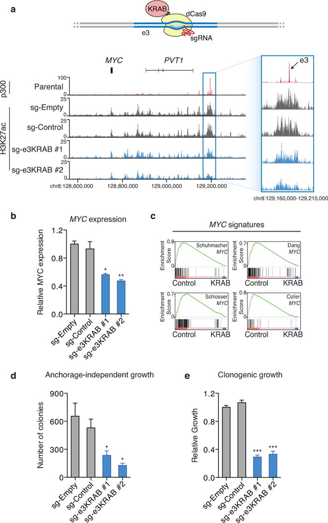
Whole-genome analysis approaches are identifying recurrent cancer-associated somatic alterations in noncoding DNA regions. We combined somatic copy number analysis of 12 tumor types with tissue-specific epigenetic profiling to identify significant regions of focal amplification harboring super-enhancers. Copy number gains of noncoding regions harboring super-enhancers near KLF5, USP12, PARD6B and MYC are associated with overexpression of these cancer-related genes. We show that two distinct focal amplifications of super-enhancers 3' to MYC in lung adenocarcinoma (MYC-LASE) and endometrial carcinoma (MYC-ECSE) are physically associated with the MYC promoter and correlate with MYC overexpression. CRISPR/Cas9-mediated repression or deletion of a constituent enhancer within the MYC-LASE region led to significant reductions in the expression of MYC and its target genes and to the impairment of anchorage-independent and clonogenic growth, consistent with an oncogenic function. Our results suggest that genomic amplification of super-enhancers represents a common mechanism to activate cancer driver genes in multiple cancer types.


Figures

Fig. 1
Fig. 1. Pan-cancer analysis identifying focally amplified super-enhancers
(a) Schematic flow chart of pan-cancer GISTIC analysis of 10,534 tumors from 29 tumor types identifying non-coding focal amplifications of super-enhancers. (b) List of non-coding focal amplification regions harboring super-enhancers. UCEC: uterine corpus endometrial carcinoma, HNSC: head and neck squamous cell carcinoma, LUAD: lung adenocarcinoma, CRC: colorectal carcinoma, LIHC: liver hepatocellular carcinoma, CESC: cervical squamous cell carcinoma, ESCA: esophageal carcinoma. (c) The focal amplification on chr13q identified in HNSC. ChIP-seq profile of H3K27ac and super-enhancer regions from the HNSC cell line BICR-31. Log2 transformed expression level (RPKM) of KLF5 in HNSC tumors with focal amplification of KLF5-HNSE alone (n = 14) and tumors without the amplification (n = 288). Box plot: Middle bar, median; lower and upper box limits, 25th and 75th percentiles, respectively; whiskers, min and max. The P-value is derived from a t-test; (**) p≤0.01; (***) p≤0.001. (d) The focal amplification region on chr13q identified in CRC. ChIP-seq profile of H3K27ac and super-enhancer regions from colon crypt. Log2 transformed expression level (RPKM) of USP12 in CRC tumors with focal amplification of USP12-CCSE alone (n = 6) and tumors without the amplification (n = 127). (e) The focal amplification on chr20q identified in LIHC. ChIP-seq profile of H3K27ac and super-enhancer regions from the LIHC cell line HepG2. Log2 transformed expression level (RPKM) of PARD6B in LIHC tumors with focal amplification of PARD6B-LCSE alone (n = 7) and tumors without the amplification (n = 245).
Fig. 2
Fig. 2. Lineage-specific focal amplification of super-enhancers adjacent to the MYC gene
(a) Focal amplification peaks adjacent to MYC identified by GISTIC in lung adenocarcinoma (n = 11/515) and UCEC (n = 20/539). (b) Whole genome sequencing rearrangement analysis of two lung adenocarcinomas reveals tandem duplications, indicated by the curves. H3K27ac ChIP-seq profile and super-enhancer regions of the LUAD cell lines A549, NCI-H2009 and NCI-H358 (c) and the UCEC cell line Ishikawa (d) in the MYC region. (e) 3C interaction frequency ± SEM measured by chromosome conformation capture assays (n = 3) in A549 and Ishikawa cells. The 3C ‘anchor’ primer targets the MYC promoter region, while the 3C ‘bait’ primers target the non-coding regions 3′ to MYC. The P-value is derived from a t-test; (**) p≤0.01; (***) p≤0.001. (f) Left: Log2 transformed expression level (RPKM) of MYC in LUAD tumors with focal amplification of either MYC alone (n = 7) or MYC-LASE alone (n = 11) and tumors without these amplifications (n = 235). Right: UCEC tumors with focal amplification of either MYC alone (n = 10) or MYC-ECSE (n = 14) and tumors without these amplifications (n = 250). Box plot: Middle bar, median; lower and upper box limits, 25th and 75th percentiles, respectively; whiskers, min and max. The P-value is derived from a t-test; (***) p≤0.001.
Fig. 3
Fig. 3. The activity of the MYC-LASE is predominantly driven by the e3 constituent enhancer
(a) H3K27ac, p300 binding and DNase I hypersensitivity profiles in A549, NCI-H2009 and NCI-H358 cells reveal the constituent enhancers e1-e5 within the super-enhancer region. (b) Luciferase reporter assay (n = 3) measuring enhancer activity of e1-e5 in A549, NCI-H358 and NCI-H2009 lung adenocarcinoma cells. The pGL3 plasmid without the enhancer region (Empty) is used as a negative control. (Y-axis) Relative Luciferase units are normalized to Renilla signal ± SEM. The P-value is derived from a t-test; (**) p≤ p0.01; (***) p≤0.001. (c) Enhancer activity of a duplicated e3 enhancer (2×e3) ± SEM as measured by luciferase reporter assay (n = 3). The P-value is derived from a t-test; (***) p≤0.001.
Fig. 4
Fig. 4. Identification of transcription factors required for the activity of the e3 enhancer
(a) Enhancer activity ± SEM of small fragments (a-f) of the e3 enhancer as assessed by luciferase reporter assays (n = 3) in A549 LUAD cells. The fragments c and d show comparable enhancer activity relative to the intact e3 enhancer, while other fragments show minimal enhancer activity. The P-value is derived from a t-test; (***) p≤0.001. (b) Transcription factor DNA recognition motifs are identified in the mini-e3 enhancer region that is defined by the c and d fragments overlap. (c) The luciferase reporter expression level ± SEM after deletion of individual transcription factor motif sequence in the e3 regions. The P-value is derived from a t-test (n = 3); (**) p≤0.01; (***) p≤0.001. (d) Luciferase reporter expression level ± SEM after silencing NFE2L2 or CEBPB by siRNA (n = 3). The P-value is derived from a t-test; (*) p≤0.05; (**) p≤0.01. (e) ChIP-seq profile of NFE2L2 and CEBPB in the e1–e5 enhancer regions in A549 cells.
Fig. 5
Fig. 5. KRAB-dCas9 mediated repression of the e3 enhancer reveals MYC as a direct target
(a) Upper: the design of KRAB-dCas9 mediated repression of the e3 enhancer. Bottom: ChIP-seq of H3K27ac in NCI-H2009 cells with and without KRAB-dCas9 enhancer repression. p300 ChIP-seq profile in parental NCI-H2009 cells indicates the e3 enhancer region. sg-Empty: no sgRNA; sg-Control: sgRNA that is predicted to not recognize any genomic regions; sg-e3KRAB #1 and sg-e3KRAB #2: two separate sgRNAs recognizing the e3 enhancer region. (b) The expression level of MYC ± SEM as measured by quantitative PCR in NCI-H2009 cells with and without KRAB-dCas9 mediated repression of the e3 enhancer (n = 2). (c) GSEA analysis of RNA-seq data generated in NIC-H2009 cells with and without KRAB-dCas9 mediated e3 enhancer repression reveals that genes regulated by e3 repression are enriched in MYC target genes identified by previous studies. The cellular transformation efficiency ± SEM as measured by anchorage-independent growth (n = 3) (d) and the cellular proliferation rate ± SEM as measured by clonogenic assays (n = 3) (e) in NCI-H2009 cells with and without KRAB-dCas9 mediated repression of the e3 enhancer. The P-value is derived from a t-test; (*) p≤0.05; (**) p≤0.01; (***) p≤0.001.
Fig. 6
Fig. 6. CRISPR/Cas9 mediated deletion of the e3 enhancer impairs the oncogenic effect of the e3 enhancer amplification
(a) Upper: design of CRISPR/Cas9 mediated deletion of the e3 enhancer. Primers used to validate the e3 enhancer deletion are indicated. Bottom: Gel pictures of PCR amplification of genomic DNA using primers outside and inside the e3 enhancer region. sg-Empty: no sgRNA; sg-Control: a pair of sgRNAs that are predicted to not recognize any genomic regions; sg-e3del #1 and sg-e3del #2: two separate pairs of sgRNAs recognizing boundaries of the e3 enhancer region. PCR products were cloned into individual vectors and sequenced. Sequencing results represent the deletions induced by sg-e3del #1 (b) and sg-e3del #2 (c). The expression level of MYC ± SEM as measured by quantitative PCR (n = 2) (d), the cellular transformation efficiency ± SEM as measured by anchorage-independent growth (n = 3) (e) and the cellular proliferation rate ± SEM as measured by clonogenic assays (n = 3) (f) in NCI-H2009 cells with and without CRISPR/Cas9 mediated deletion of the e3 enhancer. The P-value is derived from a t-test; (*) p≤0.05; (**) p≤0.01. (g) Schematic representation of genomic structural variants activating MYC expression in cancer.

References

    1. Stratton MR, Campbell PJ, Futreal PA. The cancer genome. Nature. 2009;458:719–724. - PMC - PubMed
    1. Beroukhim R, et al. The landscape of somatic copy-number alteration across human cancers. Nature. 2010;463:899–905. - PMC - PubMed
    1. Bignell GR, et al. Signatures of mutation and selection in the cancer genome. Nature. 2010;463:893–898. - PMC - PubMed
    1. Stephens PJ, et al. Complex landscapes of somatic rearrangement in human breast cancer genomes. Nature. 2009;462:1005–1010. - PMC - PubMed
    1. Weir BA, et al. Characterizing the cancer genome in lung adenocarcinoma. Nature. 2007;450:893–898. - PMC - PubMed

Publication types

MeSH terms