. 2016 Jan;23(1):67-73.
doi: 10.1038/nsmb.3141. Epub 2015 Dec 14.

Exocytotic fusion pores are composed of both lipids and proteins

Affiliations

Exocytotic fusion pores are composed of both lipids and proteins

Huan Bao et al. Nat Struct Mol Biol. 2016 Jan.

Abstract

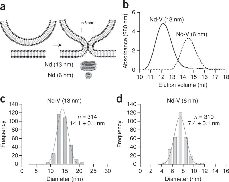
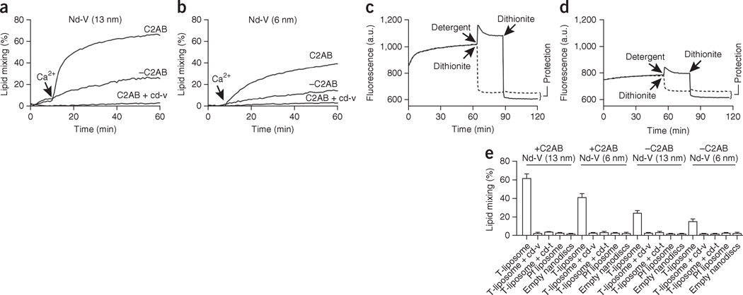
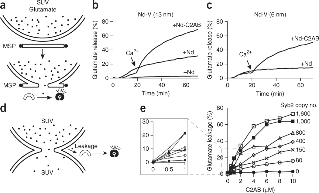
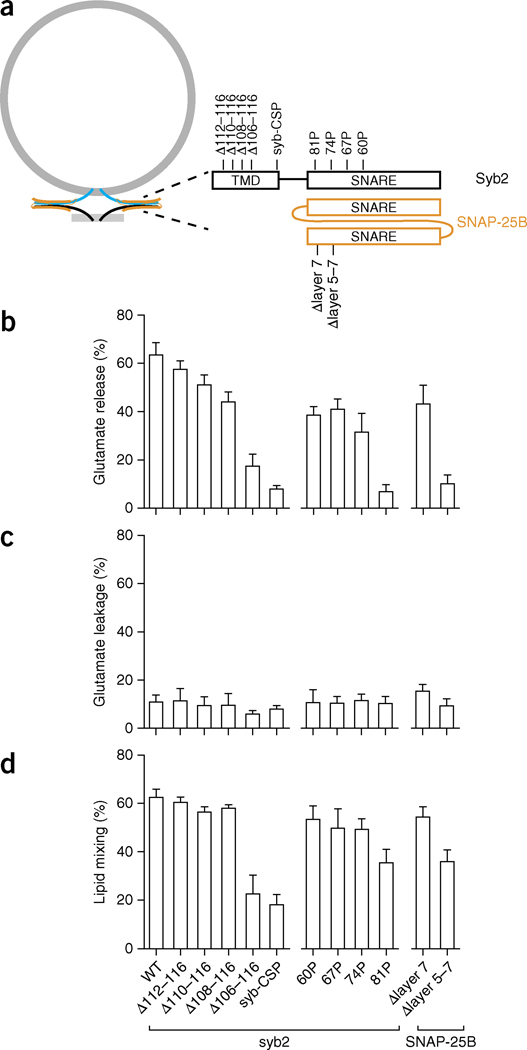
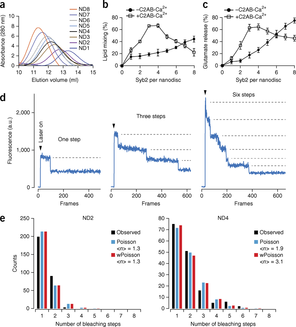
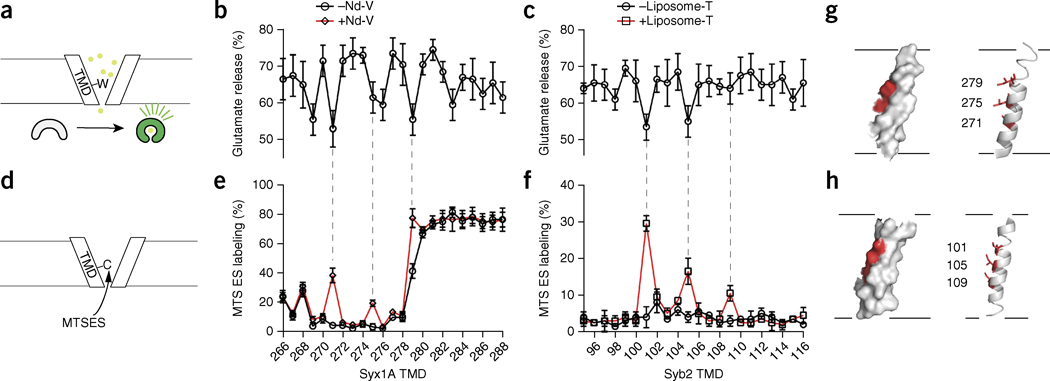
During exocytosis, fusion pores form the first aqueous connection that allows escape of neurotransmitters and hormones from secretory vesicles. Although it is well established that SNARE proteins catalyze fusion, the structure and composition of fusion pores remain unknown. Here, we exploited the rigid framework and defined size of nanodiscs to interrogate the properties of reconstituted fusion pores, using the neurotransmitter glutamate as a content-mixing marker. Efficient Ca(2+)-stimulated bilayer fusion, and glutamate release, occurred with approximately two molecules of mouse synaptobrevin 2 reconstituted into ∼6-nm nanodiscs. The transmembrane domains of SNARE proteins assumed distinct roles in lipid mixing versus content release and were exposed to polar solvent during fusion. Additionally, tryptophan substitutions at specific positions in these transmembrane domains decreased glutamate flux. Together, these findings indicate that the fusion pore is a hybrid structure composed of both lipids and proteins.


Conflict of interest statement

COMPETING FINANCIAL INTERESTS

The authors declare no competing financial interests.

Figures

Figure 1
Figure 1
Reconstitution of syb2 into 6- and 13-nm nanodiscs. (a) Illustration of a lipidic fusion pore and the relative sizes of the two kinds of nanodiscs used in this study. (b) SEC of 6- and 13-nm nanodiscs. (c,d) Diameter distributions of 6-nm nanodiscs (d) and 13-nm nanodiscs (c) determined by AFM imaging. Data represent means ± s.d. (n = number of particles analyzed).
Figure 2
Figure 2
Bilayer fusion between v-SNARE nanodiscs (Nd-V) and t-SNARE vesicles. (a,b) Time courses of lipid mixing with 13-nm Nd-V (a) and 6-nm Nd-V (b), in the presence or absence of C2AB and cd-V; Ca2+ was added as indicated. (c,d) Dithionite quenching after fusion between t-SNARE vesicles and 13-nm Nd-V (c) or 6-nm Nd-V (d). A.u., arbitrary units. (e) Nd-SUV lipid mixing assays performed with t-SNARE liposomes, pf liposomes or empty nanodiscs; in each case, assays were run in the presence or absence of cd-v, cd-t and C2AB, and in the presence of 1 mM Ca2+. Data represent means ± s.d. (n = 3 technical replicates).
Figure 3
Figure 3
Reconstitution of glutamate release through nanodiscs. (a) Illustration summarizing the glutamate release assay. During fusion between Nd-V and t-SNARE vesicles, glutamate is released through the fusion pore, thus resulting in increases in the fluorescence signal of the glutamate sensor (iGluSnFR). (b,c) Time courses of glutamate release with 13-nm Nd-V (b) and 6-nm Nd-V (c). (d) Illustration summarizing the membrane leakage assay using v- and t-SNARE vesicles. Glutamate leakage during fusion between v-SNARE and t-SNARE liposomes is detected via iGluSnFR. (e) Glutamate leakage as a function of Ca2+-C2AB concentration and v-SNARE copy number. Data represent means ± s.d. (n = 3 technical replicates).
Figure 4
Figure 4
Distinct structural elements of SNAREs differentially affect bilayer fusion, membrane leakage and glutamate release. (a) Illustration of the alterations in syb2 and SNAP-25B used to dissociate their functions. Constructs: syb-CSP, cytoplasmic domain of syb2 fused to the cysteine-rich segment of the SV protein CSP (cysteine string protein); Δlayer 5–7, deletion from layers 5 to 7 in the second SNARE motif of SNAP-25B; Δlayer 7, deletion of layer 7 in the same motif of SNAP-25B. (bd) Characterization of SNARE mutants through glutamate release (b), glutamate leakage (c) and lipid mixing assays (d). Data represent means ± s.d. (n = 3 technical replicates). WT, wild type.
Figure 5
Figure 5
Two molecules of syb2 mediate efficient membrane fusion and content release. (a) SEC profiles of nanodiscs containing 1 to 8 molecules of syb2. (b,c) Lipid mixing (b) and glutamate release (c), in the absence or presence of Ca2+-C2AB, plotted versus the syb2 copy number per nanodisc. Data represent means ± s.d. (n = 3 technical replicates). (d) Examples of single-molecule photobleaching traces. (e) Histograms of the number of observed photobleaching steps fit to either a Poisson distribution or a binomially weighted (w) Poisson distribution. <n>, average number of syb2 molecules per nanodisc.
Figure 6
Figure 6
SNARE TMDs are present in the fusion pore. (a) Illustration of the scanning tryptophan assay. (b,c) Glutamate release efficiency mediated by tryptophan-mutant forms of syx1A (b) and syb2 (c). Data represent means ± s.d. (n = 5 technical replicates). (d) Illustration of the substituted cysteine-accessibility assay to probe the TMDs of syb2 and syx1A during fusion. The accessibility of single-cysteine substitutions in syx1A (e) and syb2 (f), with (red trace) or without (black trace) fusion, as measured by the degree of MTSES labeling. Data represent means ± s.d. (n = 3 technical replicates). (g,h) Solvent-exposed residues (red) mapped onto the structure of syx1A (g) and syb2 (h).

Comment in

  • The mystery of the fusion pore.
    Sharma S, Lindau M. Sharma S, et al. Nat Struct Mol Biol. 2016 Jan;23(1):5-6. doi: 10.1038/nsmb.3157. Nat Struct Mol Biol. 2016. PMID: 26733219 Free PMC article. No abstract available.

References

    1. Wickner W, Schekman R. Membrane fusion. Nat Struct Mol Biol. 2008;15:658–664. - PMC - PubMed
    1. Rothman JE. The principle of membrane fusion in the cell (Nobel lecture) Angew Chem Int Edn Engl. 2014;53:12676–12694. - PubMed
    1. Bolger AP, et al. Neurohormonal activation and the chronic heart failure syndrome in adults with congenital heart disease. Circulation. 2002;106:92–99. - PubMed
    1. Todde V, Veenhuis M, van der Klei IJ. Autophagy: principles and significance in health and disease. Biochim Biophys Acta. 2009;1792:3–13. - PubMed
    1. Westermann B. Mitochondrial fusion and fission in cell life and death. Nat Rev Mol Cell Biol. 2010;11:872–884. - PubMed

Publication types