. 2015 Dec 11;10(12):e0141655.
doi: 10.1371/journal.pone.0141655. eCollection 2015.

IFN-λ Inhibits MiR-122 Transcription through a Stat3-HNF4α Inflammatory Feedback Loop in an IFN-α Resistant HCV Cell Culture System

Affiliations

IFN-λ Inhibits MiR-122 Transcription through a Stat3-HNF4α Inflammatory Feedback Loop in an IFN-α Resistant HCV Cell Culture System

Fatma Aboulnasr et al. PLoS One. .

Abstract

Background: HCV replication in persistently infected cell culture remains resistant to IFN-α/RBV combination treatment, whereas IFN-λ1 induces viral clearance. The antiviral mechanisms by which IFN-λ1 induces sustained HCV clearance have not been determined.

Aim: To investigate the mechanisms by which IFN-λ clears HCV replication in an HCV cell culture model.

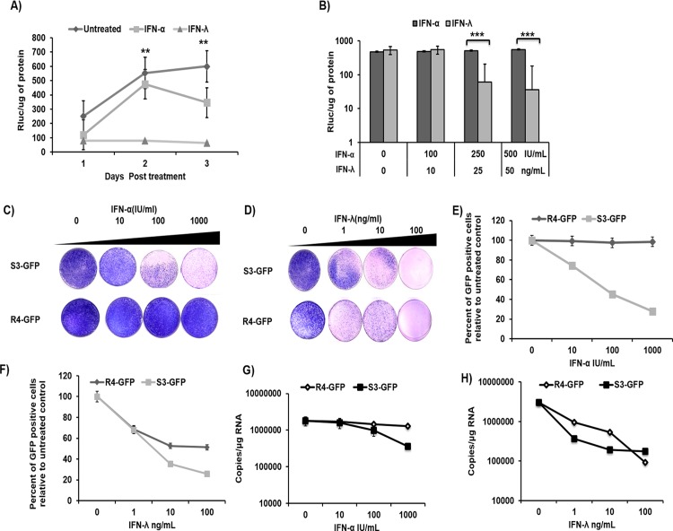
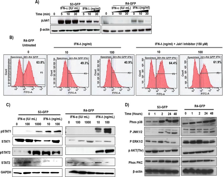
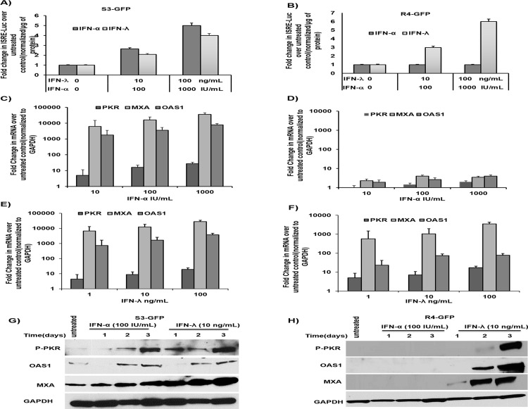
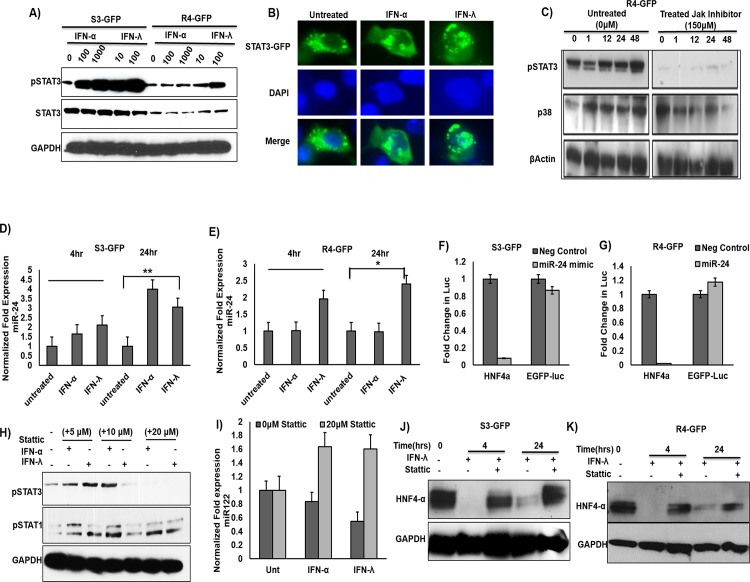
Methods: IFN-α sensitive (S3-GFP) and resistant (R4-GFP) cells were treated with equivalent concentrations of either IFN-α or IFN-λ. The relative antiviral effects of IFN-α and IFN-λ1 were compared by measuring the HCV replication, quantification of HCV-GFP expression by flow cytometry, and viral RNA levels by real time RT-PCR. Activation of Jak-Stat signaling, interferon stimulated gene (ISG) expression, and miRNA-122 transcription in S3-GFP and R4-GFP cells were examined.

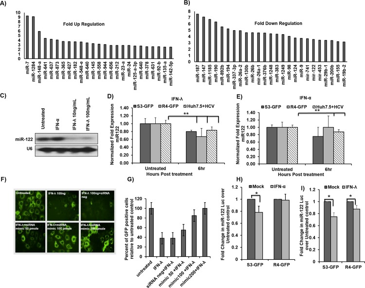
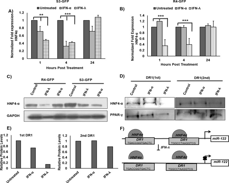
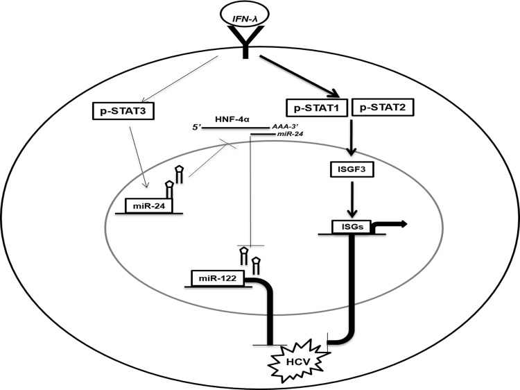
Results: We have shown that IFN-λ1 induces HCV clearance in IFN-α resistant and sensitive replicon cell lines in a dose dependent manner through Jak-Stat signaling, and induces STAT 1 and STAT 2 activation, ISRE-luciferase promoter activation and ISG expression. Stat 3 activation is also involved in IFN-λ1 induced antiviral activity in HCV cell culture. IFN-λ1 induced Stat 3 phosphorylation reduces the expression of hepatocyte nuclear factor 4 alpha (HNF4α) through miR-24 in R4-GFP cells. Reduced expression of HNF4α is associated with decreased expression of miR-122 resulting in an anti-HCV effect. Northern blot analysis confirms that IFN-λ1 reduces miR-122 levels in R4-GFP cells. Our results indicate that IFN-λ1 activates the Stat 3-HNF4α feedback inflammatory loop to inhibit miR-122 transcription in HCV cell culture.

Conclusions: In addition to the classical Jak-Stat antiviral signaling pathway, IFN-λ1 inhibits HCV replication through the suppression of miRNA-122 transcription via an inflammatory Stat 3-HNF4α feedback loop. Inflammatory feedback circuits activated by IFNs during chronic inflammation expose non-responders to the risk of hepatocellular carcinoma.


Conflict of interest statement

Competing Interests: The authors have declared that no competing interests exist.

Figures

Fig 1
Fig 1. Sustained antiviral activity of IFN-λ HCV cell culture.
(A) Persistently infected HCV (JFH1ΔV3-Rluc) cultures were treated with equivalent concentrations of IFN-λ1 (25 ng/ml) or IFN-α (250 IU/mL). Aliquot of infected cells were collected at every 24 hours and HCV replication was measured by Renilla luciferase activity. (B) Persistently infected HCV cultures were treated with increasing concentrations of IFN-α (100–500 I.U/mL) or IFN-λ (10-50ng/mL), HCV replication was measured at 24 hours. (C) R4-GFP and S3-GFP cells (2X10⁵/ 10-cm plate) were treated with IFN-α (10,100 or 1000 I.U/ml) in growth media supplemented with G-418 (1000ng/mL) for 6 weeks. The success of antiviral treatment was determined by the decrease in the number of the resistant colony. (D) The colony assay in the presence of IFN-λ (1, 10, or 100 ng/ml) treatment of S3-GFP and R4-GFP cells. (E) Quantification of HCV-GFP fusion protein by flow cytometry of S3-GFP and R4-GFP cells after IFN-α treatment. (F) Quantification of HCV-GFP fusion protein by flow cytometry of S3-GFP and R4-GFP cells after IFN-λ treatment. (G) HCV positive strand RNA levels in S3-GFP and R4-GFP cells by real-time RT-PCR after IFN-α treatment. (H) HCV positive strand RNA levels in S3-GFP and R4-GFP cells by real-time RT-PCR after IFN-λ treatment, **p≤0.01, ***p≤0.001.
Fig 2
Fig 2. IFN-λ activates the JAK-STAT pathway in R4-GFP and S3-GFP cells.
(A) R4-GFP and S3-GFP cells treated with IFN-α (1000 I.U/ml) or IFN-λ (100 ng/ml) for 5, 10 or 30 minutes and Jak phosphorylation was measured by Western blot analysis. (B) R4-GFP cells were treated with a pan-Jak inhibitor (6-Pyridone) for 1 hour prior to IFN-λ (100ng/mL) treatment and after 72 hours the antiviral effect was measured by FACs analysis. (C) STAT1 and STAT2 phosphorylation was determined by 30 minutes treatment of R4-GFP and S3-GFP cells with increasing doses of IFN-α or IFN-λ. (D) S3-GFP and R4-GFP cells were treated with 100ng/mL of IFN-λ and cells were collected at different time points after treatment. Western blotting was performed using antibodies targeting the p38, p-JNK, p-ERK and p-αKT and PKC and beta-αctin.
Fig 3
Fig 3. IFN-λ activates the ISRE promoter and induces antiviral ISGs in S3GFP and R4GFP cells.
(A) S3-GFP cells were transfected with either an ISRE-Luc reporter plasmid or a control EGFP-Luc plasmid and treated with increasing doses of IFN-α or IFN-λ. Cells were collected 24 hours post transfection and Firefly luciferase activity was measured. Values were normalized with one microgram of protein and expressed as fold increase over the control. (B) R4-GFP cells were transfected with pISRE-Luc plasmid and treated with increasing doses of IFN-α or IFN-λ. After 24 hours, firefly luciferase activity was measured. (C and D) S3-GFP and R4-GFP cells were treated with increasing doses of IFN-α for 24 hours then the expression of ISGs mRNA level was quantified by qRT-PCR. The value of each sample was normalized to GAPDH and the expression levels relative to the untreated control were calculated. (E and F) S3-GFP and R4-GFP cells were treated with IFN-λ for 24 hours; the expression of ISG mRNA was measures by real-time RT-PCR. The value of each sample was normalized to GAPDH and the expression levels relative to the untreated control were calculated (G and H). The protein levels of the ISGs were evaluated by Western blot in S3GFP and R4GFP cells treated with 100 I.U/mL of IFN-α or 10ng/mL of IFN-λ and collected every 24 hours for 72 hours.
Fig 4
Fig 4. IFN-λ decreases miR-122 transcription in HCV cells.
(A) Up regulated microRNAs expression in R4-GFP cells after IFN-λ treatment. (B) Down regulated MicroRNAs expression in R4-GFP cells after IFN-λ treatment. (C) Northern blot for miR-122 in R4-GFP cells treated with either IFN-α or IFN-λ for 6 hours. (D and E) S3-GFP, R4-GFP and persistently HCV infected Huh 7.5 cells were treated with either IFN-α (1000 IU/mL) or IFN-λ1 (100 ng/mL) for six hours. One microgram of total RNA was used for the measurement of miR-122 by real-time RT-PCR. (F and G) R4-GFP cells were transfected with increasing concentrations (25–100 pmole) of miR-122 mimic for 24 hours and then treated with IFN-λ for 48 hours. The effect of the mimic on the antiviral action of IFN-λ was assessed by fluorescence microscopy and then GFP positive cells were quantified by flow cytometry. (H and I) MiR-122-Luc promoter construct was transfected into S3-GFP and R4-GFP cells and following the transfection step, cells were treated with either IFN-α (1000 IU/mL) or IFN-λ (100ng/mL) for 6 hours. The effect of the treatment on the promoter activity was measured by comparing the fold change of firefly luciferase in the treated cells over the untreated control, *p≤0.05, **p≤0.001.
Fig 5
Fig 5. IFN-λ1 decreases miR-122 transcription through down regulation of HNF4α.
(A and B) S3-GFP and R4-GFP cells were treated either IFN-α (1000 I.U/mL) or IFN-λ (100ng/mL). Cells were harvested at 1, 4 and 24 hours and then HNF4α mRNA levels were measured by real-time RT-PCR. (C) Protein levels of HNF4-α measured by Western blot in R4-GFP and S3-GFP after 4 hours of treatment with either 1000 IU/mL of IFN-α or 100ng/mL of IFN-λ. (D) Equal amount of cell lysates from R4-GFP cells were incubated with biotinylated double-stranded oligonucleotides corresponding to the two DR1 motifs in miR-122 promoter and with sterptavidin-agarose beads. The precipitated complexes were subjected to SDS-PAGE and Western blotting for HNF4α and PPAR-γ. (E) Quantification of the bands in Fig 5D (F) Schematic representation of putative HNF4α binding sites in human miR-122 gene promoters and the mechanisms for how IFN-λ1 decreases miR-122 transcription through reduced binding of HNF4α to DR1 element, **p≤0.01, ***p≤0.001.
Fig 6
Fig 6. IFN-λ decreases HNF4α through STAT3 mediated expression of miR-24.
(A) S3-GFP and R4-GFP cells treated with either IFN-α or IFN-λ for 30 minutes. Cell lysates were measured for pStat3 and total Stat3 activation by Western blot analysis. (B) Huh-7 cells were transfected with pSTAT3-GFP plasmid and cells were treated with either of IFN-α (1000 IU/mL) or IFN-λ (100ng/mL) for 1 hour. Nuclear translocation of the Stat3-GFP fusion protein was measured by fluorescence microscopy. (C) Treatment of R4-GFP cells with a Jak inhibitor prevented the effect of IFN-λ on STAT3 activation and but not the p38 activation. (D and E) S3-GFP and R4-GFP cells were treated with either with IFN-α (1000 IU/mL) or IFN-λ (100ng/mL). Quantification of miR-24 levels was done using qRT-PCR. (F and G) miR-24-3p mimic inhibited the luciferase activity of a reporter plasmid harboring the 3’UTR of HNF4α, while it had no effect on the control plasmid EGFP-Luc. (H). A STAT3 inhibitor (Stattic) specifically inhibits STAT3 phosphorylation in R4-GFP cells but has no effect on STAT1 phosphorylation. (I) Stattic prevents the down regulation of miR-122 by IFN-α and IFN-λ in S3-GFP cells 6 hours after treatment as determined by qRT-PCR. (J and K) Treatment of R4-GFP and S3GFP cells with Stattic prevented the inhibitory effect of IFN-λ on HNF4α, *p≤0.05, **p≤0.01.
Fig 7
Fig 7. Schematic illustration of the anti-HCV mechanisms of IFN-λ involves two distinct pathways (The Jak-Stat and Stat3/HNF4α loop).
IFN-λ first binds to cell surface receptor that activates the Stat1, Stat2 and Stat3 activation. (i). The Jak-Stat pathway activation involves the heterodimerization of Stat1/Stat2 which complexes with IRF9 to form ISGF3. The ISGF3 complex binds to the ISRE element to induce interferon stimulated gene expression and inhibit HCV. (ii). The Stat3 activation by IFN-λ induced the expression of miR-24. MiR-24 decreases HNF4α expression through binding to the 3’UTR of HNF4α mRNA. Reduced expression of HNF4α decreases the expression of miR-122 which inhibits HCV replication.

References

    1. Lavanchy D. The global burden of hepatitis C. Liver Int. 2009;29 Suppl 1:74–81. 10.1111/j.1478-3231.2008.01934.x - DOI - PubMed
    1. Yang JD, Roberts LR. Hepatocellular carcinoma: A global view. Nat Rev Gastroenterol Hepatol. 2010;7(8):448–58. 10.1038/nrgastro.2010.100 - DOI - PMC - PubMed
    1. Hajarizadeh B, Grebely J, Dore GJ. Epidemiology and natural history of HCV infection. Nat Rev Gastroenterol Hepatol. 2013;10(9):553–62. 10.1038/nrgastro.2013.107 - DOI - PubMed
    1. Webster DP, Klenerman P, Dusheiko GM. Hepatitis C. Lancet. 2015;385(9973):1124–35. 10.1016/S0140-6736(14)62401-6 - DOI - PMC - PubMed
    1. Liang TJ, Ghany MG. Current and future therapies for hepatitis C virus infection. N Engl J Med. 2013;368(20):1907–17. 10.1056/NEJMra1213651 - DOI - PMC - PubMed

Publication types

MeSH terms