

Meningeal Solitary Fibrous Tumors with Delayed Extracranial Metastasis
- PMID: 26657311
- PMCID: PMC4804146
- DOI: 10.4132/jptm.2015.10.30
Meningeal Solitary Fibrous Tumors with Delayed Extracranial Metastasis
Abstract
Background: The term solitary fibrous tumor (SFT) is preferred over meningeal hemangiopericytoma (HPC), because NAB2-STAT6 gene fusion has been observed in both intracranial and extracranial HPCs. HPCs are now considered cellular variants of SFTs.
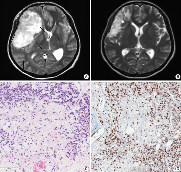
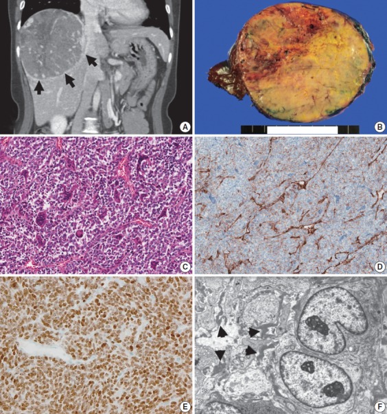
Methods: This study analyzes 19 patients with STAT6-confirmed SFTs, who were followed for over 11 years in a single institution. Ten patients (10/19, 56.2%) had extracranial metastases (metastatic group), while the remainder (9/19) did not (non-metastatic group). These two groups were compared clinicopathologically.
Results: In the metastatic group, the primary metastatic sites were the lungs (n = 6), bone (n = 4), and liver (n = 3). There was a mean lag time of 14.2 years between the diagnosis of the initial meningeal tumor to that of systemic metastasis. The median age at initial tumor onset was 37.1 years in the metastatic group and 52.5 in the non-metastatic group. The 10-year survival rates of the metastatic- and non-metastatic groups were 100% and 33%, respectively. The significant prognostic factors for poor outcomes on univariate analysis included advanced age (≥45 years) and large initial tumor size (≥5 cm). In contrast, the patients with higher tumor grade, high mitotic rate (≥5/10 high-power fields), high Ki-67 index (≥5%), and the presence of necrosis or CD34 positivity showed tendency of poor prognosis but these parameters were not statistically significant poor prognostic markers.
Conclusions: Among patients with SFTs, younger patients (<45 years) experienced longer survival times and paradoxically had more frequent extracranial metastases after long latent periods than did older patients. Therefore, young patients with SFTs require careful surveillance and follow-up for early detection of systemic metastases.
Keywords: Central nervous system; Hemangiopericytoma; NAB2-STAT6 gene fusion; Neoplasm metastases; Solitary fibrous tumors.
Conflict of interest statement
No potential conflict of interest relevant to this article was reported.
Figures

References
-
- Klemperer P, Coleman BR. Primary neoplasms of the pleura: a report of five cases. Am J Ind Med. 1992;22:1–31. - PubMed
-
- Fletcher CD, Bridege JA, Hogendoorn P, Mertens F. WHO classification of tumours of soft tissue and bone. Lyon: IARC Press; 2013.
-
- Tihan T, Viglione M, Rosenblum MK, Olivi A, Burger PC. Solitary fibrous tumors in the central nervous system: a clinicopathologic review of 18 cases and comparison to meningeal hemangiopericytomas. Arch Pathol Lab Med. 2003;127:432–9. - PubMed
-
- Yu HC, Cho BH, Kim YK, Noh SJ, Moon WS. Solitary fibrous tumor of the liver: a case report. Korean J Pathol. 2010;44:536–9.
LinkOut - more resources
Full Text Sources
Other Literature Sources
Research Materials
Miscellaneous
