. 2016 Jun 1;19(6):pyv130.
doi: 10.1093/ijnp/pyv130. Epub 2015 Dec 12.

The Omega-3 Polyunsaturated Fatty Acid Docosahexaenoic Acid (DHA) Reverses Corticosterone-Induced Changes in Cortical Neurons

Affiliations

The Omega-3 Polyunsaturated Fatty Acid Docosahexaenoic Acid (DHA) Reverses Corticosterone-Induced Changes in Cortical Neurons

Matteo M Pusceddu et al. Int J Neuropsychopharmacol. .

Abstract

Background: Chronic exposure to the glucocorticoid hormone corticosterone exerts cellular stress-induced toxic effects that have been associated with neurodegenerative and psychiatric disorders. Docosahexaenoic acid is a polyunsaturated fatty acid that has been shown to be of benefit in stress-related disorders, putatively through protective action in neurons.

Methods: We investigated the protective effect of docosahexaenoic acid against glucocorticoid hormone corticosterone-induced cellular changes in cortical cell cultures containing both astrocytes and neurons.

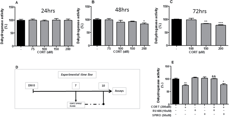
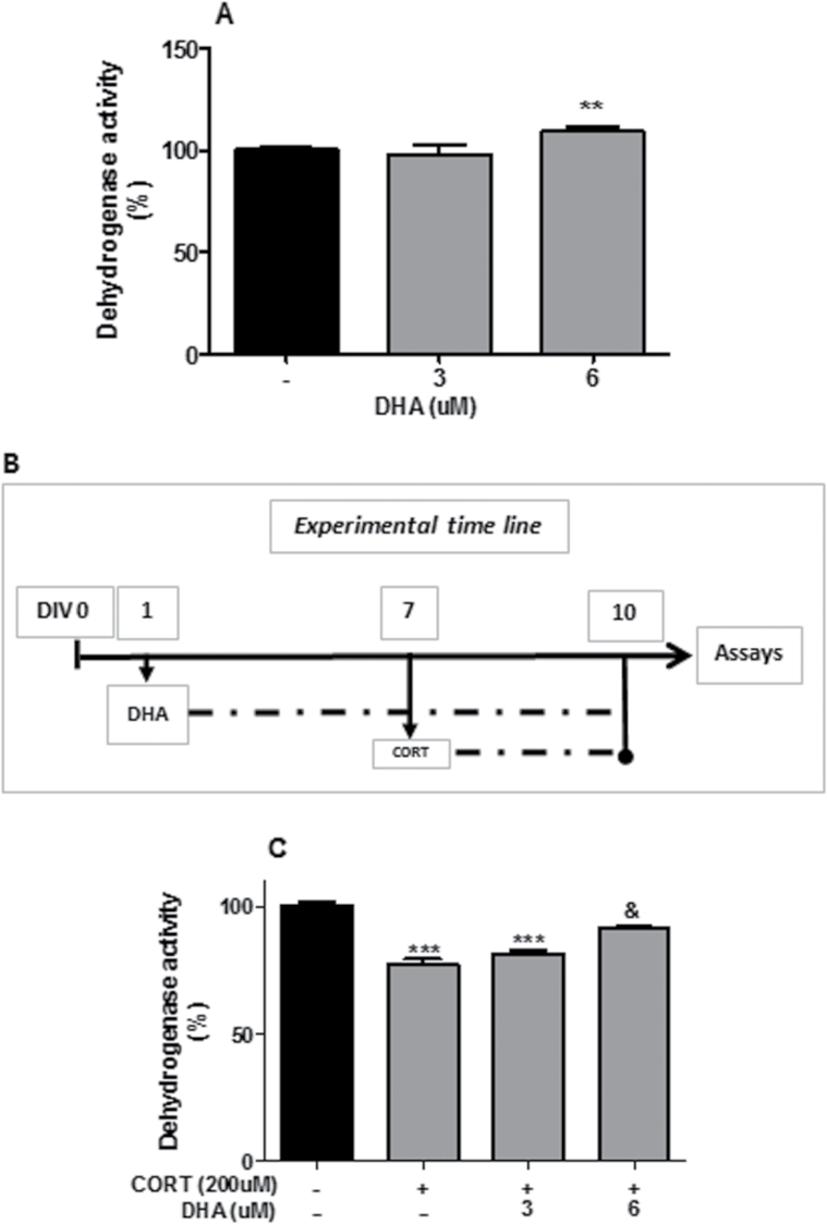
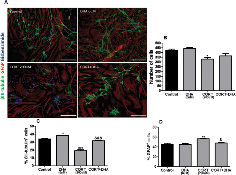
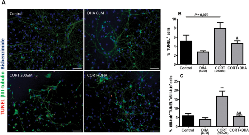
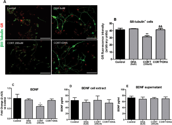
Results: We found that glucocorticoid hormone corticosterone (100, 150, 200 μM) at different time points (48 and 72 hours) induced a dose- and time-dependent reduction in cellular viability as assessed by methyl thiazolyl tetrazolium. Moreover, glucocorticoid hormone corticosterone (200 μM, 72 hours) decreased the percentage composition of neurons while increasing the percentage of astrocytes as assessed by βIII-tubulin and glial fibrillary acidic protein immunostaining, respectively. In contrast, docosahexaenoic acid treatment (6 μM) increased docosahexaenoic acid content and attenuated glucocorticoid hormone corticosterone (200 μM)-induced cell death (72 hours) in cortical cultures. This translates into a capacity for docosahexaenoic acid to prevent neuronal death as well as astrocyte overgrowth following chronic exposure to glucocorticoid hormone corticosterone. Furthermore, docosahexaenoic acid (6 μM) reversed glucocorticoid hormone corticosterone-induced neuronal apoptosis as assessed by terminal deoxynucleotidyl transferase-mediated nick-end labeling and attenuated glucocorticoid hormone corticosterone-induced reductions in brain derived neurotrophic factor mRNA expression in these cultures. Finally, docosahexaenoic acid inhibited glucocorticoid hormone corticosterone-induced downregulation of glucocorticoid receptor expression on βIII- tubulin-positive neurons.

Conclusions: This work supports the view that docosahexaenoic acid may be beneficial in ameliorating stress-related cellular changes in the brain and may be of value in psychiatric disorders.

Keywords: Docosahexaenoic acid; brain derived neurotrophic factor; corticosterone; glucocorticoid receptors; stress.


Figures

Figure 1.
Figure 1.
Corticosterone (CORT)-induced reduction in dehydrogenase activity in rat mixed cortical cultures is prevented by glucocorticoid receptor (GR) antagonist RU486. (A-C) CORT dose-dependent reduction in dehydrogenase activity at 24, 48, and 72 hours, respectively. (D) Schematic representing CORT, GR antagonist RU486, and mineralcorticoid receptor (MR) antagonist spironolactone (SPIRO) treatments. (E) RU486 (10 μM), but not SPIRO (50 μM), blocks the decrease in dehydrogenase activity upon treatment with CORT. Intergroup comparisons: *P < 0.05, **P < 0.01, and ***P < 0.001 compared with control (-); &&P < 0.01 compared with CORT (200 μM). Data are presented as mean ± SEM (n = 3).
Figure 2.
Figure 2.
Docosahexaenoic acid (DHA) prevented corticosterone (CORT)-induced reduction in dehydrogenase activity in rat mixed cortical cultures. (A) DHA dose-dependent increase in dehydrogenase activity. (B) Schematic representing CORT and DHA treatments. (C) DHA prevention of CORT-induced reduction in dehydrogenase activity. Intergroup comparisons: **P < 0.01 and ***P < 0.001 compared with control (-); &P < 0.05 compared with CORT (200 μM). Data are presented as mean ± SEM (n = 3).
Figure 3.
Figure 3.
Docosahexaenoic acid (DHA) prevented corticosterone (CORT)-induced changes in the cellular composition of rat mixed cortical cultures. (A) Representative photomicrographs of cells immunocytochemically stained for βIII-tubulin (green), glial fibrillary acidic protein (GFAP) (red), and bisbenzimide (blue). (B) Quantification of total cells stained with bisbenzimide. (C) Percentage composition of βIII-tubulin+cells (D) and GFAP+cells. Scale bar=50μm. Intergroup comparisons: *P < 0.05, **P < 0.01 compared with control; &P < 0.05 and &&&P < 0.01 compared with CORT (200 μM). Data are presented as mean ± SEM (n = 3).
Figure 4.
Figure 4.
Docosahexaenoic acid (DHA) prevented corticosterone (CORT)-induced apoptosis in rat mixed cortical cultures. (A) Representative photomicrographs of cells immunocytochemically stained for terminal deoxynucleotidyl transferase–mediated nick-end labeling (TUNEL) (red), βIII-tubulin (green), and bisbenzimide (blue). (B) Percentage composition of TUNEL+cells. (C) Percentage of βIII-tub+TUNEL+/ βIII-tub+cells. Scale bar=100μm. Intergroup comparisons: **P < 0.01 compared with control; &P < 0.05 and &&P < 0.01 compared with CORT (200 μM). Data are presented as mean ± SEM (n = 3).
Figure 5.
Figure 5.
Docosahexaenoic acid (DHA) prevented corticosterone (CORT)-induced changes in the glucocorticoid receptor (GR) and in the gene expressions of rat mixed cortical cultures. (A) Representative photomicrographs of cells immunocytochemically stained for βIII-tubulin (green) and GR (red). (B) Mean densitometry analysis of GR protein in βIII-tubulin+cells. Scale bar=100μm. (C) Relative brain derived neurotrophic factor (BDNF) gene expression from RNA extracted by rat mixed cortical cultures. (D- E) BDNF protein expression levels in rat mixed cortical cultures. Intergroup comparisons: **P < 0.01 compared with control, &&P < 0.01 compared with CORT (200 μM). Data are presented as mean ± SEM (n = 3).

References

    1. Almeida OF, Conde GL, Crochemore C, Demeneix BA, Fischer D, Hassan AH, Meyer M, Holsboer F, Michaelidis TM. (2000) Subtle shifts in the ratio between pro- and antiapoptotic molecules after activation of corticosteroid receptors decide neuronal fate. FASEB J 14:779–790. - PubMed
    1. Ardayfio P, Kim KS. (2006) Anxiogenic-like effect of chronic corticosterone in the light-dark emergence task in mice. Behav Neurosci 120:249–256. - PubMed
    1. Arnsten AF. (2009) Stress signalling pathways that impair prefrontal cortex structure and function. Nat Rev Neurosci 10:410–422. - PMC - PubMed
    1. Barsegyan A, Mackenzie SM, Kurose BD, McGaugh JL, Roozendaal B. (2010) Glucocorticoids in the prefrontal cortex enhance memory consolidation and impair working memory by a common neural mechanism. Proc Natl Acad Sci U S A 107:16655–16660. - PMC - PubMed
    1. Behl C, Lezoualc’h F, Trapp T, Widmann M, Skutella T, Holsboer F. (1997) Glucocorticoids enhance oxidative stress-induced cell death in hippocampal neurons in vitro. Endocrinology 138:101–106. - PubMed

LinkOut - more resources