. 2015 Dec 10:6:10149.
doi: 10.1038/ncomms10149.

Cellobiohydrolase 1 from Trichoderma reesei degrades cellulose in single cellobiose steps

Affiliations

Cellobiohydrolase 1 from Trichoderma reesei degrades cellulose in single cellobiose steps

Sonia K Brady et al. Nat Commun. .

Abstract

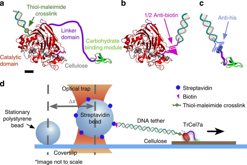
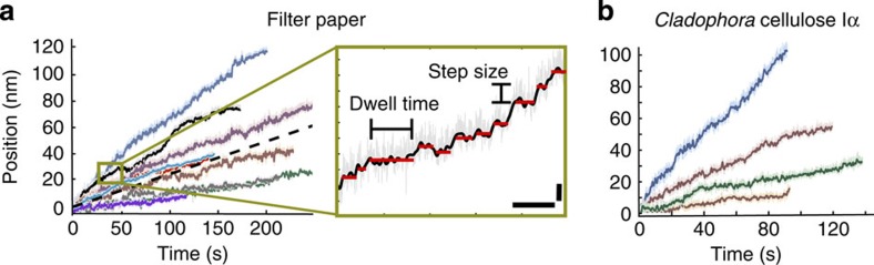
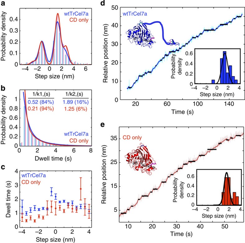
Cellobiohydrolase 1 from Trichoderma reesei (TrCel7A) processively hydrolyses cellulose into cellobiose. Although enzymatic techniques have been established as promising tools in biofuel production, a clear understanding of the motor's mechanistic action has yet to be revealed. Here, we develop an optical tweezers-based single-molecule (SM) motility assay for precision tracking of TrCel7A. Direct observation of motility during degradation reveals processive runs and distinct steps on the scale of 1 nm. Our studies suggest TrCel7A is not mechanically limited, can work against 20 pN loads and speeds up when assisted. Temperature-dependent kinetic studies establish the energy requirements for the fundamental stepping cycle, which likely includes energy from glycosidic bonds and other sources. Through SM measurements of isolated TrCel7A domains, we determine that the catalytic domain alone is sufficient for processive motion, providing insight into TrCel7A's molecular motility mechanism.


Figures

Figure 1
Figure 1. Constructs and assay schematic.
Construct details and optical trap assay schematic for (a) wtTrCel7A, where a DNA-bound sulfo-SMCC crosslinks through available surface lysines (scale bar, 1 nm), (b) isolated biotin-labelled CD ligated to DNA through a ½ anti-biotin antibody and (c) isolated CBM tethered through a DNA-bound anti-His antibody. Structures in ac are from PDB 7CEL and 2CBH. (d) A schematic of the wtTrCel7A motility assay tracks motility through a 1,010-bp tether attached to a 1.25-μm streptavidin bead held in an optical trap. Stationary fiducial beads serve to compensate for drift.
Figure 2
Figure 2. Motility traces and stepping definitions.
(a) Sample wtTrCel7A motility traces on filter paper exhibit a range of velocities with an average of 0.25 nm s−1±0.16 (s.d.) dashed line. The average is representative of 180 traces from 64 separate enzymes at 21 °C. The inset reveals an enlarged region of one trace highlighting the fine stepping motion of wtTrCel7A. Scale bars are 5 s and 2 nm, respectively. (b) Sample TrCel7a motility traces on Cladophora-derived cellulose. The average velocity is 0.25 nm s−1±0.35 (s.d.) and is representative of 68 traces from 17 separate enzymes at 21 °C.
Figure 3
Figure 3. Stepping analysis of wtTrCel7A (blue) and isolated CD (red).
(a) Step size distributions fit to Gaussian curves based on the fundamental and 2 × fundamental steps (wtTrCel7A: 1.28±0.7 nm (s.d.), N=1614; isolated CD: 1.34±0.6 nm (s.d.), N=360). (b) Dwell distributions fit to a double exponential with exponential time constants, 1/k1 and 1/k2, indicated in the figure. The mean dwell time of wtTrCel7A (N=1628) and isolated CD (N=369) are 1.6 and 1.2 s, respectively. (c) The relationship between step size and dwell time shows generally shorter dwell times associated with negative steps and isolated CD measurements (red). Error bars denote s.e.m. (d) and (e) provide sample traces with individual trace step distributions (insets) with similar behaviour between the wtTrCel7A and isolated CD constructs.
Figure 4
Figure 4. Force and temperature dependence of velocity.
(a) The force velocity relationship for wtTrCel7A. Opposing loads (negative) appear to have little effect up to 20 pN while assisting loads (positive) increase velocity. Full data set (blue, N=15666 opposing and N=5939 assisting) and an axial trace subset (red, N=5855 opposing and N=208 assisting), in which the force vector is within 18.5° of the cellulose axis. Error bars represent standard deviation (s.d.). (b) An Arrhenius fit of wtTrCel7A motility data from 21 to 34 °C, using average velocity (cellobiose units per s) as a rate, yielding an activation energy of 20 kBT (49.8 kJ mol−1). The averages are found from N=76 (21 °C), N=47 (28 °C), N=52 (34 °C).

References

    1. Kurasin M. & Väljamäe P. Processivity of cellobiohydrolases is limited by the substrate. J. Biol. Chem. 286, 169–177 (2011). - PMC - PubMed
    1. Sullivan A. C. O. Cellulose: the structure slowly unravels. Cellulose 4, 173–207 (1997).
    1. Costerton J. W. i. et al.. Bacterial biofilms in nature and disease. Ann. Rev. Microbiol. 41, 435–464 (1987). - PubMed
    1. Solano C. et al.. Genetic analysis of Salmonella enteritidis biofilm formation: critical role of cellulose. Mol. Microbiol. 43, 793–808 (2002). - PubMed
    1. Rosan B. & Lamont R. J. Dental plaque formation. Microbes Infect. 2, 1599–1607 (2000). - PubMed

Publication types

MeSH terms

Substances