. 2016 Aug;27(8):2370-81.
doi: 10.1681/ASN.2015070747. Epub 2015 Dec 11.

A Novel Three-Dimensional Human Peritubular Microvascular System

Affiliations

A Novel Three-Dimensional Human Peritubular Microvascular System

Giovanni Ligresti et al. J Am Soc Nephrol. 2016 Aug.

Abstract

Human kidney peritubular capillaries are particularly susceptible to injury, resulting in dysregulated angiogenesis, capillary rarefaction and regression, and progressive loss of kidney function. However, little is known about the structure and function of human kidney microvasculature. Here, we isolated, purified, and characterized human kidney peritubular microvascular endothelial cells (HKMECs) and reconstituted a three-dimensional human kidney microvasculature in a flow-directed microphysiologic system. By combining epithelial cell depletion and cell culture in media with high concentrations of vascular endothelial growth factor, we obtained HKMECs of high purity in large quantity. Unlike other endothelial cells, isolated HKMECs depended on high vascular endothelial growth factor concentration for survival and growth and exhibited high tubulogenic but low angiogenic potential. Furthermore, HKMECs had a different transcriptional profile. Under flow, HKMECs formed a thin fenestrated endothelium with a functional permeability barrier. In conclusion, this three-dimensional HKMEC-specific microphysiologic system recapitulates human kidney microvascular structure and function and shows phenotypic characteristics different from those of other microvascular endothelial cells.

Keywords: angiogenesis; endothelial cells; fenestrae; kidney; microphysiological system; peritubular microvessels.


Figures

Figure 1.
Figure 1.
HKMECs were characterized in human kidney peritubular microvessels in vivo. (A and B) Histologic images of (A) human fetal and (B) adult kidney tissues containing (A.1 and B.1) established nephron structures. In the interstitial stroma, microvascular endothelial cells strongly expressed (A.2 and B.2) CD31 (red) and (A.3 and B.3) PV1 (red) but not (A.3 and B.3) vWF (green). Blue indicates nuclei. (C and D) From flow cytometric analysis, roughly 1.67% and 0.77% of the total cell population are endothelial cells (CD31+CD45−) in (C) fetal and (D) adult kidneys, respectively. G, glomeruli; S, stroma; T, tubule. Scale bar, 50 μm.
Figure 2.
Figure 2.
HKMECs were isolated, purified, and characterized in vitro. (A) Summarized procedure for HKMEC enrichment. (B) Flow cytometric analysis of a single-cell suspension of isolated kidney cells after 5 days of culture indicating the proportion of endothelial cells in three distinct culture conditions: without VEGF-A (top panel), with 40 ng/ml VEGF-A (middle panel), and with VEGF-A and prior depletion of epithelial cells from the single-cell suspension (bottom panel). (C) RT-PCR confirmed the endothelial cell expression of PECAM, vWF, VE Cadherin, VEGFR2, TIE2, PDGF-BB, and the microvascular markers CD146 and ROBO4 and the absence of CD45 and E Cadherin. ECs: HKMECs; M: DNA marker; NC: Negative control. (D and E) Isolated cultured HKMECs show uniform morphology with purity >98%, and strongly express (D.1 and E.1) CD31, (D.2 and E.2) PV1, and (D.3 and E.3) VE Cadherin but not (D.4 and E.4) vWF in both (D) fetal and (E) adult kidneys. Scale bar, 50 μm.
Figure 3.
Figure 3.
HKMECs had different tubulogenic and angiogenic characteristics compared with HUVECs. (A and C) Confocal images of vascular tube formation (tubulogenesis assay) for (A) HKMECs and (C) HUVECs: xy plane (left panel) and (A.1, A.2, C.1, and C.2) two yz cross–sectional planes at the dashed lines. Arrows indicate the enclosed lumen. Scale bar, 50 μm. (B and D) 3D reconstruction of vessel tubes in A and C with a 200-μm depth in the z direction, showing the (B) connected microvascular network formed by HKMECs and (D) disconnected tubes formed by HUVECs. (E) Quantification of vessel diameters indicated that HKMECs formed connective networks with an average vessel diameter of approximately 25 μm, significantly larger than in HUVEC networks (approximately 10 μm). ***P<0.001. (F) Quantification of vessel density within HKMEC networks was around fourfold higher than that in HUVEC networks. (G and H) Confocal images of (G) HKMEC and (H) HUVEC monolayers remaining on the surface of a 2 mg/ml collagen gel in the xy plane (left panel) and yz cross-sections at the dashed lines (right panel) after 72 hours of culture. HUVECs readily sprouted into matrix, whereas HKMECs did not. (I) Quantification of the number of sprouts per area for HKMECs and HUVECs.
Figure 4.
Figure 4.
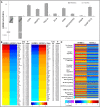
Molecular signatures were intrinsically different between HKMEC and HUVECs. (A) Real–time quantitative PCR on selective genes (bold) verified similar trends in downregulated genes MMP2 and RGS5 and upregulated genes CD34, ANGPT2, CXCL12, DLL4, JAM2, KDR, PDGFB, and PV1. (B) Ingenuity pathway analysis for angiogenesis function with significantly changed gene expression comparing HKMECs versus HUVECs. Red indicates upregulation, and blue indicates downregulation. (C) Heat map of canonical pathway analysis of HKMECs compared with HUVECs showing the most significantly differentiated genes that belong to specific ontologies.
Figure 5.
Figure 5.
Human kidney peritubular microvessels were reconstructed in vitro. (A and B) Schematic diagram of 3D MPS set up with (A) 3D views and (B) cross-sections. (C) Example of kidney microvessel networks generated in a 3D MPS. Red indicates CD31, and blue indicates nuclei. (D) z-Stack projection of confocal image of engineered human kidney microvessel in xy plane (left panel) and cross-sectional yz plane at the dashed line (right panel). Red indicates CD31, green indicates vWF, and blue indicates nuclei. Scale bar, 50 μm. (E) z-Stack projection of confocal image of human kidney microvessels at a junction of the network. Red indicates VE Cadherin, and blue indicates nuclei. Scale bar, 25 μm. (F) Quantification of the amount of vWF per area for both HKMECs and HUVECs and in both 2D static culture and 3D flow-based microvessel cultures. (G) Quantification of SI (4πA/P2) for HKMECs in 3D microvessels and 2D static cultures and HUVECs in 3D microvessels. (H) Immunofluorescence image of a cryosectioned HKMEC vessel network (thickness of 7 μm). Red indicates CD31, green indicates collagen IV, and blue indicates nuclei. Scale bar, 100 μm. (I) Fluorescence image of 40-kD FITC-dextran perfused through the HKMECs after 1 minute of perfusion. Arrows show good barrier, and stars show focal leakage. Scale bar, 100 μm.
Figure 6.
Figure 6.
Reconstructed human kidney peritubular microvessels were fenestrated in vitro. (A) z-Stack projection of confocal image of engineered human kidney microvessel at a junction of vessel network (left panel) and in a zoomed view (right panel). Red indicates F-actin, green indicates PV1, and blue indicates nuclei. (B–D) Transmission electron microscopy reveals the ultrastructure of HKMECs microvessels containing proper junctions (red arrows) formed at cell-cell contacts between (B and C) two adjacent cells C1 and C2 and (B and D) numerous fenestrae (black arrows) throughout the peripheral regions.

References

    1. Himmelfarb J, Ikizler TA: Acute kidney injury: Changing lexicography, definitions, and epidemiology. Kidney Int 71: 971–976, 2007 - PubMed
    1. Coresh J, Selvin E, Stevens LA, Manzi J, Kusek JW, Eggers P, Van Lente F, Levey AS: Prevalence of chronic kidney disease in the United States. JAMA 298: 2038–2047, 2007 - PubMed
    1. Chawla LS, Eggers PW, Star RA, Kimmel PL: Acute kidney injury and chronic kidney disease as interconnected syndromes. N Engl J Med 371: 58–66, 2014 - PMC - PubMed
    1. Brenner BM, Falchuk KH, Keimowitz RI, Berliner RW: The relationship between peritubular capillary protein concentration and fluid reabsorption by the renal proximal tubule. J Clin Invest 48: 1519–1531, 1969 - PMC - PubMed
    1. LeBrie SJ: Renal peritubular capillary permeability to macromolecules. Am J Physiol 213: 1225–1232, 1967 - PubMed

Publication types