. 2016 Jun;23(6):1038-48.
doi: 10.1038/cdd.2015.157. Epub 2015 Dec 11.

Cdk12 is essential for embryonic development and the maintenance of genomic stability

Affiliations

Cdk12 is essential for embryonic development and the maintenance of genomic stability

H-C Juan et al. Cell Death Differ. 2016 Jun.

Abstract

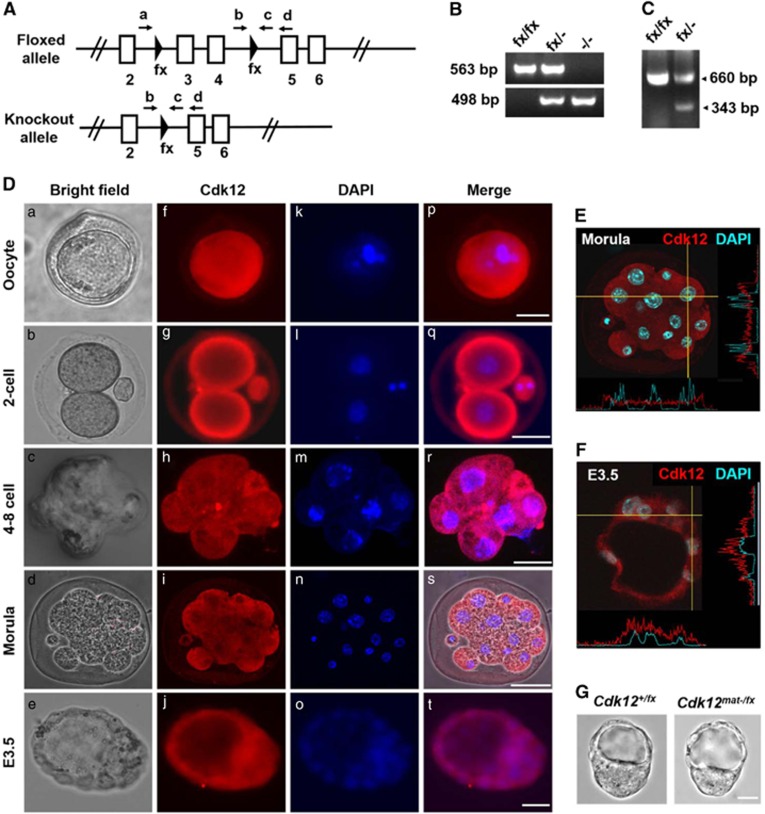
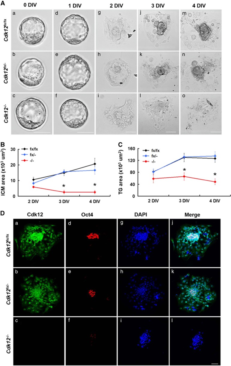
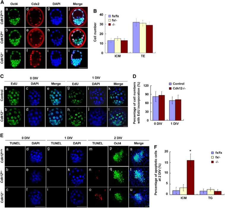
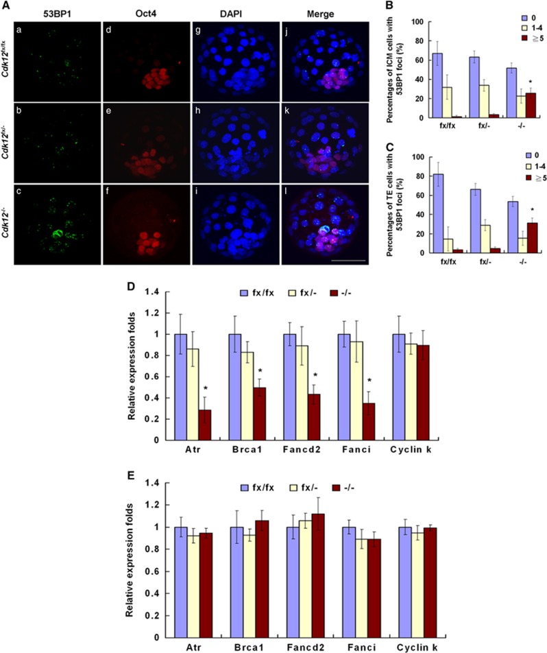
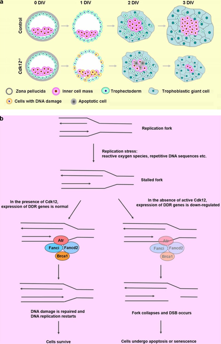
The maintenance of genomic integrity during early embryonic development is important in order to ensure the proper development of the embryo. Studies from cultured cells have demonstrated that cyclin-dependent kinase 12 (Cdk12) is a multifunctional protein that maintains genomic stability and the pluripotency of embryonic stem cells. Perturbation of its functions is also known to be associated with pathogenesis and drug resistance in human cancers. However, the biological significance of Cdk12 in vivo is unclear. Here we bred mice that are deficient in Cdk12 and demonstrated that Cdk12 depletion leads to embryonic lethality shortly after implantation. We also used an in vitro culture system of blastocysts to examine the molecular mechanisms associated with the embryonic lethality of Cdk12-deficient embryos. Cdk12(-/-) blastocysts fail to undergo outgrowth of the inner cell mass because of an increase in the apoptosis of these cells. Spontaneous DNA damage was revealed by an increase in 53BP1 foci among cells cultured from Cdk12(-/-) embryos. Furthermore, the expression levels of various DNA damage response genes, namely Atr, Brca1, Fanci and Fancd2, are reduced in Cdk12(-/-) embryos. These findings indicate that Cdk12 is important for the correct expression of some DNA damage response genes and indirectly has an influence on the efficiency of DNA repair. Our report also highlights that DNA breaks occurring during DNA replication are frequent in mouse embryonic cells and repair of such damage is critical to the successful development of mouse embryos.


Figures

Figure 1
Figure 1
Generation of Cdk12 knockout mice. (A) A schematic diagram of Cdk12 floxed and knockout alleles is shown. Arrows represent the locations of the genotyping primers. (B) E3.5 blastocysts were collected after the Cdk12fx/− intercross and genotyped by nested-PCR. The bands representing floxed and knockout alleles are 563 and 498 bp, respectively. (C) mRNA was extracted from Cdk12fx/fx and Cdk12fx/−E14.5 embryos and transcripts derived from the floxed and knockout Cdk12 allele were detected by RT-PCR. The bands representing the normal transcript and the knockout transcript are 660 and 343 bp, respectively. (D) Oocyte and embryos were examined using Cdk12 antibody. Nuclei are indicated by DAPI staining (E and F) Intensity profiles of Cdk12 expression in the cytoplasm and the nucleus along the indicated lines of interest were obtained by confocal sectioned images that reveal the cellular localization of Cdk12 protein at morula stage and in E3.5 embryos. (G) Photographs of E3.5 embryos derived from mating between Zp3-Cre+/0; Cdk12fx/+ female mice with Cdk12fx/fx male mice. Scale bar, 20 μm
Figure 2
Figure 2
Cdk12 deficiency results in impaired outgrowth of the blastocyst. (A) Blastocysts were collected by Cdk12fx/− intercrossing and cultured in vitro for 4 days. At the indicated day, embryos were photographed under bright-field conditions. The surface areas of the ICM (B) and TG (C) outgrowth at daily intervals are showed as mean±S.E.M. *P<0.05, by one-way ANOVA. Cdk12fx/fx, n=10; Cdk12fx/−, n=21; Cdk12−/−, n=8. (D) E3.5 embryos were cultured in vitro for 4 days and then immunostained with anti-Cdk12 and anti-Oct4 antibodies. Nuclei are marked by DAPI staining. Scale bar, 50 μm
Figure 3
Figure 3
Cdk12 deficiency induces apoptosis of ICM cells. (A) E3.5 embryos were collected and immunostained with anti-Oct4 and anti-Cdx2 antibodies. Nuclei were indicated by DAPI staining. Individual confocal sections of whole-mount embryos are shown. (B) The cell numbers of ICM and TE lineages in E3.5 embryos were quantified. Cdk12fx/fx, n=5; Cdk12fx/−, n=8; Cdk12−/−, n=4. (C) Cell proliferation of 0 and 1 DIV embryos was measured using a 45-min EdU incorporation assay. (D) Quantitative results of (C). At 0 DIV, control, n=8; Cdk12−/−, n=3. At 1 DIV, control, n=11; Cdk12−/−, n=3. (E) Apoptotic cells were detected by TUNEL assay at 0, 1 and 2 DIV. (F) Quantitative results of (E) at 2 DIV. Cdk12fx/fx, n=3; Cdk12fx/, n=5; Cdk12−/−, n=3. Results are shown as mean±S.E.M. *P<0.05, by one-way ANOVA. Scale bar, 20 μm
Figure 4
Figure 4
Cdk12 deficiency leads to an increase in the number of DSBs and reduces the expression of DDR genes. (A) Blastocysts were cultured for 1 day and then immunostained with anti-53BP1 and anti-Oct4 antibodies. Nuclei were marked by DAPI staining. The percentage of cell numbers with 53BP1 foci in ICM cells (B) and TE cells (C) were quantified. ICM and TE cells were defined as Oct4+ and Oct4, respectively. Cdk12fx/fx, n=4; Cdk12fx/−, n=6; Cdk12−/−, n=5. Relative mRNA levels of the indicated genes were determined by real-time PCR at 1 DIV ((D), Cdk12fx/fx, n=4; Cdk12fx/−, n=8; Cdk12−/−, n=5) and morula stage ((E), Cdk12fx/fx, n=6; Cdk12fx/, n=11; Cdk12−/−, n=4). Results are shown as mean±S.E.M. *P<0.05, by one-way ANOVA
Figure 5
Figure 5
A schematic diagram demonstrates that Cdk12 is required to sustain the viability of the ICM and TE through the maintenance of genome integrity. (a) In the absence of active Cdk12, blastocysts failed to outgrow because of an increase in DNA DSBs in ICM and TE cells. (b) Cdk12 has a crucial role in the regulation of expression of DDR genes that preserve the replication fork structure and repair lesions. In the absence of Cdk12, the fork collapses and as a consequence of this DSBs occur

References

    1. Papaioannou VE. Lineage analysis of inner cell mass and trophectoderm using microsurgically reconstituted mouse blastocysts. J Embryol Exp Morphol 1982; 68: 199–209. - PubMed
    1. de Waard H, de Wit J, Gorgels TG, van den Aardweg G, Andressoo JO, Vermeij M et al. Cell type-specific hypersensitivity to oxidative damage in CSB and XPA mice. DNA Repair (Amst) 2003; 2: 13–25. - PubMed
    1. Van Sloun PP, Jansen JG, Weeda G, Mullenders LH, van Zeeland AA, Lohman PH et al. The role of nucleotide excision repair in protecting embryonic stem cells from genotoxic effects of UV-induced DNA damage. Nucleic Acids Res 1999; 27: 3276–3282. - PMC - PubMed
    1. Khanna KK, Jackson SP. DNA double-strand breaks: signaling, repair and the cancer connection. Nat Genet 2001; 27: 247–254. - PubMed
    1. Li X, Heyer WD. Homologous recombination in DNA repair and DNA damage tolerance. Cell Res 2008; 18: 99–113. - PMC - PubMed

Publication types

MeSH terms

Substances