. 2016 May;18(5):639-48.
doi: 10.1093/neuonc/nov292. Epub 2015 Dec 11.

MiR-138 exerts anti-glioma efficacy by targeting immune checkpoints

Affiliations

MiR-138 exerts anti-glioma efficacy by targeting immune checkpoints

Jun Wei et al. Neuro Oncol. 2016 May.

Abstract

Background: Antibody therapeutic targeting of the immune checkpoints cytotoxic T-lymphocyte-associated molecule 4 (CTLA-4) and programmed cell death 1 (PD-1) has demonstrated marked tumor regression in clinical trials. MicroRNAs (miRNAs) can modulate multiple gene transcripts including possibly more than one immune checkpoint and could be exploited as immune therapeutics.

Methods: Using online miRNA targeting prediction algorithms, we searched for miRNAs that were predicted to target both PD-1 and CTLA-4. MiR-138 emerged as a leading candidate. The effects of miR-138 on CTLA-4 and PD-1 expression and function in T cells were determined and the therapeutic effect of intravenous administration of miR-138 was investigated in both immune-competent and -incompetent murine models of GL261 glioma.

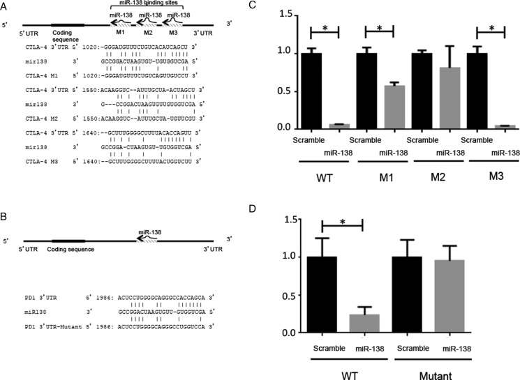
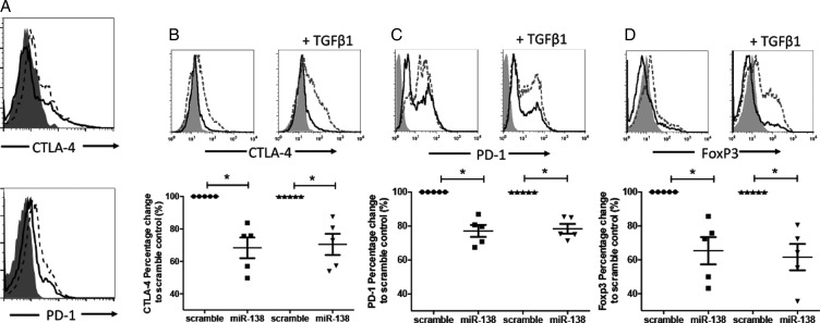
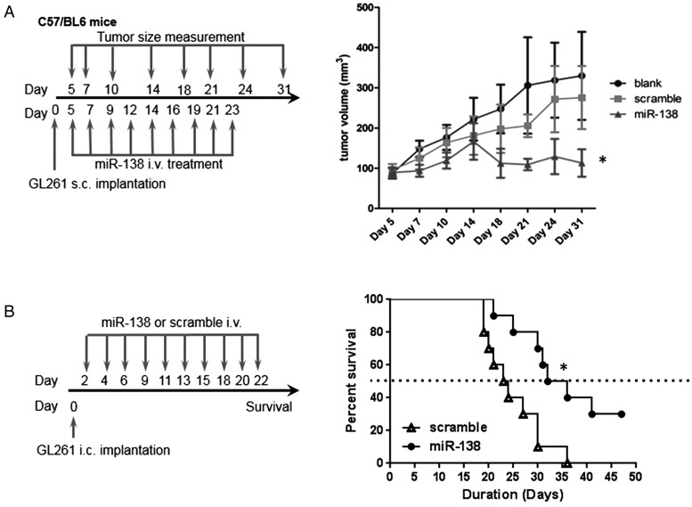
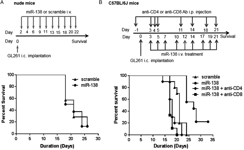
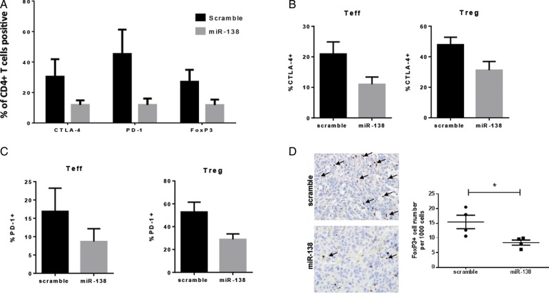
Results: Target binding algorithms predicted that miR-138 could bind the 3' untranslated regions of CTLA-4 and PD-1, which was confirmed with luciferase expression assays. Transfection of human CD4+ T cells with miR-138 suppressed expression of CTLA-4, PD-1, and Forkhead box protein 3 (FoxP3) in transfected human CD4+ T cells. In vivo miR-138 treatment of GL261 gliomas in immune-competent mice demonstrated marked tumor regression, a 43% increase in median survival time (P = .011), and an associated decrease in intratumoral FoxP3+ regulatory T cells, CTLA-4, and PD-1 expression. This treatment effect was lost in nude immune-incompetent mice and with depletion of CD4+ or CD8+ T cells, and miR-138 had no suppressive effect on glioma cells when treated directly at physiological in vivo doses.

Conclusions: MiR-138 exerts anti-glioma efficacy by targeting immune checkpoints which may have rapid translational potential as a novel immunotherapeutic agent.

Keywords: CTLA-4; PD-1; glioblastoma; miR-138; microRNAs.


Figures

Fig. 1.
Fig. 1.
MiR-138 binds the 3′ UTRs of CTLA-4 and PD-1. (A) The 3 predicted miR-138 binding sites in the 3′ UTR of CTLA-4 are noted with their sequences. The mutational alterations are noted for each luciferase expression construct. (B) The miR-138 binding site sequence of the 3′ UTR of PD-1 and its mutant in the luciferase expression construct are noted. (C) The relative luciferase expression in HeLa cells transfected with miR-138 pre-miR versus scramble control pre-miR is shown. A significant decrease in luciferase expression is seen when the cells are cotransfected with a reporter plasmid containing the wild-type 3′ UTR of CTLA-4 and miR-138 pre-miR. The M3 mutation appears to have no effect, whereas the M1 mutation reduces the modulation of the miR-138 pre-miR, and the M2 mutation completely abolishes the significant effect of the miR-138 pre-miR, implying that the M2 binding site is the most critical. *P < .05. (D) A significant decrease in luciferase expression is seen when the cells are cotransfected with a reporter plasmid containing the wild-type 3′ UTR of PD-1 and miR-138, whereas this difference is abolished when the mutant 3′ UTR reporter plasmid is evaluated. *P < .05.
Fig. 2.
Fig. 2.
MiR-138 inhibits human checkpoint expression in Tregs. (A) CD4+CD25+ Tregs isolated from healthy donor murine splenocytes by fluorescence activated cell sorting were observed to have both CTLA-4 and PD-1 downregulation when transfected with miR-138 compared with scramble control (n = 4). Summary data dot plots are shown below the T-cell histograms. (B–D) Healthy donor human CD4+ T cells were stimulated by anti-CD3/CD28 antibodies for 48 h in the absence or presence of TGF-β to induce CTLA-4, PD-1, and FoxP3+ Tregs and subsequently transfected with miR-138 pre-miR or scramble control. MiR-138 downmodulated the expression of (B) CTLA-4, (C) PD-1, and (D) FoxP3 in CD4+ T cells. The solid-gray histogram is the isotype control; the dashed-line histogram is the scramble control; and the solid black-line histogram is miR-138. The percent change refers to the frequency of the positive cell population. Representative histograms are shown as above, and summary data dot plots are shown below the T-cell histograms, in which each dot represents the analysis of one human donor's peripheral CD4+ T cells (n = 5). *P < .05.
Fig. 3.
Fig. 3.
MiR-138 suppresses GL261 tumors in a syngeneic mouse model. (A) The treatment schema and the volumes of subcutaneous GL261 tumors in C57BL/6J mice treated intravenously with either miR-138 or scramble control, or left untreated, starting on day 5 (n = 10/group/experiment). The figure is the result of a single experiment but was repeated with identical results. In the miR-138 group, *P < .01 compared with both the untreated and scramble control tumors. Standard deviations are shown. Arrows indicate days of treatment and tumor size measurements. (B) Treatment schema and graph of the Kaplan–Meier estimate demonstrating survival of C57BL/6J mice with intracranial GL261 tumors that were treated intravenously with miR-138 versus scramble control. MiR-138 treatment results in a marked increase in median survival time relative to that in the scramble control group (33.5 d and 23.5 d, respectively; P = .01). This experiment was repeated with similar results.
Fig. 4.
Fig. 4.
The therapeutic effect of miR-138 is immune mediated. (A) Treatment schema and graph of the Kaplan–Meier survival estimate demonstrating the lack of therapeutic effect of miR-138 in nude mice with intracerebral (i.c.) GL261 gliomas (n = 8 in scramble control group, n = 7 in miR-138 treatment group, P = .87). (B) Treatment schema and graph of the Kaplan–Meier survival estimate demonstrating that the therapeutic efficacy of miR-138 is ablated in the setting of CD4 or CD8 T-cell depletions in C57BL/6J mice with i.c. GL261. MiR-138 treatment (n = 9) resulted in a median survival of 27 days in comparison with the scramble control (n = 10), which had a median survival of 21 days (P = .0001). When either CD4 (n = 9) or CD8 (n = 10) T cells were depleted, the median survival was 17 or 18 days, respectively. Ab, antibody.
Fig. 5.
Fig. 5.
MiR-138 downregulates immune checkpoints in the tumor microenvironment. (A) Ex vivo analysis of the expression of PD-1 and CTLA-4 on CD4+ cells from the tumor microenvironment of miR-138- and scramble-control-treated C57BL/6J mice implanted with subcutaneous GL261 tumors, as measured by flow cytometry. (B) Bar graphs showing downregulation of CTLA-4 expression on glioma-infiltrating effector T cells (Teff, CD4+FoxP3−) and glioma-infiltrating regulatory T cells (Treg, CD4+FoxP3+) in C57BL/6J mice harboring subcutaneous GL261 treated with miR-138 (n = 5/group). (C) Bar graphs showing PD-1 expression on the same cells as panel (B). (D) Photomicrograph (400×) showing FoxP3-expressing lymphocytes in GL261 tumors treated with scramble control and in those treated with miR-138. The dot plot graph summarizes the number of FoxP3+ cells per 1000 observed in scramble- and miR-138-treated intracerebral GL261 tumors. *P < .05.

Comment in

Similar articles

Cited by

References

    1. Stupp R, Mason WP, van den Bent MJ et al. . Radiotherapy plus concomitant and adjuvant temozolomide for glioblastoma. N Engl J Med. 2005;352(10):987–996. - PubMed
    1. Wen PY, Kesari S. Malignant gliomas in adults. N Engl J Med. 2008;359(5):492–507. - PubMed
    1. Bartel DP. MicroRNAs: genomics, biogenesis, mechanism, and function. Cell. 2004;116(2):281–297. - PubMed
    1. Chan JA, Krichevsky AM, Kosik KS. MicroRNA-21 is an antiapoptotic factor in human glioblastoma cells. Cancer Res. 2005;65(14):6029–6033. - PubMed
    1. Ciafre SA, Galardi S, Mangiola A et al. . Extensive modulation of a set of microRNAs in primary glioblastoma. Biochem Biophys Res Commun. 2005;334(4):1351–1358. - PubMed

Publication types

MeSH terms