

Structural and Functional Brain Remodeling during Pregnancy with Diffusion Tensor MRI and Resting-State Functional MRI
- PMID: 26658306
- PMCID: PMC4675543
- DOI: 10.1371/journal.pone.0144328
Structural and Functional Brain Remodeling during Pregnancy with Diffusion Tensor MRI and Resting-State Functional MRI
Abstract
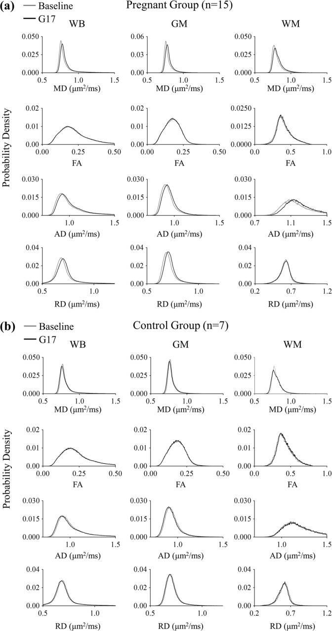
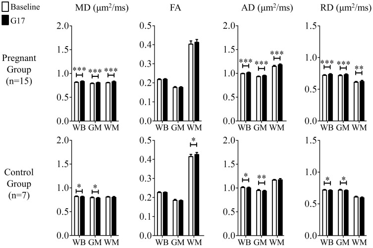
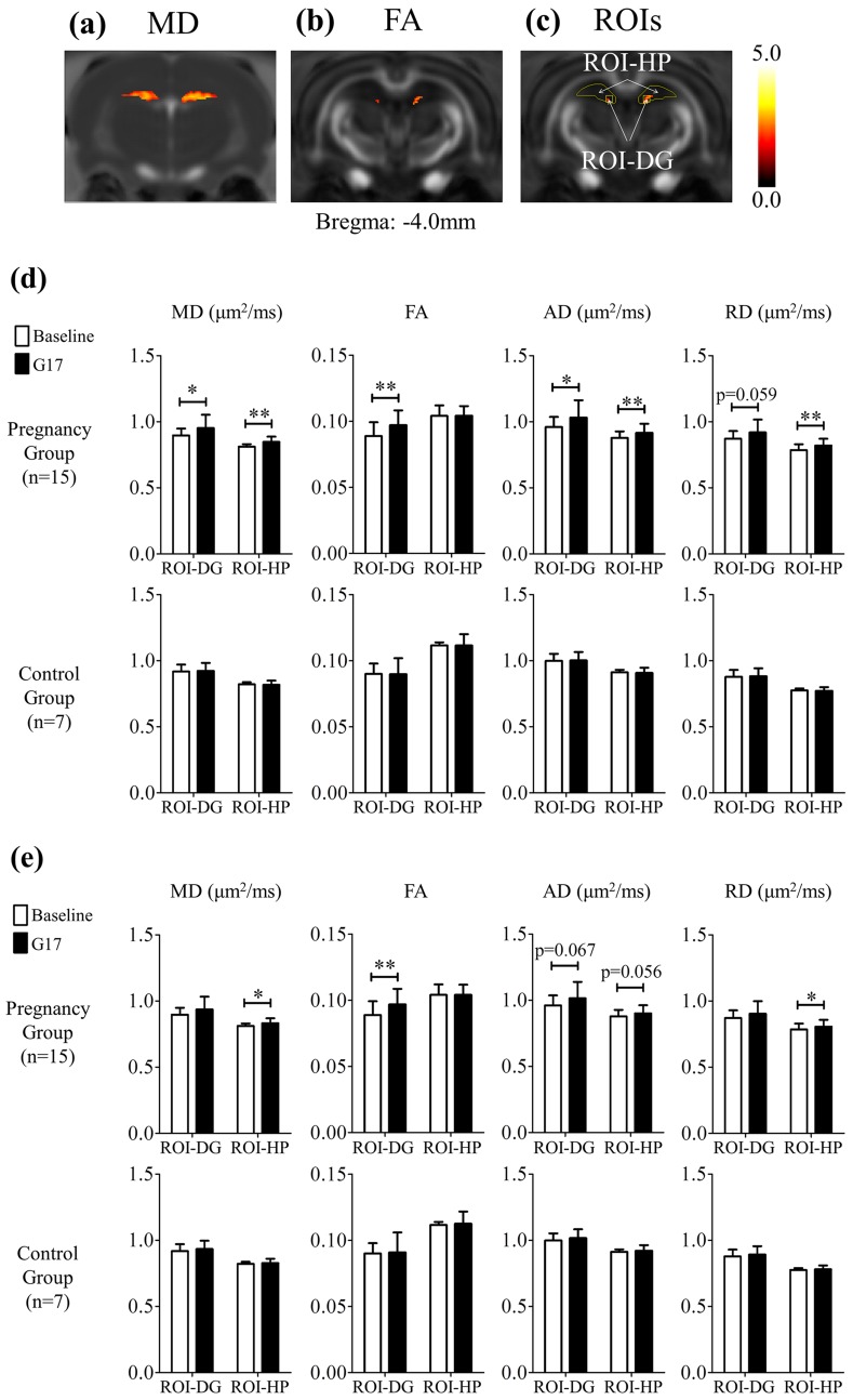
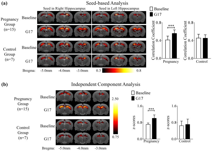
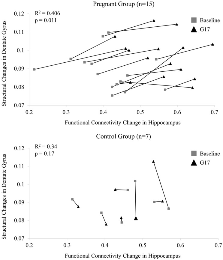
Although pregnancy-induced hormonal changes have been shown to alter the brain at the neuronal level, the exact effects of pregnancy on brain at the tissue level remain unclear. In this study, diffusion tensor imaging (DTI) and resting-state functional MRI (rsfMRI) were employed to investigate and document the effects of pregnancy on the structure and function of the brain tissues. Fifteen Sprague-Dawley female rats were longitudinally studied at three days before mating (baseline) and seventeen days after mating (G17). G17 is equivalent to the early stage of the third trimester in humans. Seven age-matched nulliparous female rats served as non-pregnant controls and were scanned at the same time-points. For DTI, diffusivity was found to generally increase in the whole brain during pregnancy, indicating structural changes at microscopic levels that facilitated water molecular movement. Regionally, mean diffusivity increased more pronouncedly in the dorsal hippocampus while fractional anisotropy in the dorsal dentate gyrus increased significantly during pregnancy. For rsfMRI, bilateral functional connectivity in the hippocampus increased significantly during pregnancy. Moreover, fractional anisotropy increase in the dentate gyrus appeared to correlate with the bilateral functional connectivity increase in the hippocampus. These findings revealed tissue structural modifications in the whole brain during pregnancy, and that the hippocampus was structurally and functionally remodeled in a more marked manner.
Conflict of interest statement
Figures
Similar articles
-
Diffusion tensor imaging during recovery from severe traumatic brain injury and relation to clinical outcome: a longitudinal study.Brain. 2008 Feb;131(Pt 2):559-72. doi: 10.1093/brain/awm294. Epub 2007 Dec 14. Brain. 2008. PMID: 18083753
-
New insights into the developing rabbit brain using diffusion tensor tractography and generalized q-sampling MRI.PLoS One. 2015 Mar 23;10(3):e0119932. doi: 10.1371/journal.pone.0119932. eCollection 2015. PLoS One. 2015. PMID: 25798595 Free PMC article.
-
Longitudinal metabolic changes in the hippocampus and thalamus of the maternal brain revealed by proton magnetic resonance spectroscopy.Neurosci Lett. 2013 Oct 11;553:170-5. doi: 10.1016/j.neulet.2013.08.041. Epub 2013 Aug 27. Neurosci Lett. 2013. PMID: 23994391
-
Structural abnormalities in bipolar euthymia: a multicontrast molecular diffusion imaging study.Biol Psychiatry. 2014 Aug 1;76(3):239-48. doi: 10.1016/j.biopsych.2013.09.027. Epub 2013 Oct 4. Biol Psychiatry. 2014. PMID: 24199669
-
Neuroimaging effects of prenatal alcohol exposure on the developing human brain: a magnetic resonance imaging review.Acta Neuropsychiatr. 2015 Oct;27(5):251-69. doi: 10.1017/neu.2015.12. Epub 2015 Mar 17. Acta Neuropsychiatr. 2015. PMID: 25780875 Review.
Cited by
-
Peripartum sertraline impacts maternal neurobehavioral and neurodegenerative mechanisms in pregnant and postpartum mice.Mol Psychiatry. 2025 Jul 18. doi: 10.1038/s41380-025-03094-x. Online ahead of print. Mol Psychiatry. 2025. PMID: 40681843
-
Sex Hormones, Sleep, and Memory: Interrelationships Across the Adult Female Lifespan.Front Aging Neurosci. 2022 Jul 14;14:800278. doi: 10.3389/fnagi.2022.800278. eCollection 2022. Front Aging Neurosci. 2022. PMID: 35912083 Free PMC article. Review.
-
The transition to motherhood: linking hormones, brain and behaviour.Nat Rev Neurosci. 2023 Oct;24(10):605-619. doi: 10.1038/s41583-023-00733-6. Epub 2023 Aug 23. Nat Rev Neurosci. 2023. PMID: 37612425 Review.
-
Disrupted Spontaneous Neural Activity Related to Cognitive Impairment in Postpartum Women.Front Psychol. 2018 May 3;9:624. doi: 10.3389/fpsyg.2018.00624. eCollection 2018. Front Psychol. 2018. PMID: 29774003 Free PMC article.
-
Structural Alterations in a Rat Model of Short-Term Conductive Hearing Loss Are Associated With Reduced Resting State Functional Connectivity.Front Syst Neurosci. 2021 Aug 12;15:655172. doi: 10.3389/fnsys.2021.655172. eCollection 2021. Front Syst Neurosci. 2021. PMID: 34456689 Free PMC article.
References
-
- Kinsley CH, Lambert KG. The maternal brain. Scientific American. 2006;294(1):72–9. Epub 2006/02/14. . - PubMed
-
- Moya J, Phillips L, Sanford J, Wooton M, Gregg A, Schuda L. A review of physiological and behavioral changes during pregnancy and lactation: Potential exposure factors and data gaps. Journal of exposure science & environmental epidemiology. 2014;24(5):449–58. Epub 2014/01/16. 10.1038/jes.2013.92 . - DOI - PubMed
-
- Brunton PJ, Russell JA. Hypothalamic-pituitary-adrenal responses to centrally administered orexin-A are suppressed in pregnant rats. Journal of neuroendocrinology. 2003;15(7):633–7. Epub 2003/06/06. . - PubMed
Publication types
MeSH terms
LinkOut - more resources
Full Text Sources
Other Literature Sources
