

High-Magnitude and/or High-Frequency Mechanical Strain Promotes Peripapillary Scleral Myofibroblast Differentiation
- PMID: 26658503
- PMCID: PMC4682490
- DOI: 10.1167/iovs.15-17848
High-Magnitude and/or High-Frequency Mechanical Strain Promotes Peripapillary Scleral Myofibroblast Differentiation
Abstract
Purpose: To determine the effects of altered mechanical strain on human peripapillary scleral (ppSc) fibroblast-to-myofibroblast differentiation.
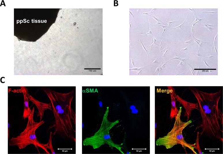
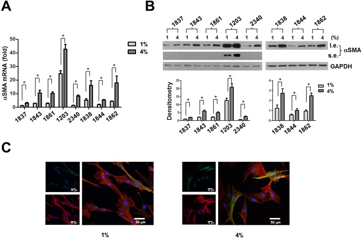
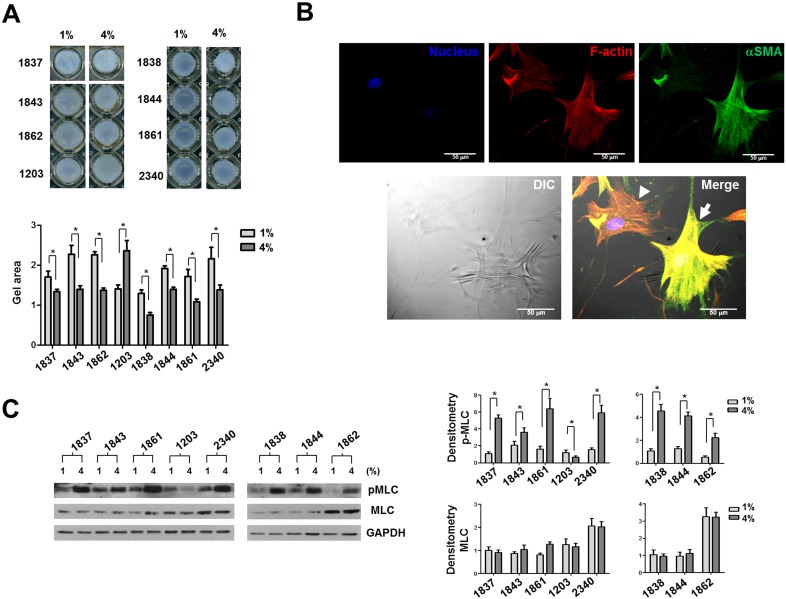
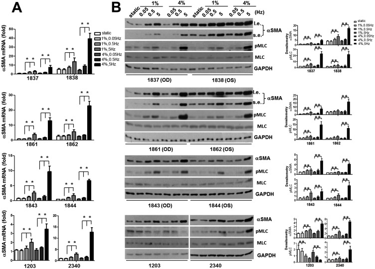
Methods: Eight human ppSc fibroblast cultures were isolated from three paired eyes and two unilateral eyes of five donors using an explant approach. Human ppSc fibroblast isolates were subjected to 1% and 4% cyclic strain at 0.05 to 5 Hz for 24 hours. Levels of α smooth muscle actin (αSMA) mRNA and protein were determined by real-time PCR and immunoblot. Incorporation of αSMA into actin stress fibers was evaluated by confocal immunofluorescent microscopy. Myofibroblast contractility was measured by fibroblast-populated three-dimensional collagen gel contraction assay and phosphorylation of myosin light chain (MLC20).
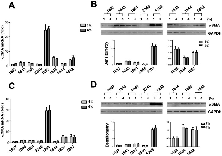
Results: Human ppSc fibroblasts contained 6% to 47% fully differentiated myofibroblasts before strain application; 4% cyclic strain increased αSMA mRNA and protein expression in ppSc fibroblasts compared with 1% strain applied at 5 Hz, but not at lower frequencies. Seven of eight ppSc fibroblast isolates responded to high-magnitude and high-frequency strain with increased cellular contractility and increased MLC20 phosphorylation. In addition, increasing strain frequency promoted αSMA expression in ppSc fibroblasts under both 1% and 4% strain conditions.
Conclusions: High-magnitude and/or high-frequency mechanical strain promotes differentiation of human ppSc fibroblasts into contractile myofibroblasts, a fibroblast phenotypic change known to be key to tissue injury-repair responses. These findings suggest that the cellular constituent of ppSc may play an important role in the regulation of optic nerve head biomechanics in response to injurious IOP fluctuations.
Figures
Similar articles
-
The Role of the RhoA/ROCK Signaling Pathway in Mechanical Strain-Induced Scleral Myofibroblast Differentiation.Invest Ophthalmol Vis Sci. 2018 Jul 2;59(8):3619-3629. doi: 10.1167/iovs.17-23580. Invest Ophthalmol Vis Sci. 2018. PMID: 30029249
-
Dasatinib inhibits peripapillary scleral myofibroblast differentiation.Exp Eye Res. 2020 May;194:107999. doi: 10.1016/j.exer.2020.107999. Epub 2020 Mar 13. Exp Eye Res. 2020. PMID: 32179077 Free PMC article.
-
Wnt/β-catenin pathway forms a negative feedback loop during TGF-β1 induced human normal skin fibroblast-to-myofibroblast transition.J Dermatol Sci. 2012 Jan;65(1):38-49. doi: 10.1016/j.jdermsci.2011.09.012. Epub 2011 Oct 17. J Dermatol Sci. 2012. PMID: 22041457
-
Cyclic mechanical stretch reduces myofibroblast differentiation of primary lung fibroblasts.Biochem Biophys Res Commun. 2011 Jan 7;404(1):23-7. doi: 10.1016/j.bbrc.2010.11.033. Epub 2010 Nov 20. Biochem Biophys Res Commun. 2011. PMID: 21094632
-
A model for positive feedback control of the transformation of fibroblasts to myofibroblasts.Prog Biophys Mol Biol. 2019 Jul;144:30-40. doi: 10.1016/j.pbiomolbio.2018.08.004. Epub 2018 Aug 30. Prog Biophys Mol Biol. 2019. PMID: 30174171 Free PMC article. Review.
Cited by
-
Cell-Matrix Interactions in the Eye: From Cornea to Choroid.Cells. 2021 Mar 20;10(3):687. doi: 10.3390/cells10030687. Cells. 2021. PMID: 33804633 Free PMC article. Review.
-
Rho-Kinase Inhibition Reduces Myofibroblast Differentiation and Proliferation of Scleral Fibroblasts Induced by Transforming Growth Factor β and Experimental Glaucoma.Transl Vis Sci Technol. 2018 Nov 14;7(6):6. doi: 10.1167/tvst.7.6.6. eCollection 2018 Nov. Transl Vis Sci Technol. 2018. PMID: 30479877 Free PMC article.
-
Lamina cribrosa in glaucoma.Curr Opin Ophthalmol. 2017 Mar;28(2):113-119. doi: 10.1097/ICU.0000000000000354. Curr Opin Ophthalmol. 2017. PMID: 27898470 Free PMC article. Review.
-
Racial Differences in the Rate of Change in Anterior Lamina Cribrosa Surface Depth in the African Descent and Glaucoma Evaluation Study.Invest Ophthalmol Vis Sci. 2021 Apr 1;62(4):12. doi: 10.1167/iovs.62.4.12. Invest Ophthalmol Vis Sci. 2021. PMID: 33844828 Free PMC article.
-
Analysis of 2-dimensional regional differences in the peripapillary scleral fibroblast cytoskeleton of normotensive and hypertensive mouse eyes.Sci Rep. 2025 Jul 1;15(1):21207. doi: 10.1038/s41598-025-08251-4. Sci Rep. 2025. PMID: 40595239 Free PMC article.
References
-
- Burgoyne CF,, Downs JC,, Bellezza AJ,, Suh JK,, Hart RT. The optic nerve head as a biomechanical structure: a new paradigm for understanding the role of IOP-related stress and strain in the pathophysiology of glaucomatous optic nerve head damage. Prog Retin Eye Res. 2005; 24: 39–73. - PubMed
-
- Campbell IC,, Coudrillier B, Ross Ethier C. Biomechanics of the posterior eye: a critical role in health and disease. J Biomech Eng. 2014; 136: 021005. - PubMed
-
- Sigal IA,, Ethier CR. Biomechanics of the optic nerve head. Exp Eye Res. 2009; 88: 799–807. - PubMed
Publication types
MeSH terms
Substances
Grants and funding
LinkOut - more resources
Full Text Sources
Other Literature Sources
