. 2015 Dec;56(13):7847-58.
doi: 10.1167/iovs.15-17278.

LIM Kinase, a Newly Identified Regulator of Presynaptic Remodeling by Rod Photoreceptors After Injury

LIM Kinase, a Newly Identified Regulator of Presynaptic Remodeling by Rod Photoreceptors After Injury

Weiwei Wang et al. Invest Ophthalmol Vis Sci. 2015 Dec.

Abstract

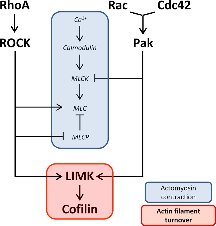
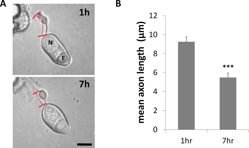
Purpose: Rod photoreceptors retract their axon terminals and develop neuritic sprouts in response to retinal detachment and reattachment, respectively. This study examines the role of LIM kinase (LIMK), a component of RhoA and Rac pathways, in the presynaptic structural remodeling of rod photoreceptors.

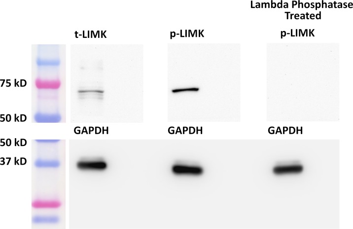
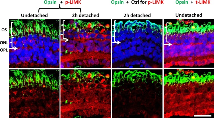
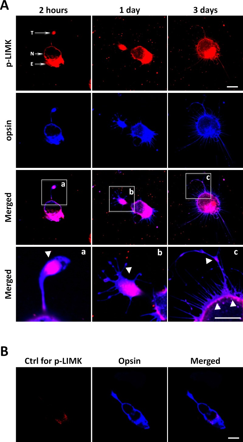
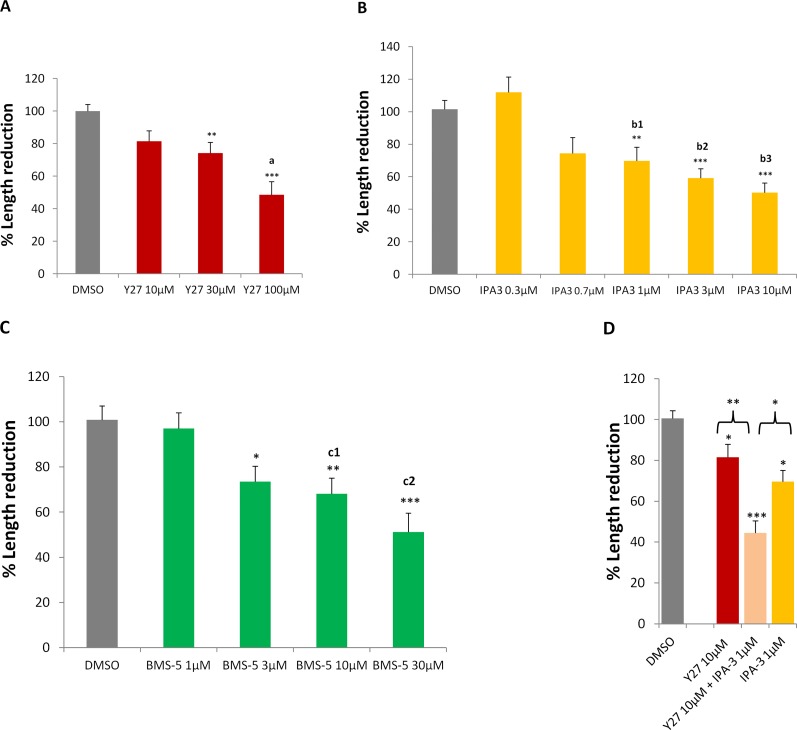
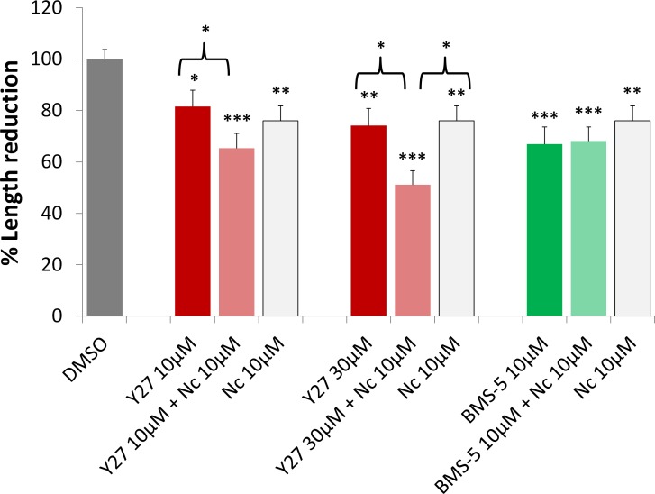
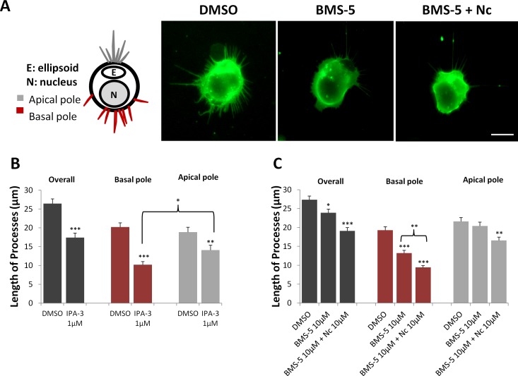
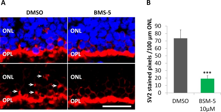
Methods: Phosphorylated LIMK (p-LIMK), the active form of LIMK, was examined in salamander retina with Western blot and confocal microscopy. Axon length within the first 7 hours and process growth after 3 days of culture were assessed in isolated rod photoreceptors treated with inhibitors of upstream regulators ROCK and p21-activated kinase (Pak) (Y27632 and IPA-3) and a direct LIMK inhibitor (BMS-5). Porcine retinal explants were also treated with BMS-5 and analyzed 24 hours after detachment. Because Ca2+ influx contributes to axonal retraction, L-type channels were blocked in some experiments with nicardipine.

Results: Phosphorylated LIMK is present in rod terminals during retraction and in newly formed processes. Axonal retraction over 7 hours was significantly reduced by inhibition of LIMK or its regulators, ROCK and Pak. Process growth was reduced by LIMK or Pak inhibition especially at the basal (axon-bearing) region of the rod cells. Combining Ca2+ channel and LIMK inhibition had no additional effect on retraction but did further inhibit sprouting after 3 days. In detached porcine retina, LIMK inhibition reduced rod axonal retraction and improved retinal morphology.

Conclusions: Thus structural remodeling, in the form of either axonal retraction or neuritic growth, requires LIMK activity. LIM kinase inhibition may have therapeutic potential for reducing pathologic rod terminal plasticity after retinal injury.


Figures

Figure 1
Figure 1
Diagram of proposed pathways involved in rod cell axonal and neuritic plasticity. Two activities, actomyosin contraction and actin filament turnover, are suggested to contribute to plasticity in the axon and axon terminal after injury. Both RhoA-ROCK and Cdc42/Rac-Pak pathways converge on LIMK, which promotes actin filament turnover through regulation of cofilin. However, while RhoA-ROCK promotes actomyosin contraction through activating MLC and inhibiting MLCP, Cdc42/Rac-PAK inhibits it through inhibiting MLCK. Ca2+-calmodulin promotes actomyosin contraction through activating MLCK. RhoA, Rac, and Cdc42 are Rho GTPases; ROCK, Rho kinase; Pak, p21-activated kinase; LIMK, LIM kinase; MLC, myosin light chain; MLCP, myosin light chain phosphatase; MLCK, myosin light chain kinase.
Figure 2
Figure 2
Active LIMK is present in salamander retina 2 hours after detachment. Antibodies against total-LIMK (t-LIMK, upper left lane) and phosphorylated LIMK (p-LIMK, upper middle lane) labeled a 72-kDa band. Lambda phosphatase–treated blot showed no band (upper right lane). GAPDH, from the same SDS-PAGE gel, served as an internal loading control (lower lanes). n = 3 animals, 6 retinal explants.
Figure 3
Figure 3
Confocal microscopy demonstrates distribution of LIMK in salamander retina. The outer segments (OS) of rod photoreceptors are labeled for opsin (green). The outer nuclear layer (ONL) contains the photoreceptor cell bodies; nuclei are labeled with propidium iodide (blue). The outer plexiform layer (OPL) is located immediately beneath the ONL as a thin layer where nuclei are absent. Normal and 2-hour detached retinas, in the first and second columns, respectively, are labeled for p-LIMK (red); third column, a labeling control, anti–p-LIMK is omitted in 2-hour detached retina; fourth column, normal retina labeled for t-LIMK (red). Labeling of t-LIMK occurs throughout the normal undetached retina; p-LIMK signal is present but spotty in the normal retina and more diffuse, with an apparent increase, in 2-hour detached retina. Optical sections, 1 μm. Scale bar: 50 μm. n = 3 animals, 2 retinal explants per animal (retina from 1 eye was detached and cultured for 2 hours, retina from other eye was not detached and fixed immediately); 9 cryosections per group (3 cryosections per retinal explant), 2 or 3 images per cryosection.
Figure 4
Figure 4
Active LIMK is present in isolated salamander rod cells in culture. (A) Rod photoreceptors, which had lost their outer segments, double labeled for p-LIMK (red) and rod opsin (blue) and examined with confocal microscopy. Labeling occurs throughout the cell except in the nucleus, both during retraction, which occurs during day 1, and after growth of new processes, seen at 3 days. (ac) Enlarged images show the presence of opsin in the cell membrane and p-LIMK in the cytosol and directly under the plasmalemma (arrowheads). (B) Control for p-LIMK labeling. Rod photoreceptor in 2-hour culture labeled with anti-opsin but without anti–p-LIMK, T, axon terminal; N, nucleus; E, ellipsoid, a collection of mitochondria, in the inner segment. Optical sections, 1 μm. Scale bars: 10 μm. n = 3 animals, 3 or 4 cultures per animal, 10 to 15 cells imaged per dish (∼39 cells per group).
Figure 5
Figure 5
Retraction of rod photoreceptor axon terminals begins during the first 7 hours in culture. (A) Representative isolated rod photoreceptor in culture after 1 and 7 hours. Axon length was measured from the base of the cell body to the tip of the axon terminal, shown with red bars. N, nucleus; E, ellipsoid. Scale bar: 10 μm. (B) Mean length of axons was reduced by approximately 40% over 7 hours. n = 3 animals, 6 cultures, 59 rod cells, ∼30 cells per group; ***P < 0.001; Student's t-test.
Figure 6
Figure 6
Retraction of the axon is reduced by inhibiting the LIMK pathway. (AC) Inhibiting ROCK or Pak, two different LIMK regulators, or inhibiting LIMK directly, with Y27632 (Y27), IPA-3, or BMS-5, respectively, reduced axonal retraction in a dose-dependent manner. (D) Concomitant inhibition of ROCK and Pak with 10 μM Y27632 and 1 μM IPA-3 resulted in significantly more effect on axonal retraction than either treatment alone. % Length reduction = (L1L7) ÷ L1 × 100%; L1, axon length in 1-hour culture; L7, axon length in 7-hour culture. All data were normalized to control (DMSO) group, which is set at 100%. (A) n = 9 animals, 22 cultures, 411 rod cells, ∼100 cells per group. (B) n = 7 animals, 26 cultures, 483 rod cells, ∼80 cells per group. (C) n = 5 animals, 34 cultures, 430 rod cells, ∼86 cells per group. (D) n = 9 animals, 40 cultures, 502 rod cells, ∼126 cells per group. One-way ANOVA, *P < 0.05, **P < 0.01, ***P < 0.001; post hoc (Tukey's test), a: Y27 10 vs. 100 μM, a < 0.05; b1–b3: IPA3 0.3 vs. 1, 3, or 10 μM, respectively, b1 < 0.01, b2 < 0.001, b3 < 0.001; c1, c2: BMS-5 1 vs. 10 or 30 μM, respectively, c1 < 0.05, c2 < 0.01.
Figure 7
Figure 7
Blockage of Ca2+ influx increases the effect of ROCK inhibition on axonal retraction over 7 hours, but does not affect LIMK inhibition. 10 μM nicardipine (Nc), an L-type Ca2+ channel blocker, reduced axonal retraction; 10 μM Nc plus Y27632 (Y27), a ROCK inhibitor, at 10 or 30 μM, had significantly more effect on reducing axonal retraction than Y27632 treatment alone. Ten μM Nc plus 10 μM BMS-5, a LIMK inhibitor, did not demonstrate an additional effect on retraction over Nc or BMS-5 treatment alone. Analysis of length was the same as in Figure 6. n = 10 animals, 64 cultures, 957 rod cells, ∼95 cells per group; *P < 0.05, **P < 0.01, ***P < 0.001; 1-way ANOVA between DMSO and (Y2710 μM, Y2710 μM + Nc10 μM, Nc 10μM), or (Y27 30 μM, Y27 30 μM + Nc 10 μM, Nc 10 μM), or (BMS-5 10 μM, BMS-5 10 μM + Nc 10 μM, Nc 10 μM).
Figure 8
Figure 8
Growth of processes by rod cells is reduced with Pak or LIMK inhibition; blockage of Ca2+ influx furthers the effect of LIMK inhibition. (A) Left: Cartoon of regional designations for rod cell processes. In the intact retina, axonal processes grow from the basal/nuclear pole, whereas calycal processes that surround the outer segment grow from the apical/ellipsoidal pole. Right: Representative rod cells with rod opsin labeling from DMSO control, 10 μM BMS-5-, and 10 μM Nc plus 10 μM BMS-5-treated cultures after 3 days. Cells in control, untreated cultures show the most growth. Scale bar: 10 μm. (B) Length of the longest process from three regions (overall, longest process regardless of region of origin; basal, from the cell surface close to the nucleus; apical, from the cell surface close to the ellipsoid) of each rod cell. Pak inhibition with 1 μM IPA-3 reduced process growth of rod cells after 3 days in culture from all regions, compared to control. In control cells, length of apical and basal processes is similar; in IPA-3-treated group, basal processes shortened significantly more than apical processes. n = 3 animals, 12 cultures, 360 rod cells, ∼180 cells per group; *P < 0.05, **P < 0.005. (C) LIM kinase inhibition with 10 μM BMS-5 reduced process growth overall and from the basal but not the apical pole. Blockage of Ca2+ influx increased the effect of LIMK inhibition and reduced process growth from basal and apical regions. n = 3 animals, 18 cultures, 540 rod cells, ∼180 cells per group; *P < 0.05, **P < 0.01, ***P < 0.001; (B) Student's t-test; (C) 1-way ANOVA.
Figure 9
Figure 9
Inhibition of LIMK reduces axonal retraction in the porcine retina maintained in vitro for 24 hours after detachment. (A) Representative control and treated retinas after 24 hours in vitro. The outer plexiform layer (OPL) with rod synaptic terminals is labeled for synaptic vesicles, SV2 (red). The outer nuclear layer (ONL) contains the photoreceptor cell bodies; nuclei are labeled with propidium iodide (blue). Left: With no treatment, SV2 label is present among the photoreceptor cell bodies (arrows) indicating that the rod terminals have retracted. Right: Treatment with 10 μM BMS-5, a LIMK inhibitor, reduced the level of SV2 in the ONL indicating an inhibition of retraction. Scale bar: 10 μm. (B) Retraction is measured using the area of SV2 label in the ONL. Treatment with 10 μM BMS-5 reduced SV2 labeling in the ONL significantly, compared to untreated retinas. Optical sections, 1 μm. n = 3 animals, 2 retinal explants per animal (1 explant with BMS-5 treatment from one eye, the other explant with DMSO as control from the other eye); 7 cryosections per group (2 or 3 cryosections per retinal explant), 3 or 4 images per cryosection. ***P < 0.001; Student's t-test.

Similar articles

Cited by

References

    1. Erickson PA,, Fisher SK,, Anderson DH,, Stern WH,, Borgula GA. Retinal detachment in the cat: the outer nuclear and outer plexiform layers. Invest Ophthalmol Vis Sci. 1983; 24: 927–942. - PubMed
    1. Lewis GP,, Linberg KA,, Fisher SK. Neurite outgrowth from bipolar and horizontal cells after experimental retinal detachment. Invest Ophthalmol Vis Sci. 1998; 39: 424–434. - PubMed
    1. Li ZY,, Kljavin IJ,, Milam AH. Rod photoreceptor neurite sprouting in retinitis pigmentosa. J Neurosci. 1995; 15: 5429–5438. - PMC - PubMed
    1. Milam AH,, Li ZY,, Cideciyan AV,, Jacobson SG. Clinicopathologic effects of the Q64ter rhodopsin mutation in retinitis pigmentosa. Invest Ophthalmol Vis Sci. 1996; 37: 753–765. - PubMed
    1. Lewis GP,, Charteris DG,, Sethi CS,, Fisher SK. Animal models of retinal detachment and reattachment: identifying cellular events that may affect visual recovery. Eye (Lond). 2002; 16: 375–387. - PubMed

Publication types