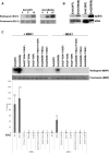
Intrinsically active variants of Erk oncogenically transform cells and disclose unexpected autophosphorylation capability that is independent of TEY phosphorylation
- PMID: 26658610
- PMCID: PMC4791124
- DOI: 10.1091/mbc.E15-07-0521
Intrinsically active variants of Erk oncogenically transform cells and disclose unexpected autophosphorylation capability that is independent of TEY phosphorylation
Abstract
The receptor-tyrosine kinase (RTK)/Ras/Raf pathway is an essential cascade for mediating growth factor signaling. It is abnormally overactive in almost all human cancers. The downstream targets of the pathway are members of the extracellular regulated kinases (Erk1/2) family, suggesting that this family is a mediator of the oncogenic capability of the cascade. Although all oncogenic mutations in the pathway result in strong activation of Erks, activating mutations in Erks themselves were not reported in cancers. Here we used spontaneously active Erk variants to check whether Erk's activity per se is sufficient for oncogenic transformation. We show that Erk1(R84S) is an oncoprotein, as NIH3T3 cells that express it form foci in tissue culture plates, colonies in soft agar, and tumors in nude mice. We further show that Erk1(R84S) and Erk2(R65S) are intrinsically active due to an unusual autophosphorylation activity they acquire. They autophosphorylate the activatory TEY motif and also other residues, including the critical residue Thr-207 (in Erk1)/Thr-188 (in Erk2). Strikingly, Erk2(R65S) efficiently autophosphorylates its Thr-188 even when dually mutated in the TEY motif. Thus this study shows that Erk1 can be considered a proto-oncogene and that Erk molecules possess unusual autoregulatory properties, some of them independent of TEY phosphorylation.
© 2016 Smorodinsky-Atias, Goshen-Lago, et al. This article is distributed by The American Society for Cell Biology under license from the author(s). Two months after publication it is available to the public under an Attribution–Noncommercial–Share Alike 3.0 Unported Creative Commons License (http://creativecommons.org/licenses/by-nc-sa/3.0).
Figures


Similar articles
-
Isolation of intrinsically active (MEK-independent) variants of the ERK family of mitogen-activated protein (MAP) kinases.J Biol Chem. 2008 Dec 12;283(50):34500-10. doi: 10.1074/jbc.M806443200. Epub 2008 Oct 1. J Biol Chem. 2008. PMID: 18829462 Free PMC article.
-
Variants of the yeast MAPK Mpk1 are fully functional independently of activation loop phosphorylation.Mol Biol Cell. 2016 Sep 1;27(17):2771-83. doi: 10.1091/mbc.E16-03-0167. Epub 2016 Jul 13. Mol Biol Cell. 2016. PMID: 27413009 Free PMC article.
-
Active ERK2 is sufficient to mediate growth arrest and differentiation signaling.FEBS J. 2015 Mar;282(6):1017-30. doi: 10.1111/febs.13197. Epub 2015 Feb 3. FEBS J. 2015. PMID: 25639353 Free PMC article.
-
Mutations That Confer Drug-Resistance, Oncogenicity and Intrinsic Activity on the ERK MAP Kinases-Current State of the Art.Cells. 2020 Jan 6;9(1):129. doi: 10.3390/cells9010129. Cells. 2020. PMID: 31935908 Free PMC article. Review.
-
Interactions between Ras and Raf: key regulatory proteins in cellular transformation.Mol Reprod Dev. 1995 Dec;42(4):493-9. doi: 10.1002/mrd.1080420418. Mol Reprod Dev. 1995. PMID: 8607981 Review.
Cited by
-
Phenotypic Characterization of a Comprehensive Set of MAPK1/ERK2 Missense Mutants.Cell Rep. 2016 Oct 18;17(4):1171-1183. doi: 10.1016/j.celrep.2016.09.061. Cell Rep. 2016. PMID: 27760319 Free PMC article.
-
Role of extracellular signal-regulated kinase 1/2 signaling underlying cardiac hypertrophy.Cardiol J. 2021;28(3):473-482. doi: 10.5603/CJ.a2020.0061. Epub 2020 Apr 24. Cardiol J. 2021. PMID: 32329039 Free PMC article. Review.
-
Copper dependent ERK1/2 phosphorylation is essential for the viability of neurons and not glia.Metallomics. 2022 Apr 1;14(4):mfac005. doi: 10.1093/mtomcs/mfac005. Metallomics. 2022. PMID: 35150272 Free PMC article.
-
Navigating the ERK1/2 MAPK Cascade.Biomolecules. 2023 Oct 20;13(10):1555. doi: 10.3390/biom13101555. Biomolecules. 2023. PMID: 37892237 Free PMC article. Review.
-
Conformation Selection by ATP-competitive Inhibitors and Allosteric Communication in ERK2.bioRxiv [Preprint]. 2023 Nov 6:2023.09.12.557258. doi: 10.1101/2023.09.12.557258. bioRxiv. 2023. Update in: Elife. 2024 Mar 27;12:RP91507. doi: 10.7554/eLife.91507. PMID: 37745518 Free PMC article. Updated. Preprint.
References
-
- Adams JA, McGlone ML, Gibson R, Taylor SS. Phosphorylation modulates catalytic function and regulation in the cAMP-dependent protein kinase. Biochemistry. 1995;34:2447–2454. - PubMed
-
- Askari N, Beenstock J, Livnah O, Engelberg D. P38alpha is active in vitro and in vivo when monophosphorylated at threonine 180. Biochemistry. 2009;48:2497–2504. - PubMed
-
- Askari N, Diskin R, Avitzour M, Capone R, Livnah O, Engelberg D. Hyperactive variants of p38alpha induce, whereas hyperactive variants of p38gamma suppress, activating protein 1-mediated transcription. J Biol Chem. 2007;282:91–99. - PubMed
-
- Askari N, Diskin R, Avitzour M, Yaakov G, Livnah O, Engelberg D. MAP-quest: could we produce constitutively active variants of MAP kinases. Mol Cell Endocrinol. 2006;252:231–240. - PubMed
-
- Avruch J, Khokhlatchev A, Kyriakis JM, Luo Z, Tzivion G, Vavvas D, Zhang XF. Ras activation of the Raf kinase: tyrosine kinase recruitment of the MAP kinase cascade. Recent Prog Horm Res. 2001;56:127–155. - PubMed
Publication types
MeSH terms
Substances
Grants and funding
LinkOut - more resources
Full Text Sources
Other Literature Sources
Molecular Biology Databases
Research Materials
Miscellaneous

